Week 3 - respiratory system Flashcards

1
Q

Anatomically, how is the respiratory system divided?

A
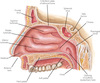
  • Upper respiratory tract, consisting of:
    • Nostrils
    • Nasal cavity
    • Pharynx
    • Epiglottis
    • Larynx
  • Lower respiratory tract, consisting of:
    • Trachea
    • Bronchi
    • Bronchioles
    • Lungs
2
Q

Where does air enter the respiratory system?

Outline the structure

A
  • Air enters the respiratory system through the nostrils
  • Nostrils are divided by the nasal septum cartilage
3
Q

Describe the entrance of the nasal cavity

A
  • The surface wall consists of:
    • Stratified epithelial cells
    • Sebaceous glands
    • Nose hairs
  • The above serve to filter out inhaled particles
4
Q

Describe the nasal passage

A
  • The walls of the nasal passage are lined with respiratory mucosa
  • Respiratory mucosa is covered by pseudostratified ciliated columnar epithelium which is interspersed with goblet cells
  • Cilia cover the surface of epithelial cells which have rythmic movement (mocucillary action)
  • Goblet cells and mucosal glands are responsible for secreting mucous
5
Q

What is the function of mucous in the respiratory system?

A
  • Mucous traps inhaled particles and the mucocillary action moves the airborne contaminants towards the pharynx (throat)
6
Q

How is the inahled air warmed by the nasal cavity?

A
  • Inspired (inahaled) air is warmed and conditioned by a network of thin walled veins that are located under the nasal epithelium
  • There are several blood vessels in this area which causes the transfer of heat to the colder inspired air
7
Q

What are conchae?`

A
  • Conchae, aka turbinate, are present in the nasal cavity
  • Conchae are lined with mucosa which increases the surface area over which inspired air passes through in the nasal cavity
  • The inspired air is warmed and humidified as it passes over the conchae
8
Q

What else is present in the mucosa of the nasal cavity?

A
  • Nerve endings are located in the nasal mucosa which detect smell (olfactory nerve), pressure, temperature and pain
9
Q

What are paranasal sinuses?

A
  • Paranasal sinuses are located close to the nose inside the frontal, maxillary, sphenoidal and ethmoid bones
  • The paranasal sinuses are lined with a mucous membrane that drains into the nasal cavities which assist in the production of mucous from the respiratory tract
10
Q

Besides the paranasal sinuses, what else drains into the respiratory tract?

A
  • The lacramal sacs also drain into the nasal cavity
    • They are responsible for collecting tears from each eyelid and drain them into the nasal cavity
11
Q

What is sneezing?

A
  • Sneezing generally occurs when an irritant passes through the nasal hairs to reach the nasal mucosa
  • This triggers the release of a histamine which irritates the nerve cells in those
  • This results in an electrical impulse being sent to the brain which initiates the sneeze reflex
  • The brain also simulates the pharyngeal, tracheal and chest muscle to expel a large volume of air from the lungs through the nose and mouth
    • Sneezing can also be triggered by allergies and sinus infection
12
Q

Give some examples of sino-nasal pathologies

A
  • Turbinate / mucosa hypertrophy:
    • terbinate hypertrophy (enlargement) may arise due to inflammation or abnormal developement which results in an increase in volume of the turbinate mucosa
  • Deviated septum:
    • Occurs when the septum has drastically deviated from the midline between the nostrils
  • Nasal polyps:
    • Abnormal nonconcerous growth of tissues projecting from the mucous membrane in the nasal sinus
13
Q

What is the pharynx and what can it be divided into?

A
  • The pharynx (throat) is a tube-like structure that connects the posterior oral and nasal cavities to the larynx and oesophagus
  • Divided into three anatomical portions according to location:
  1. Nasopharynx: posterior to the nasal chambers and located between the internal nares and the soft plate
  2. Oropharynx: posterior to the mouth
  3. Laryngopharynx: posterior to the pharynx
14
Q

Describe the nasopharynx

A
  • At the walls of the nasopharynx are the left and the right Eustachian (auditory) tubes connected to the middle ear
  • The lining of the Eustachian tubes is continuous with the lining of the nasopharynx
  • The connection between the nasopharynx and the middle ear permits the equilisation of air pressure between the middle ear and the exterior ear
  • Tonsils are located in the pharynx and pharygneal tonsils are situated in the nasopharynx
15
Q

What is the purpose of the pharynx?

A
  • Serves as a passageway for both the digestive system and the respiratory system because food and air travels through it
  • Air enters the pharynx from the two nasal cavities and food enters from the mouth
16
Q

How is food and air directed through the pharynx?

Describe this structure

A
  • The epiglottis controls the direction of food from the pharynx down the oesophagus and the direction of air down the trachea
  • The epiglottis is a flap of elastic cartilage that acts as a lid which covers the trachea when food is swallowed to prevent it from entering the larynx
  • During swallowing breathing is momentarily stopped because the soft palate and its uvula point upwards so that neither food nor air can pass through it
17
Q

What is the nasopharynx lined by?

A
  • The surface of the nasopharynx is covered by pseudo-stratified columnar epithelium
  • They also have a simular mechanism of mucous secretion from goblet cells in the epithelium to warm, humidify and filter the inahled air
18
Q

What is the orthopharynx and the laryngopharynx lined with?

A
  • The surface is lined with non-keratinising stratified squamous epithelium which is required as it is exposed to the passage of food unlike the nasopharynx
19
Q

Describe some pathologies of the pharynx

A
  • Pharyngeal cancer: can arises in the squamous epithelial cells of the pharynx which can restrict the airway and alter the natural flow of air in the pharynx
  • Pharyngitis: the inflammation of the pharynx can be acute or chronic with different causes.
    • Can cause swelling of the oropharynx and elnarged tonsils restricting the passageway for breathing and swallowing
  • Tonsillitis: inflammation of the tonsils can be caused by bacteria or viral infection
    • Tonsils become enlarged and this restricts the opening of the airways from the oral cavity to the oropharynx causing breathing and swallowing difficulties
20
Q

Describe the larynx

A
  • The larynx is commonly referred to as the voice box and is situated below the pharynx
  • It is composed of several cartilages (thryoid, cricoid, epiglottis, arytenoid, corniculate and cuneiform) connected by ligaments and membranes
  • The larynx houses the vocal folds that are responsible for phonation (sound production)
  • The space between the vocal cords is the glottis
21
Q

Besides phonation, what else is the larynx responsible for?

A
  • The larynx serves as a sphincter in transmitting the inspired air from the oropharynx to the trachea (and also increating sound for speach)
22
Q

How is the larynx responsible for phonation?

A
  • The expired air released from the lungs passes through the glottis and the vocal cords induces phonation at the larynx
  • Similar to guitar strings, vocal cords in the larynx produce a vibrating sound and the final sound depends on the pharynx, nose, mouth, tongue and lips
23
Q

What is the larynx lined with?

A
  • The larynx is lined with a ciliated mucous that further contributes to the removal of particulates and to warm and humidify the air
24
Q

Describe the larynx during swallowing

A
  • Food is prevented from entering the larynx during swallowing becuase the back of the tongue that is attached to the top of the larynx pushes upwards, forcing the epinglottis to cover the glottis
  • If food items do enter the larynx and come into contact with the vocal cords, the larynx muscles become stimulated and induce a cough reflex in attempt to expel the items to prevent choking
25
Q

Describe some pathologies of the larynx

A
  • Laryngitis: acute and chronic laryngitis can occur due to inflammation and swelling of the larynx, which is generally caused by excessive shouting, viruses or inhalation of contaminated air
  • Laryngomalacia: common during infancy where immature cartilage of the upper larynx collapses inwardly during inhalation causing airway obstruction
  • Presbylarynx: condition involving age related atrophy (degeneration) of the vocal cord tissues in the larynx, causing a weak voice, restricted vocal range and sometime aspiration (swallowing difficulty)
26
Q

Describe the trachea

A
  • The trachea (windpipe), the main airway, is a hollow tube that extends from the larynx in the back of the neck to the bronchi in the chest
    • Trachea is lined with mucosa that consists of pseudo-stritified ciliated columnar epithelium and its submucosa contains cartilage, smooth muscle and seromucous glands
27
Q

What does the trachea consist of?

A
  • Consists of several C-shaped cartilagenous rings and the posterior tracheal membrane
  • Tracheal rings hold and support the trachea in place to prevent it from collapsing in on itself
28
Q

What does the trachea divide into?

A
  • Trachea divides into two bronchi, each bronchus enters the lungs and branches many times into progressivley smaller bronchi which form the bronchioles
29
Q

What are the three main functions of the trachea, bronchi and bronchioles?

A
  1. To warm and humidify the air
  2. To distribute air evenly to the deeper parts of the lungs
  3. To function as part of the body’s defence system by removing noxious gasses, dust and bacteria from the lungs
30
Q

Describe differences in structure as we progress from trachea to bronchi to brochioles

A
  • The cartilage support becomes progressively smaller and less complete
  • As the bronchi become smaller the cartilaginous rings turn into irregular plates of cartilage and they are absent from the bronchioles
  • The epithelium also changes from pseudo-stratified columnar to columnar and then to cuboidal in the terminal bronchioles
  • The amount of smooth muscle in the airway walls increases as the tubes become smaller
31
Q

Describe the bronchioles comapared to the bronchi and the trachea

A
  • The bronchioles do not contain any cilia or mucous producing cells, therefore macrophages are required to ingest any microbes that reach the alveoli
  • Bronchioles are supported by elastic tissue in the lung parenchyma and the elasticity of the lung tissue keeps the airways open
32
Q

Describe the conducting zone of the respiratory system

A
  • The conducting zone has its own distinct circulation called the bronchial circulation, which originates from the descending aorta and drains into the pulmonary veins
  • Gas exchange does not occur in the conducting zone
    *
33
Q

What is the respiratory zone?

A
  • The respiratory zone is the site of gas exchange
  • It consists of respiratory bronchioles which subdivide into alveolar ducts
  • Each alveolar duct ends in several thin-walled air sacs called the alveoli
  • The respiratory zone has its own distinct circulation called the pulmonary circulation
34
Q

Describe the pulmonary circulation of the respiratory zone

A
  • Lungs have the most extensive capillary network relative to any organ in the body
  • Each alveolus is surrounded by a network of capillaries that brings the blood into close proximity with the air inside the alveolus
35
Q

What are alveoli and what are the structural characteristics?

A
  • Alveoli are described as the units of respiration and are the site of gas exchange between the respiratory and the circulatory system
  • There are two structural characteristics of the alveoli which assist in performing this function:
  1. Walls of each alveolus consists of a single layer of squamous epithelial cells and so are the capillaries that surround them
    1. This thin barrier is the respiratory membrane
  2. Each adult lung contains ~300-500 million alveoli which has an enormous combined surface area where large amounts of CO2 and O2 can be exchanged by diffusion
36
Q

What is each alveolus coated with?

A
  • Each alveolus is coated with a substance called pulmonary surfactant which is a lipoprotein
  • Pulmonary surfactant serves to lower the surface tension at the gas-lipid interface and it also helps promote alveolar stability at low lung volumes by preventing them from collapsing as air moves in and out during respiration
37
Q

Describe the lungs

A
  • The lungs are large organs and each lung is completely enclosed in a sack called the pleural sac.
    • The parietal pleura and visceral pleura contribute to the formation of this sack
  • The parietal pleura is attached to the thoracic wall and the visceral pleura covers the lungs
  • Intrapleural fluid lies between the two pleural membranes
    • Intrapleural fluid encloses the lungs and lubricates the two surfaces
    • Changes in pressure of the intrapleural fluid allow the lungs and thoracic wall to move together during ventilation
  • The lungs are housed in the thoracic cavity and are separated from the abdomen by the diaphragm
38
Q

What is the main difference between the left and the right lungs?

A
  • The left lungs has two lobes and the right lung has three lobes
39
Q

Outline some differences in breathing capactiy

A
  • Male breathing capacity > female breathing capacity
  • Tall people breathing capacity > short people breathing capacity
  • People living at higher altitudes have higher breathing capacity than people living at low altitudes
  • Non-smokers breathing capacity > smokers breathing capacity
40
Q

What is the total surface area of the lungs and what is the total length of the airways?

A
  • Lungs have a total SA of ~70 M2
  • The lungs have ~1500 miles of airways
41
Q

What is pulmonary ventilation and what are the differnet phases?

A
  • Pulmonary ventilation refers to the exchange of air between the external environment and the alveoli
  • Inspired air will move in or out of the lungs depending on the pressure of the alveoli
  • Changes in volume of the lungs changes the pressure in the alveoli
  • There are two phases to pulmonary ventilation:
    1. Inspiration
    2. Expiration
42
Q

What is the ventilatory system composed of?

A
  • Composed of the lungs, chest wall, intercostal muscles, pleural lining and diaphragm
43
Q

What is inspiration initiated by?

A
  • Inspiration is initated by contraction of the diaphragm
  • Two phrenic nerves innervate the diaphragm
  • During normal resting inspiration nerve impulses pass through the phrenic nerves which stimulate the diaphragm to contract
  • The contaction of the diaphragm causes the volume of the thoracic cavity to increase
44
Q

How is the volume of the thoracic cavity increased?

A
  • The volume of the thoracic cavity is increased by two ways:
  1. First, when the diaphragm contacts it moves down in the abodomen which causes the abdominal contents to be pushed downwards, enlarging the thorax
  2. Secondly, when the diaphragm contracts the inferior and the thoracic muscles pull the chest outwardly (pushing the rib cage outwardly) causing further enlargement of the thoracic cavity
45
Q

What is the effectiveness of the diaphragm in manipulating the thoracic cavity related to?

A
  • It is related to the diaphragms dome-shaped conformation
46
Q

Explain how changes in the thoracic volume cause inspiration

A
  • During the expansion of the thoracic cavity the lungs are held to the thoracic wall by negative pressure in the pleural cavity
  • As the thoracic cavity enlarges in volume, this also causes the lungs to expand and causes a decrease of pressure inside the lungs
  • The decrease in pressure causes air from the external environment to follow down the pressure gradient intothe alveoli of the lungs where oxygen diffuses into the blood and carbon dioxide diffuses into the alveoli
  • NOTE: if the pressure of the alveoli is lower than the atmospheric pressure air will continue to move into the airways
47
Q

What happens in forces inspiration?

A
  • During forced inspiration accessory muscles such as external intercostal muscles are also used which causes the rib cage to be pulled upwards and outwards
48
Q

Outline expiration

A
  • During resting ventilation, expiration is a passive process and no muscles are contracted
  1. When the lungs are expanded and stretched, stretch receptors in the alveoli send inhibitory impulses to the medulla oblongata
  2. Medulla oblongata stops sending impulses to the diaphragm to contract
  3. The lungs are elastic, therefore when the diaphragm and intercostal muscles relax this causes elastic recoil which creates a positive pressure
  4. Pressure in the lungs becomes greater than atmospheric pressure and air moves down the pressure gradient and is expelled from the lungs
49
Q

Describe how forces expiration is an example of voluntary control

A
  • The respiratory system is essentially involuntary and is regulated by the medulla oblongata
  • During exercise, expiratory muscles become active - forced expiration
  • These muscles include the diaphragm, the intercostal muscles and muscles in the abdominal wall
  • Acessory respiratory muscles are essential for functions such as: defecating, straining, coughing, sneezing and vomiting
  1. Contraction of the abdominal muscles causes the abdominal pressure to increase which forces the relaxed diaphragm up into the chest
  2. Intercostal muscles pull the rib cage down to reduce the thoracic volume causing air to be forced out of the lungs
50
Q

What is external respiration?

A
  • It is the exchange of gasses between the inpsired air of the alveoli and the blood within the pulmonary capillaries
  • Oxygen diffuses from the alveolar air into the blood and carbon dioxide diffuses out of the blood into the alveolar air
51
Q

What is barometric pressure (PB)?

A
  • Barometric pressure is the term used to describe the pressure exerted by the atmosphere
52
Q

What is the Dalton Law?

A
  • The Dalton law governs the relationship between total pressure produced by a mixture of gasses and the pressure of individual gasses
  • The law states that:
    • Total barometric pressure = sum of partial pressurs of the individual pressures
53
Q

What is the measurement of partial pressures used for?

A
  • The measurement of partial pressures is critical in the diagnosis and treatment of many disease conditions
54
Q

Describe the significance of partial pressure gradients regarding external respiration

A
  • Diffusion of gasses in external respiration is a passive process and gasses move from an area of high partial pressure to an area of low partial pressure down the partial pressure gradient
  • Oxygen is continuously consumed by body cells during cellular respiration therefore when doexygenated blood flows into the pulmonary arteries it has a low partial pressure relative to the alveolar air
    • Oxygen diffuses down the partial pressure gradient from the alveolar air to the pulmonary capillary blood
  • Blood in the pulmonary capillaries has a higher Pco2 (produced during cellular respiration) relative to alveolar air therefore CO2 diffuses from the pulmonary capillaries to the alveolar air
55
Q

What is internal respiration?

A
  • Internal respiration is the exchange of gasses at the cellular level
  • During this process oxygen diffuses out of the oxygenated blood in the systemic capillaries into the interstitial fluid down a partial pressure gradient
  • The supply of oxgen to cells is paramount for aerobic cellular respiration
  1. During internal respiration oxygenated blood is changed to deoxygenated blood in the systemic capillaries as oxygen diffuses into the cell
  2. Carbon dioxide, a by-product of cellular respiration, diffuses from an area of high partial pressure to an area of low partial pressure from the cells to the systemic capillaries
56
Q

Briefly outline the transport of gasses

A
  • Oxygen and carbon dioxide are transported in the blood in either a dissovled state or combined with other chemicals
  • Upon entering the blood both oxygen and carbon dioxide are dissolved in the plasma but most of the O2 and CO2 rapidly binds with haemoglobin
    • This is because fluids, such as blood plasma, can only hold small amounts of gas in solution, therefore once oxygen and carbon dioxide are bound to haemoglobin the partial pressure decreases and more gas can diffuse into the plasma
57
Q

How is oxygen transported to tissues?

A
  • Oxygen is transported to tissues in two forms: physically dissolved in blood (2%) or combined with haemoglobin (98%)
  • RBCs contain haemoglobin which binds to oxygen for transport from lungs to other organs and tissues
58
Q

Describe haemoglobin

A
  • It exists in one of two forms:
    • Deoxyhaemoglobin (Hb) - not bound to oxygen
    • Oxyhaemoglobin (HbO2) - bound to oxygen
  • Each haem molecule contains four oxygen binding haem sites and a globular protein chain
  • Oxygen binds reversibly to haemoglobin
    • O2 + Hb ↔ HbO2.
  • In pulmonary capillaries the PO2 is high so the reaction shifts to the right
  • In systemic capillaries the PO2 is low and shifts to the left
59
Q

With regards haemoglobin, what does oxygen content and oxygen-carrying capacity refer to?

A
  • Oxygen content - the amount of oxygen bound to haemoglobin
  • Oxygen-carrying capactiy - the amount of oxygen that can potentially bind to haemoglobin
60
Q

What is the oxyhaemoglobin equilibration curve?

A
  • AKA oxygen dissociation curve
  • Depicts how plasma PO2 affects the loading and unloading of oxygen from haemoglobin
  • The oxyhaemoglobin equilibration curve is an S shapred curve over a range of arterial oxygen tensions
61
Q
A
62
Q

What are the different phases of the oxyhaemoglobin equilibration curve?

A
  • Loading phase: the plateau region of the curve in which oxygen is loaded onto haemoglobin to form oxyhaemoglobin in the pulmonary capillaries
  • Unloading phase: the steep region of the curve in which large quantities of oxygen is unloaded from haemoglobin in the systemic capillaries
63
Q

What is the significance of the S shape of the oxyhaemoglobin equilibration curve regarding the saturation of haemoglobin?

A
  • The S shape enables oxygen to saturate haemoglobin under high partial pressures in the lungs and release large amounts of oxygen with lower capillary PO2 at the tissue level
64
Q

What factors affect the binding of oxygen to haemoglobin?

How does this affect the oxyhaemoglobin equilibration curve?

A
  • Arterial pH
  • Blood pressure
  • Arterial carbon dioxide tension (Bohr Effect)
  • A change in the binding affinity of haemoglobin for oxygen shifts the oxyhaemoglobin-equilibrium to the left or to the left
65
Q

How is carbon dioxide removed from the body?

What are the three ways of achieving this?

A
  • CO2 is removed from the body during expiration, for this to happen the CO2 must be transported back to the lungs by one of three ways:
  1. Dissolved in the blood plasma
  2. Combined with haemoglobin to form carboxyhaemoglobin (HbCO2), CO2 competes with O2 for the same binding site on Hb
    1. Hb + CO2 ↔ HbCO2
  3. Majority is transported to the lungs as HCO3- ions. When the blood enters the pulomary capillaries HCO3- and H+ are converted to carbonic acid H2CO3 which dissociates to CO2 and H2O
    1. This consumes H+ and raises blood pH permitting Hb to bind to more oxygen molecules
66
Q

What is used to measure the amount of air exchanged in breathing?

What is the recording known as?

A
  • A spirometer - a volume recorder
  • The recording is known as a spirogram
67
Q

What is lung space?

A
  • Lung space is defined by using four volumes and four capacities:
  1. Tidal volume: volume of air leavingthe lungs during 1 breath
  2. Inspiritory reserve volume: max volume that can be inhaled above the tidal volume
  3. Expiratory reserve volume: max volume that can be expired after the expiration of a tidal volume
  4. Residual volume: volume left after the lungs have reached maximum expiration
  5. Functional residual capacity: vol air remaining in lungs at the end of expiration
  6. Inspiritory capacity: volume of max inhalation
  7. Vital capacity; max volume of air that can be exhaled after a max inspiration
  8. Total lung capacity: max amount of air in the lungs at the end of max inhalation
68
Q

What are chemoreceptors and how do they affect ventilation?

A
  • The carotid and aortic bodies are distinct structures located in the carotid sinus and aortic arch which detect changes in blood oxygen, pH and carbon dioxide
  • These structures are referred to as chemoreceptors
  • The carotid and arotic chemoreceptors influence ventilation and affect the cardiovascular system through neurogenic reflexes
  • Peripheral chemoreceptors send impulses to the respiratory centres and cause an increase in firing of these impulses when there is a decrease in PO2 of arterial blood, a decrease in PCO2 of arterial blood or an increase in blood acidity
  • Respiratory centres, located in the pons and medulla, modify respiratory rates
69
Q

What does chronic obstructive pulmonary disease (COPD) refer to?

A
  • It is a term used to refer to a large group of lung diseases characterisd by an obstruction of airflow which interferes with ventilation.
    • Emphysema and chronic bronchitis are important conditions that impose COPD and they can coexist
      • Asthma is also a type of COPD
70
Q

Outline asthma including causes, mechanism and symptoms

A
  • Asthma is a respiratory disease of the bronchi and bronchioles and is also classified as an allergic disorder
  • The airways are extremely sensitive to irritants including tobacco, pollen and dust
  • When exposed to an irritant the smooth muscle in the bronchioles undergoes muscle spasm
  • Several asthma patiens have some degree of bronchial inflammation that reduced the diameter of the airways and contributes to the seriousness of the attack
  • Symptoms include: wheezing, shortness of breath and coughing up mucous
71
Q

What is emphysema and what is it caused by?

A
  • It is a condition of the lung characterised by abnormal and permanent enlargement of the alveoli accompanied by alveolar cell wall death and/or failure of alveolar wall maintainance
  • Chronic inflammation and oxidative stress contributes to increased destruction and/or impaired lung maintainance and repair
72
Q

What does emphysema stem from and what is the result of this?

A
  • It stems from the loss of elastic recoil and over-distention of alveoli
  • Destruction of the alveoli leads to a decrease in blood oxygen levels and an increase in blood CO2 levels
    • This causes shortness of breath which becomes more pronounced as the condition progresses
  • The destruction of elasticity makes it diffucult to exhale
73
Q

What are some causes of emphysema?

A
  • The most common cause of emphysema is cigarette smoking and exposure to environmental toxins
  • Some patients with emphysema inherit a form of the disease due to a deficiency in a protein called alpha-1-antitrypsin
74
Q

How is emphysema treated?

A
  • Emphysema is a permanent condition which is irreversible although there are some medications available to relieve the symptoms
75
Q

What is bronchitis?

A
  • Bronchitis is a respiratory condition that results in inflammation of the bronchial tubes and bronchioles
  • Chronic bronchitis refers to chronic inflammation of the bronchi and bronchioles that causes persistant coughing and production of phlegm (sputum) and mucus
76
Q

What does bronchitis result in and what is it caused by?

A
  • It results in excessive secretions of mucus by goblet cells and tissue swelling that reduces the diameter of the bronchial tubes making it extremely difficult to breathe
  • It is caused by an interaction between noxious inhaled agents and host factors, such as resiratory infections or genetic predispositions, which stimulates injury or irritation of the respiratory epithelium of the walls and lumen of the bronchi and bronchioles
77
Q

How are environmental toxins important in bronchitis and what is seen regarding cilia?

A
  • Exposure to environmental toxins is thought to stimulate an immune response which stimulates inflammation against allergens and excessive mucous production
  • A decrease in number and length in cilia is seen in chronic bronchitis
    • Without functioning cilia, a continuous blanket of mucous lines the respiratory tract which is difficult to mobilise and swallow
78
Q

What changes occur when chronic bronchitis progresses?

A
  • Ciliated cells are replaced with goblet cells which results in more mucus being produced