Week 4: Neuroanatomy Flashcards
What are the three principlal tissue layers that protect the CNS? Describe each of them.
The meninges include:
Dura mater: outermost and toughest
Arachnoid mater: middle, spiderweb-like layer
Pia mater: innermost and most delicate (direct contact with brain tissue)
What is between the pia and arachnoid mater and what does it contain?
The subarachnoid space is the space between these layers, anc contains CSF
What are the four main lobes of the brain and what is their function?
Frontal lobe: execution, function, cognitive control, movement
Temporal lobe: memory
Parietal lobe: sensory information processing
Occipital lobe: vision

What are the sections of the brainstem and what are the main functions of the brainstem as a whole?
The thalamus, midbrain, pons and medulla oblongata are the sections of brainstem from top to bottom.
The general function of the brainstem is:
1) Pathway for sensory and motor information to and from the cortex/body
2) Provides main motor and sensory nerve supply to the face and neck via cranial nerves
3) Pathway for information entering and exiting the cerebellum (via cerebellar peduncles)

What are the nerve segments of the spinal cord?
Cervical nerves
Thoracic nerves
Lumbar nerves
Sacral nerves
Why do we conduct spinal taps at the L3-L5 segments of the spinal cord? What is the name for the region of nervous tissue in this area?
The end of the spinal cord cuts off at the T12 vertebra, with the nerves below that area belonging to the cauda equina (horse’s tail). These are free nerves attached to the spinal cord, and a lumbar puncture will not “nick” the cord if done at this level.
What are the main shapes found in the brain? When do they occur?
Gyri (ridges)formduring fetal and neonatal development, and thesulci (depressions/grooves) form simultaneously.
What are the four main folding structures on the temporal and parietal lobes that are connected, and what do they do?
The Sylvian fissure, superior temporal gyrus and angular gyrus are all found temporally on the cerebellum. The supramarginal gyrus (the other curve in the photo) is also key.
Angular gyrus = language, reading and writing interpretation
Supramarginal gyrus = somatosensory cortex, limb location and interpretation of tactile sensory data

How do you find the central sulcus from a sagittal section of brain?
Find the cingulate gyrus (pink), and trace the cingulate sulcus (above the gyrus) to the marginal branch of the cingulate sulcus. Then, move one sulcus anteriorly to find the central sulcus.

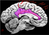
What are the functions of the gyri near the central sulcus?
Precentral (red) and postcentral (blue) gyri are the primary motor and primary sensory cortices in the brain
What is the paracentral lobule? Where is it located?
The paracentral lobule is where sensory and motor information are integrated, and it bridges the central sulcus deep to the sulcus itself (red curve)

What is the cortical homunculus?
It is the concept that sensory and motor information is “mapped” to certain areas of the brain, for example the feet and toes are within the central region, whereas the eyes, nose, face, lips and tongue are located laterally

What is the importance of the insular cortex?
It is an ancient structure, deep to the lateral sulcus, responsible for everything from compassion and empathy to consciousness, emotion and homeostasis
What is the primary auritory cortex, what is another name for it, and where is it located?
The primary auditory complex, or Heschl’s gyrus, is located posteriorly to the insula (insular cortex), and is responsible for integration of auditory information from the environment. It sits deep to the temporal lobe.

Which aspect of the cortex is “deepest,” and what special cells lie within this aspect? What layer do we find them in?
The fifth layer (V) of the primary cortex contains Betz cells, specialized pyramidal cortical neurons that send motor signals down the spinal cord

Which components of the primary sensory cortex receive sensory information from the skin?
3b and 1

Which components of the primary sensory cortex receive sensory input from the joints and muscles?
3a and 2

What aspects of Brodmann’s areas take in sensory information, and what occurs after that information is processed?
Sensory information is taken in at the primary somatic sensory cortex (BAs) 3, 1 and 2, and is then sent to the posterior parietal cortex (areas 5/7).
Then, that information travels all the way to the premotor cortex (area 8), and the supplemental motor cortex (area 6), then into the primary motor cortex (area 4), and down to the spinal cord to initiate the motor response.

What are the main parts of the limbic system, and how do we remember them?
Limb-ic (like a bird on a limb) “HATCH”
Hippocampus
Amygdala
Thalamus
Cingulate gyrus
Hypothalamus
+ Mammillary body and Fornix

What is the Circuit of Papez and what is the path followed?
The Circuit of Papez is the path information takes through the limbic system. Information comes in through the amygdala, moves through the hippocampus, then through the fornix, through the mamillary bodies, then to the thalamus (it goes further than this, but that’s all we need to know)
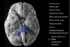
What is the cerebellum and where is it located?
The cerebellum regulates motor movement, controlling posture, balance, coordination and speech. This results in smooth and fluid motor movement.
It is located posteriorly in the brain, in the inferior aspect of the hindbrian.

What is the basis pedunculi and where is it located?
The basis pedunculi (cerebral peduncle) attach the cerebrum to the brainstem, and are located medially, inferiorly, and centrally in the brain (near the midbrain).

What is the pons and where is it located?
Located medially and at the top of the brainstem, the pons relays information from the forebrain to the cerebellum. The pons works to control basic functions including sleep, respiration, swallowing, bladder control, hearing, equilibrium, taste, eye movement, facial expressions, facial sensation, and posture.

What are the pyramids of the brain and what do they do?
The pyramids are located on the anterior side of the brainstem and contain motor fibers that pass from the brain to the medulla oblongata and spinal cord.

What are the white and gray matter areas of the brain and what are their respective functions?
Gray matter (mostly external): contains cell bodies, dendrites, and axon terminals of neurons
White matter (mostly internal): made of axons, connecting parts of gray matter to one another
What are the key white matter bundles in the brain and what are their functions?
The corpus callosum connects the left and right hemispheres of the brain
The anterior commissure connects the anterior parts of the temporal lobe (amygdala, etc.), contributing to the role of memory, speech, and hearing.
The posterior commissure is the inferior element of the pineal gland, and connects the language processing elements of the brain

What key white matter bundle connects the spinal cord with key gray areas of the brain?
The internal capsule moves through the mid anterior and posterior elements of the brain (central portion), and fans out to connect the spinal cord with gray matter regions of the brain.

Identify regions A-F

A - Medulla
B - Pons
C - Midbrain
D - Thalamus
E - Anterior commissure
F - Corpus callosum

What is the septum pellucidym and what are the septum nuclei? Where are they located?
The septum pellucidum separates the left and right ventricles of the brain.
The septal nuclei assist with the generation of theta waves in the hippocampus, and play a role in reward and reinforcement in animals.

Identify brain regions A-J

A - Occipital lobe
B - Cingulate cortex
C - Cerebellum
D - Corpus callosum
E - Thalamus (major sensorimotor relay point)
F - Hypothalamus (homeostasis, ANS, temperature, regulates pituitary
G - Anterior commissure
H - Tectum
I - Tegmentum
J - Pons

Identify A - G

A - Corpus callosum
B - Temporal lobe
C - Frontal lobe
D - Internal capsule
E - Lateral ventricles
F - Cingulate gyrus
G - Caudate (medial) and putamen (lateral)

What meets at the “bottom” of the “crab claw” formed by the caudate and putamen, and what does it do?
The nucleus accumbens (ventral striatum) meets at this point, and plays a central role in the reward circuit. Its operation is based chiefly on two essential neurotransmitters: dopamine, which promotes desire, and serotonin, whose effects include satiety and inhibition.

What gray matter usually contacts the ventricles?
The caudate nucleus

What is this structure and what is it used for?

The globus pallidus is used for voluntary movements, with primary input from the dorsal striatum and moving towards the thalamus.

What is this structure and what is it’s significance?

The anterior commissure connects the temporal lobes

Name this structure

Hypothalamus
Name A - D

A - Lateral ventricle
B - Caudate
C - Putamen
D - Globus pallidus

Name A - E

A - Thalamus
B - Hypothalamus
C - Amygdala
D - Hippocampi
E - Mammillary bodies

What is the structure referenced here, and what is it’s function?

The caudate nucleus is involved in planning the execution of movement. It starts more anteriorly and loops around towards the posterior, and can be seen twice in some coronal sections
What are the purple, pink, and turquoise structures referenced here, and what are their functions?
Purple - the thalamus is a combination of nuclei that work together to synthesize conscious perceptions and alertness
Pink - the medial geniculate nucleus (MGN) of the thalamus processes auditory sensory information
Turquoise - the lateral geniculate nucleus (LGN) of the thalamus processes visual sensory information

What structure is referenced here and what is it’s function?

The posterior commissure is important in language processing and connects the language processing centres of both cerebral hemispheres.
How do we identify the hippocampus, most commonly?
They are squiggly structures lateral to the midbrain that act as elements of the limbic relay system. It plays a major role in learning and memory. “Hippocampus” derives from the Greek word for seahorse (similar shape-wise)
Identify structures A - D

A - Lateral ventricles (atrium (post) + anterior horn)
B - 3rd ventricle
C - Occipital lobe
D - Cingulate gyrus

Identify structures A - D

A - Cerebellum
B - Globus pallidus
C - Putamen
D - Caudate nucleus

Identify structures A - C

A - Insular cortex
B - Internal capsule (anterior and posterior limbs + genu)
C - Corpus callosum (genu)
Genu = bend in CC and internal capsule components (literally, “knee”)

What are the main functions of the ventricular system?
To produce and circulate cerebrospinal fluid
What is CSF? Where is it made?
Cerebrospinal fluid (CSF) is produced by the choroid plexus within the ventricles, and acts as a buffer that protects the brain, regulates cerebral blood flow, and clears waste
What are the main ventricles of the brain?
2 lateral ventricles
3rd ventricle
cerebral aqueduct
4th ventricle
What occurs when cerebral fluid flow is blocked? What are the two kinds of issues that arise?
Hydrocephalus occurs, which is when the ventricles fill with too much CSF.
Communicating hydrocephalus occurs when normal flow of CSF is occurring, but CSF is not being reabsorbed
Obstructive hydrocephalus occurs when normal flow of CSF is impaired due to congenital malformations, tumors, etc. (VERY dangerous)
Name structures A - C

A - Internal carotid arteries (brain)
B - Vertebral arteries (brain)
C - External carotid arteries (head/neck)

Identify A - L

A - Vertebral artery
B - Anterior inferior cerebellar artery (AICA)
C - Basilar artery
D - Pontine arteries
E - Superior cerebellar artery
F - Posterior cerebral artery
G - Posterior communicating artery
H - Internal carotid artery
I - Middle cerebral artery
J - Anterior communicating artery
K - Anterior cerebral artery
L - Posterior inferior cerebellar artery (PICA)

Identify structures A - K

A - Vertebral artery
B - Posterior inferior cerebellar artery (PICA)
C - Basilar artery
D - Anterior inferior cerebellar artery (AICA)
E - Superior cerebellar artery
F - Posterior cerebral artery
G - Internal carotid artery
H - Middle cerebral artery
I - Anterior communicating artery
J - Anterior cerebral artery
K - Circle of Willis

What is the significance of the Circle of Willis?
The Circle of Willis connects the vessels of the anterior cerebrum, and allows fluid “communication” of blood. This prevents occlusion issues, such that if blood is blocked from one route, it can be supplied via other routes.
What arteries supply the yellow, red, and blue areas of the brain?

Yellow - anterior cerebral artery
Red - middle cerebral artery
Blue - posterior cerebral artery

What creates the blood brain barrier?
Tight junctions, rather than pore passages lined with water (as in normal BVs), as well as glial cells
Glucose, nucleic acids, vitamins, AA precursors, hormones and other small lipophilic molecules can pass through the BBB