Week 4 Flashcards

1
Q

What are three reasons for a kidney transplantation rather than dialysis?

What are the indications for a kidney transplant?

What are contraindications for a kidney transplant (3)?

A
  • Rationale: They live longer, better quality of life, and cheaper than dialysis (cost is taken out in three years)
    • Best survival: DR > B > A
  • Indications: Creatinine clearance < 20 ml/min
  • Contraindications for receiving: infection, malignancy within (2-5 years ago), cardiovascular issues
2
Q

What is important to do before you give a kidney transplant?

A
  • Pre-transplant immunological testing is critical (This is here because Arsh did not know this.)
3
Q

What are three decisions and factors you need to look at when figuring out how to ensure the best kidney transplant?

A

Decision-making on amount of immunosuppression

Assess non-immunological risk

Evaluate quality of origin

4
Q

For immunological risk, what are some groups who are are more immunosuppressed?

What do you do if there was a previous Calcineuirin inhibitor nephrotoxicity?

What do you do if there is a higher PRA?

A
  • Assess immunological risk
    • If previous CNI nephrotoxicity à consider belatacept
    • Degree of HA sensitization (higher PRA à more immunosuppression)
    • African Americans are more immunosuppressed
    • Older people are more immunosuppressed
5
Q

Who do yoi need to be concerned with?

Who do you not give steroids to?

Who do you not give sirolimus to?

What do you give for skin cancer to immunosuppress?

A
  • Assess non-immunological risk
    • Pre-existing kidney disease: risk of recurrence
    • Don’t give steroids to fat people
    • Do not give sirolimus to patients with pre-existing hyperlipidemia
    • If they have skin cancer à sirolimus
    • Do not give steroids to people with osteoporosis
6
Q

What are the three factors that determine the quality of an organ?

A
  • Evaluate quality of organ
    • Older people have shittier organs
    • Longer preservation time of organ
    • Reduced nephron mass in donor organ
7
Q

What is the importance of individualized immunosupression?

What is the most common cause of death?

A
  • Importance of individualized immunosuppression
    • Prevent allograft rejection and avoid over immunosuppression (i.e. old black people)
    • Most common cause of death: cardiovascular issues
8
Q

What is the short term graft survival?

What are the acute rejection rates?

What is the long-term outcome with a pre-emptive transplant or a living donor?

A
  • Short term graft survival: high (90-95%)
  • Acute rejection rates: low (10-15%)
  • Improved long-term outcome with pre-emptive transplant (transplant before dialysis)
  • Improved long-term outcome with living donors as opposed to a corpse
9
Q

What is the early cause of graft failure? The late cause of graft failure?

A
  • Causes of graft failure
    • Early: dehydration, ATN, rejection, CNI nephrotoxicity
    • Late: chronic allograft nephropathy (fibrosis), cardiovascular disease
10
Q

What are long term risks to the donor?

What are the contraindications for donating?

A
  • Long term risks to donor: HTN, proteinuria

Contraindications: Kidney disease, HTN, infections, malignancies, mental considerations

11
Q

What are the types of RBC antigens that are important to match in transplant?

What is the result of anti-ABOs? Is Rh factor important?

A
  • RBC antigens
    • Blood group and RBC antigens (Lewis, Kelly)
      • Anti-ABOs are IgM cause agglutination, complement, and hemolysis
      • Rh factor is not expressed and does not play a role in graft rejection (i.e. A+ can give organ to A-)
12
Q

What occurs with WBC antigens in a kidney transplant?

A
  • WBC antigens
    • Complement-Dependent cytoxicity: place donor lymphocytes with recipient serum (complement) to check if allograft donor’s lymphocytes attack recipient’s cells → turning on complement pathway
13
Q

What are the MHC antigens (Class I and Class II)?

A
  • MHC class I: HLA A, B, and C (found on all nucleated cells – T & B cells)
  • MHC class II: HLA DP, DQ, and DR (found only on immunological cells – only B cells)
14
Q

How does floy cytometry crossmatch occur to ensure that the match is correct?

A
  • Flow cytometry crossmatch:
    • DC3-PerCP antibody checks for T-cells (red light)
    • CD19-PE antibody checks for B-cells (orange light)
    • IgG checks for antibodies attached to the MHC class type (green light)
15
Q

How does PRA work?

A
  • Panel Reactive Antibody (PRA)
    • Check recipient’s serum with that of 100 donor lymphocyte samples to check sensitivity (0% = compatible, 100% = bad)
16
Q

What are minor histocompatibility antigens?

A
  • Minor histocompatibility antigens: peptide sequences on the MHC complexes of the organ donor
17
Q

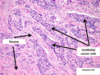
What are the three types of tranplant rejection and provide a timeline for each one?

A
  • Hyper-acute rejection (minutes to hours after revascularization)
    • Antibody-mediated, cell disruption, platelet margination, complement activation, thrombosis
  • Acute rejection (early – days to weeks after transplant)
    • Cell-mediated or antibody mediated
  • Chronic rejection (months to years after transplant)
18
Q

Provide the phases of immunosuppression.

A
19
Q

What are the events following transplant?

A
20
Q

How are T-cell activated?

A

How are T-Cells activated?

  • Antigen is recognized by the MHC complex (signal 1) → CD80/86 (antigen) binds to CD28 on T-cell to start co-stimulation (signal 2) → Calcineurin is activated and acts as a transcription factor to activate IL-2 (signal 3) → IL-2 causes T-cell proliferation
    • Can be regulated by apoptosis, when mTOR is activated
21
Q

Provide the MOA, Use, and SE for corticosteroids?

A
  • Corticosteroids
    • MOA: blocks cytokine gene expression and blocks T-cell activation
    • Use: Induction maintenance and anti-rejection
    • SE: Cushing’s, Osteoporosis, Obesity
22
Q

What are three types of antirejection treatments?

A
  • Anti-rejection treatments
    • Intravenous immunoglobulin (IVIG)
    • Plasmapheresis
    • Rituximab
      • MOA: CD20 antibody that destroys B-cells
      • SE: allergic reaction
23
Q

What is the type of drug, MOA, and SE of belatacept?

A
  • Fusion protein (CTLA-4-Ig)
    • Belatacept
      • MOA: CTLA-4-IgG → binds to CD80/86 → blocks co-stimulation → downregulation of T-Cells
      • SE: anemia, neutropenia
24
Q

What is the type of drug, MOA, and SE of Mycophenolate Mofetil (MMF)?

A
  • Anti-proliferative agents
    • Mycophenolate Mofetil (MMF)
      • MOA: blocks inositol monophosphate dehydrogenases → blocks DNA synthesis → blocks WBC synthesis
      • SE: Bone marrow suppression, diarrhea, CMV infection
25
What is the type of drug, MOA, and SE of azathioprine?
* Anti-proliferative agents * Azathioprine * MOA: purine analog that blocks purine synthesis → less DNA available for WBC synthesis * SE: skin cancer, lymphoma, pancreatitis/hepatitis * Drug interactions: interacts with allopurinol
26
What is the type of drug, MOA, and SE of sirolimus?
* mTOR inhibitor - maintenace drug * Sirolimus * MOA: Inhibits mTOR → blocking T-cell proliferation → permits apoptosis * SE: less nephrotoxicity than CNI, dyslipidemia, thrombocytopenia
27
What is the type of drug, MOA, and SE of sirolimus?
* mTOR inhibitor - maintenace drug * Sirolimus * MOA: Inhibits mTOR → blocking T-cell proliferation → permits apoptosis * SE: less nephrotoxicity than CNI, dyslipidemia, thrombocytopenia
28
What is the type of drug, MOA, and SE of cyclosporine, tacrolimus?
* Calcineurin inhibitors (CNI) - maintenance drugs * Cyclosporine, Tacrolimus * MOA: inhibit calcineurin → IL-2 transcription → blocks T-cell activation and blocks apoptosis * SE: Hyperkalemia, Hyperlipidemia (esp. cyclosporine), renal insufficiency, hypophosphatemia
29
What is the type of drug, MOA, and SE of anti Anti-CD52-antibody?
* Anti-CD52-antibody (Alemtuzumab) * * Type: inudction/immune modulation * MOA: CD-52 antibody causing lysis of T-cells and B-cells * SE: cytokine release syndrome, long-term depletion of lymphocytes
30
What is the type of drug, MOA, and SE of anti IL-2 antibodies?
* Anti-IL-2 receptor antibodies (Basiliximab) * Type: inudction/immune modulation * MOA: anti CD-25 (IL-2 receptor) antibody → blocks T-cell proliferation * SE: minimal
31
What is the type of drug, MOA, and SE of anti-thymocyte globulin?
* Anti-thymocyte globulin (rATG – Thymoglobulin): destroys T-cells * Type: inudction/immune modulation * MOA: polyclonal rabbit antibody for CD3 receptors → T-cell destruction * SE: Serum sickness, anaphylaxis, cytokine release syndrome
32
What are the three receptors in the bladder * Provide their receptor type, location, and action on the bladder
* M3 * Receptor type: Gq * Location: Detrusor (SMC surrounding bladder lumen) * Action: contraction leading to “voiding” aka peeing * Beta2/3 * Receptor type: Gs * Location: Detrusor (SMC surrounding bladder lumen) * Action: relaxation leading to “filling” aka not peeing * Alpha-1 * Receptor type: Gq * Location: Sphincter at bladder exist * Action: contraction leading to “filling” aka not peeing
33
What are the 4 types of incontinence and provide descriptions of each along with treatments?
* Urge (OAB) – overactivity of the detrusor/ inappropriate contractions during filling phase * Tx: M3 antagonists, Beta-3 agonists, alpha-1agonists * Stress – incompetent sphincter aka messed up alpha-1 receptors * Tx: Alpha-1 agonists aka pseudoephedrine or phenylephrine * Overflow – bladder is volume overextended → detrusor muscle cannot contract properly aka neurogenic bladder * Tx: alpha-1 antagonists aka -osin drugs, muscarinic agonists * Functional – couldn’t make it to the loo * Tx: run faster
34
What are two drugs for overactive bladder? Provide MOA and SE.
* Oxybutynin * MOA: muscarinic antagonist approved for OAB * SE: “Can’t see, can’t pee, can’t spit, can’t s\*it,” * Contraindications: elderly, prostate cancer, glaucoma, K+ tablets causes GI bleeds * Mirabegron * MOA: beta-3 agonist → increase storage capacity during filling phase * SE: minimal
35
For UTI, provide clinical features, lab features, risk factors, and etiology.
* Clinical feature: dysuria, frequency, urgency * Lab features: pyuria, bacteruria * Risk factors: sexually active, multiple partners, female, spermicides, preganacy * Etiology: E. coli (most common), Staphylococcus saprophyticus, Enterobacteriaceae
36
What are the three abx indicated for UTI?
* Trimethoprim-Sulfamethoxazole * Fluroquinilones * Nitrofunatoin
37
Provide MOA, SE, contraindications, and drug interactions for Trimethoprim-Sulfamethoxazole?
* Trimethoprim-Sulfamethoxazole * MOA: * Sulfa drug: acts on PABA * Trimethoprim: acts on dihydrofolate reductase * Together these drugs blocks folate synthesis in bacteria and are together are bactericidal * SE: G6PD, steven-johnson, photosensitivity, * Contraindications: G6PD deficiency, renal/hepatic impairment, pregnant/nursing (displacement of bilirubin) * Drug interactions: warfarin (bleeding), ACE/ARBs (hyperkalemia)
38
For fluoroquinilones, provide an example, MOA, SE, and contraindications?
* Fluoroquinolones * Example: Ciprofloxacin * MOA: DNA gyrase/topoisomerase IV inhibitors * SE: QT prolongations, tendon rupture * Contraindications: pregnant/nursing, seizures
39
For nitrofurantoin, provide use, MOA, SE, and contraindications
* Nitrofurantoin * Use: only for bladder infections (because it is most bioavailable here) * MOA: Interferes with carbohydrate metabolism and cell wall formation * SE: minimal but possible pulmonary issues * Contraindications: hemolytic anemia if G6PD deficient, pregnant/nursing
40
What drug should be used for a pregnant woman with a UTI?
* For pregnant women use cephalosporin
41
What are some non-prescription therapies for UTI (4)?
* Non-prescription therapies * Phenazopyridine – analgesic for urinary tract * Increase fluids * Cranberry juice * Avoid spermicides and multiple partners
42
What are the 5 risk factors for urethelial carcinoma?
* Age (median age at diagnosis: 72) * Gender (men ~4X more likely to be diagnosed) * Cigarette smoking (~half of all cases in men; a third of all cases in women) * Exposure to aromatic amines (aniline dyes) * Schistosoma hematobium infection
43
Provide the layers of the urinary tract from outer to inner? What are the different cell types at each part?
* Micoranatomy of Lower Urinary Tract * Outer to Inner * Smooth Muscle * Lamina Propria – connective tissue, immune cells, blood vessels * Urothelium – topped with umbrella cells * Cell types * Mostly pseudostratified transitional epithelium * Exception: squamous epithelium at distal urethra * Function * Stretch and barrier
44
For bladder cancer, provide the following: * incidence * presentation
Incidence: * More common and deadly in men * Likely recurrence Presentation * Painless intermittent hematuria * Common: bladder irritability, dysuria * Advanced: flank pain, weight loss
45
For bladder cancer, provide the following: * Diangosis * Treatment (based on stage)
Diagnosis * Microscopy cytology – examine urine sediment after bladder washing * Not specific for low-grade cancers * Cystoscopy – scope of the bladder * Blue light can be used to detect flat cancer Treatment * Stage 1 and below: local resections * Stage 2 and above: chemotherapy and bladder removal * Neoadjuvant: MVAC, anti PD-1, anti-CTLA-4, EGFR inhibitor
46
For upper tract urothelium cancer, provide the incidence, presentation, and diagnosis?
* Incidence: 10% of renal neoplasms * Presentation: Urothelial carcinoma of the kidney/ureter * Treatment * Nephroureterectomy * Can also use neoadjuvant chemo
47
For urethral cancer, provide the following: * Incidence * Presentation * Diagnosis * Treatment
* Incidence * Extremely rare * Men more than women * Presentation * Urothelial cell carcinoma is the most common histological type * Diagnosis * Risk factor: multiple UTI * Distal cancers present earlier at lower stage * Treatment * Tumor resection
48
What does MVAC stand for?
\*MVAC = methotrexate, vinblastine, doxorubicin, cisplatin
49
What is indicated by the stages of bladder cancer (stage pTa, carcinoma in situ, stage 1-4)
50
What are features of squamous carcinoma and small cell carcinoma?
* Squamous * Keratin Pearls * Intercellular Bridges * Small Cell * Small cytoplasm with large nuclei
51
What does papillary urothelial cell carcinoma look like on histology?
52
What does invasive adenocarcinoma look like on histology?
53
What does invasive squamous cell carcinoma look like on histology?
54
What does invasive small cell carcinoma look like on histology?
55
What doesinvasive UCC look like on histology?
56
Recognize the clinical scenarios in which voiding dysfunction may cause a danger to the kidneys. (5)
* Incontinence * Urinary tract infections (UTIs) * Bothersome voiding symptoms
 * Hydronephrosis – urine gets backed up into the kidney * CKD / ESRD
57
Passive continence - explain the anatomy and mechanisms
* Tight closure - maintained by urethral mucosa, submucosa, and smooth muscle fibers * Smooth muscle – supplied by autonomic nerves (not well understood)
58
Active continence - explain anatomy and mechanism
External sphincter – somatic nerve (nicotinic receptor) innervated by S2-4
59
Bladder compliance normal verus pathological states
* Normal: increase in volume does not change the internal pressure drastically * Pathological states: low compliance results in a volume increase with accompanied large change in pressure
60
causes (2) and complications of poor bladder compliance
* Causes of poor bladder compliance: * Fibrosis – chronic inflammation, radiation * Myelomeningocele (spina bifida) – neurological issues leading to loss of compliance (not well understood) * Complications: hydronephrosis, UTI (due to static urine), CKD, ESRD
61
Urge incontinence clinical defintion and causes (3)
* Clinical definition * Over activity of the detrusor (smooth muscle that surrounds the bladder) and inappropriate contractions during the filling phase * Causes * Bladder irritation/obstruction (UTI, stones, tumor) * Cerebral cortex lesion or legion of spinal cord *above* the sacral region * Idiopathic
62
Stress incontinence clinical definition and causes (4)
* Clinical definition * Leaking of urine due to increased intra-abdominal pressure * i.e. coughing, sneezing, jumping, lifting heavy objects * Causes * Childbirth * Fibrosis or urethra – due to decreased estrogen * Neurologic lesions *at* sacral cord level * Prostatectomy in men
63
how does normal bladder empty
Detrusor contracts (S2-4 Ach/M3) --\> proximal urethra opens --\> external sphincter opens (S2-4 Ach)
64
Mechanisms of failure to empty
Weak or absent detrusor Bladder obstruction
65
two types of messed up detrusor muscle and their causes
* Neurogenic – poor innervation * Sacral spinal cord or cauda equine * Pelvic nerves going from sacral cord to bladder * Diabetes mellitus * Myogenic – weak muscle * Diabetes mellitus * Chronic overdistention
66
two types of bladder obstruction
* Anatomic * Urethral lumen narrowed/obstructed * Prostate enlargement, stones * Functional
67
functional obstruction Detrusor-sphincter dyssynergia pathophysiology and complications
* Detrusor-sphincter dyssynergia - sphincter does not open appropriately when bladder is contracted * Neurological disconnect * Complications: high-pressure voiding, incomplete bladder emptying, recurrent UTI, hydronephrosis, CKD/ESRD
68
List the major functions of the kidney (beyond just filtration)
Filtration of waste, salt/water homeostasis, acid/base homeostasis, K+ homeostasis, Ca/PO4/VitD/PTH, RAAS, EPO
69
Compare the role of dialysis to that of transplantation
Dialysis: maintains kidney function --\> life support (increased mortality) Transplant: regain kidney function --\> new kidneys, new you (decreased mortality)
70
How does dialysis work?
* Blood is removed from body --\> entered through a semi permeable membrane (with different concentrations of ions) --\> the blood is filtered and inserted back into the body * Water is filtered by altering hydraulic pressures across the membrane * Ions are filtered by altering the concentration gradients across the membrane
71
List the indications for chronic dialysis (5)
Refractory volume overload, HTN, K+/H+ excesses Uremia (pericarditis, encephalopathy, bleeding, N/V, anorexia) Malnutrition (albumin \< 3.5 or decreased protein intake)
72
explain av fistula, temporary access, synthetic graft
* Temporary access: for patients who require transient dialysis for an acute condition * AV Fistula: * Indicated for chronic dialysis patients * Creates a direct connection from arteries to veins allowing for good blood flow * Once the fistula is mature, it can be used for hemodialysis * Synthetic graft * Indicated for chronic dialysis patients who cannot use AV fistulas * Often used for diabetic patients
73
Complications of Dialysis
* Access complications: infections, stenosis, clot, inflammation due to plastic * Non-access complications: UTI, pneumonia, peritonitis, TB * Cardiovascular complications: mortality; endothelial issues, microalbuminuria
74
Compare HD vs PD
* Hemodialysis can be done at home or outpatient (indicated more for passive patients) * Contraindicated in patients without vascular site * Peritoneal dialysis can be done while asleep (indicated for active patients * Contraindicated in patients who have had previous surgeries on peritoneum
75
uncomplicated bacterial cystitis definition, epidemiology, pathophysiology, etiology, complications, tx loong but pretty common sense
* Definition: community-acquired infection in non-pregnant, healthy person * Epidemiology: females; rare in men (anatomical) * Pathophysiology: fecal bacteria colonize vagina --\> bacteria move from vagina to urethra and bladder --\> cystitis if immune system does not succeed * Etiology: most commonly E. Coli; second most common: staph * Complications: most will resolve, but risk of progressing to pyelonephritis * Treatment: Abx (avoids progression and hastens recovery)
76
how does the femal body prevent bacteria from colonizing vagina
* Normal flora: Lactobacillis * Compete with and inhibit growth of other bacteria by maintaining acidic pH (lacto = lactic acid) * Vaginal fluid inhibits bacterial adhesion because of secretory IgA
77
how do we eliminate bacteria from entering the bladder urine and bladder factors
* Urine factors: * Inhibit or kill bacteria: Voiding, low pH, hypertonic urine, salts, urea * Inhibit bacterial adherence: low molecular weight oligosaccharides, IgA * Bladder factors * Antibacterial effects: antibodies, cytokines, PMN leukocytes * Bladder epithelium resists adherence
78
what are host factors that increase risk of cystitis
* Epithelial cell receptivity: some epithelium can bind E. Coli better (Lewis antigens more likely to get UTIs) * Altered vaginal flora due to: spermicides, abx, menopause (increases vaginal pH) * Voiding dysfunction * Diabetes: glucose promotes bacterial growth; often immunosuppressed * Immunosuppression
79
what are bacterial factors tht increase risk of getting bacterial cystitis 3 virulence factors
* Virulence factors causing infection: * Adhesins: promote bacterial attachment to tissue receptors * Type 1 pili --\> associated with cystitis * P pili --\> associated with pyelonephrits * Hemolysins * Absent ability so synthesize nutrients
80
simple cystitis symptoms, labs, treatment when is it complicated? when do you do a pelvic exam
* Symptoms of pyelonephritis: flank pain, fever, feeling ill, pain w/ voiding * Complicated UTI: pregnancy, diabetes, immunosuppression, previous GU problem/surgery, recent abx/cath/hospitalization * Pelvic exam indicated if: UA is normal (if dysuria – pain with urine), vaginitis * Labs * Dipsticks: Leukocyte esterase (produced by neutrophils) and nitrites * Treatment: 3 days of Bactrim or nitrofuranotin
81
Name the bacterial and host factors that increase the risk of cystitis progressing to pyelonephritis.
* Bacterial factors: P pili * Host factors: Pregnancy (causes ureteral dilation/urine stasis), diabetes, immunosuppression, anatomical anomaly (vesicoureteral reflux)
82
vesico-ureteral reflux what is it? epidemiology, complications who should be tested?
* Definition: regurgitation of bladder urine into the upper urinary tract * Epidemiology: \< 1% in healthy children, 29-50% in children with UTI, 33% in sibling of child with reflux (lmao Rahul’s brother), rare in African American (ayooo!) * Complications: cystitis à pyelonephritis (due to regurgitation) à renal scarring à renal failure * Can also lead to hydronephrosis à renal failure * Who should tested? * All children with UTI * All babies with hydronephrosis
83
pyelonephritis symptoms, signs, labs, imaging, tx
* Symptoms: Flank pain, fever, chills * Signs: CV tenderness * Labs * Urinalysis: pyuria, bacteriuria +/- hematuria * Imaging * Lobar nephronia – pyelonephritis with a lobar distribution * Treatment * Outpatient: fluoroquinolone * Inpatient: blood culture, IV antibiotics
84
Lobar nephronia – pyelonephritis with a lobar distribution
85
Name two major complications of pyelonephritis. difference between acute and chronic?
* Complications of acute infection * Sepsis * Premature labor if pregnant * Complications of chronic infection * Renal scarring --\> renal failure
86
what are these things? tx? Pyelonephritis plus obstruction Renal or perinephric abscess Emphysematous pyelonephritis
* Pyelonephritis plus obstruction * Bacteria builds up behind obstruction can lead to sepsis * Tx: drainage * Renal or perinephric abscess * Bacteria builds up in abscess --\> pressure forces bacteria to blood --\> sepsis * Tx: drainage * Emphysematous pyelonephritis * Necrotizing infection with gas forming bacteria --\> sepsis * Usually occurs in diabetics * Tx: nephrectomy
87
Obstructive Uropathy definition and ddx
* Definition: Structural/functional changes in urinary tract secondary to obstruction of urinary flow * DDx: Stone, stricture, uretero-pelvic junction (UPJ) obstruction, tumor, papillary necrosis
88
Obstructive Uropathy Structural Changes
* Can lead to permanent renal damage if not treated (obstructive nephropathy) * Increased collecting system pressure and dilation of urinary tract (hydronephrosis or hydroureter) * Hydronephrosis does not always indicate obstruction * Can also be caused by reflux and post chronic obstruction
89
Obstructive Uropathy Physiological Changes
* Increased intratubular pressure and decreased RBF --\> loss of tubular function * Decreased GFR --\> renal failure * Vasoconstriction/ischemia --\> tubular cell damage * Altered Na/H2O handling and impaired response to hormones (ADH/PTH) * CHECK THE NEW PHYSIOLOGY
90
Obstructive Uropathy Signs/symptoms
* Flank pain, asymptomatic (only if chronic), urinary symptoms, hematuria, nausea, vomiting, fever, chills * Variable: hydronephrosis, renal trophy (parenchyma thinning), increased creatinine, UTI, CVA tenderness, suprapubic tenderness (if bladder obstruction)
91
Unilateral vs bilateral obstruction
* Unilateral * Usually secondary to urolithiasis * Preserved renal function due to one functioning kidney * Bilateral * Usually secondary to benign prostate hyperplasia (BPH) * Compromised renal function (AKI) * Bladder distension and overflow incontinence
92
Nephrolithiasis common natural history? where do these occur?
* Common and likely to recur after first episode * Common junctions of stones (narrowest parts of ureter) * Ureteropelvic junction (UPJ) * Over iliac vessels * Ureterovescial junction (UVJ) – where ureter meets bladders
93
stones: causes, labs, tx, epidemiology Cystine
* Epidemiology: children (~12 y/o); high recurrence rate * Pathophysiology: autosomal recessive AA transport disorder --\> cystinuria * Labs: low urine pH (acidic) * Tx: aggressive fluid intake, low Na diet; alkalization of urine * Resistant to shockwave lithotripsy
94
stones: causes, labs, imaging Struvite
* Cause: UTI * Labs: high urine pH (basic) * Imaging: radiopaque on XR
95
stones: causes, tx, labs, imaging Uric acid
* Causes: hyperuricemia (gout) * Labs: low urine pH (acidic) * Imaging: radiolucent on XR * Tx: alkalinzation of urine, decreased purine intake, potassium citrate, NaHCO3
96
stones: causes, description, labs Calcium phosphate
* Description: powder-like --\> cloudy urine * Causes: distal RTA (Type I) * Labs: elevated urinary pH (basic)
97
stones: causes, imaging, labs Calcium oxalate
* Causes: hypercalcemia, anatomic, commonly idiopathic * Primary hyperparathyroidism --\> hypercalcemia * Imaging: radiopaque on XR * Labs: variable pH
98
Nephrolithiasis when is surgery indicated before 4 weeks types of surgical options?
* Indications for early surgery: * Elevated, non-responsive creatinine, bilateral stones, infected urine, uncontrolled sx, immunocompromised, DM, elderly * Types: shock wave lithotripsy, urteroscopy with laser lithotripsy, percutaneous nephrolithotomy
99
Nephrolithiasis management when do you do surgery
* Spontaneous stone passage * Tx: Fluids, pain management, alpha blockers (relaxes bladder muscles) * If 4 weeks have passed without passing --\> surgery
100
Nephrolithiasis signs/symptoms, labs, imaging, complications
* Signs/symptoms * Acute onset of back pain (only if obstructive stone), nausea, vomiting, intermittent severity of pain (sharp and dull), hematuria * Signs: CVA tenderness * Labs: Urinalysis, CBC * Imaging: * Non-contrast CT abd/pelvis: provides stone location and presence of hydronephrosis * X-ray KUB (kidney, ureter, bladder): presence of stone (opacity) * Renal ultrasound: low sensitivity for location of stone * Complications: obstructive pyelonephritis