Week 3 lectures (hearing and vision) Flashcards

1
Q

What the the stages of the process of perception

A
  1. sensory signal
  2. travels to sensory receptors
  3. Combine past experience and attention to create a
  4. Percept
  5. which leads to a perceptual understanding
  6. Which leads to a behavioural response
2
Q

How does the amplitude of the sound wave affect the sound?

A

greater amplitude=larger sound

3
Q

From what decibels can humans hear sounds?

A

0-140db

4
Q

From what Hz can humans hear sounds?

A

20Hz to 20KHz

5
Q

How does the frequency of the sounds wave influence the sound?

A

Higher frequency= higher pitch

6
Q

Whats the outer ear consist of?

A

The stuff we can easily see and touch

the pinna/auricle
the external auditory canal

7
Q

What is the tymphanic membrana also known as?

A

The eardrum

8
Q

Which parts of the ear does the tymphanic membrane seperate?

A

The outer ear from the middle ear cavity

9
Q

How many bones does the middle ear cavity have and what are they called?

A

malleus

incus

stapes

10
Q

Where are the Auditory hair cells?

A

Cochlear

11
Q

Where are the stapes?

A

In the middle ear, at the entrace to the cochlear

12
Q

What to the stapes do?

A

The stapes are bones which move in a lever like direction to transmit information from the middle ear to the cochlear

13
Q

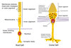
What two types of hair cells are in the cochlear?

A

The inner hair cells (main hair cells involved in detecting auditory stimuli)

the outer hair cells (plays a role in amplifying signals)

14
Q

what two nerves in the ear come from the cochlear and combine to form one of the cranial nerves?

A

the cochlear nerve

the vestibular nerve

15
Q

What are 4 functions of the outer ear?

A

Funnels sound inwards

amplifies the sound by acting as a tube for it to echo in

Shape of the pinna Helps sound localisation in the vertical plane

protection: earwax is water resistant, antibacterial and antifungal
the outer ear has an acidic environment
hairs in the outer ear prevent entry

16
Q

What are two functions of the middle ear?

A
  1. protection- middle ear reflex can lock position of bones to prevent transmission of loud sounds
  2. Acoustic impedance matching- amplifies pressure created by sound wave to prevent loss of signal as it eners fluid filled cochlear

(when sound travels from air filled middle ear to fluid filled cochlear some sound waves are reflected back, meaning signal is lost. The shape of the middle ear increases the pressure created by the sound wave to prevent loss of signal)
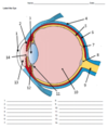
17
Q

what is 1?

A

pinna

18
Q

what is 2?

A

malleus

19
Q

what is 3?

A

The incus

20
Q

What is 4?

A

The semicircular ducts

21
Q

What is 5?

A

The vestibular nerve

22
Q

What is 6?

A

The cochlear nerve

23
Q

What is 7?

A

The cochlear

24
Q

What is 8?

A

The auditory tube

25
What is 10?
The stapes
26
What is 11?
The tympanic cavity
27
What is 12?
The eardrum
28
What is 13?
The auditory canal
29
Where in the cochlear are the inner hair cells located?
in the organ of corti
30
The inner hair cells have 'hairs' of varying lengths, what are they called and what is the tallest one called?
The stereocilia The tallest is the kinocyleium
31
What happens when the stereocilia bend towards the kinocyleium?
The cell becomes excited
32
What happens when the stereocilia bend away from the kinocyleium?
The cell is inhibited
33
When the stereocilia are at rest, what is going on with the k+ gated channels on the stereocilia?
The channels are shut, so no ions can move into the stereocilia
34
What are two types of gated channels on an inner hair cell?
Mechanically gated k+ channels on the stereocilia Voltage gated calcium channels on the main cell body
35
What neurotransmitter does the inner hair cell contain vescicles of?
Glutamate
36
What receptor does the postsynaptic neuron in the auditory nerve, connecting to the inner hair cell contain?
AMPAr receptors (glutamate receptors)
37
What happens in the inner hair cell at the arrival of a soundwave?
1. the soundwave causes fluid in the stereocilia to move 2. this causes the stereocilia to move in the direction of the kynocilium 3. This physically opens the mechanically gated k+ channes 4. There is a high concentration of k+ outside of the cell, so k+ moves into the inner hair cell through the channels (down the concentration and electrostatic gradient) 5. The charge of the cell becomes more positive leading to depolarisation 6. The depolarisation opens the voltage gated calcium channels and calcium ions flood in 7. This triggers vescicles of glutamate to be released 8. glutamate binds to the receptors on the auditory nerve and excites the cell so it triggers an action potential
38
What type of potential does the hair cell have (instead of an action potentail?)
A receptor potential
39
What is the auditory pathway from the cochlear to the brain?
Cochlea Cochlear nuclear complex (brainstem) Superior olivary complex (brainstem) Inferior colliculus Medial geniculate nucleus (thalamus) primary auditory cortex
40
At what level of the auditory pathway is information from both ears combined into both areas of the brain?
The superior olivary complex (brainstem)
41
What are two ways that the brain can deduce frequency of a sound wave?
Place code Rate code
42
What is place code?
1. specific hair cells in the cochlear respond to specific frequencies 2. The hair cells then excite specific auditory nerve cells 3. They have a tonotopic relationship meaning that two hair cells next to each other will activate two nerve fibres next to each other 4. There is a specific mapping of the hair cells and nerve cells all the way up the pathway. 5. The brain uses this specific mapping to deduce the frequency of the sound wave
43
What is rate code?
- Hair cells oscillate at the frequency of the sound wave - this means the neurotransmitters are released at the frequency of the sound wave - This means that the frequency of the auditory nerve potential is the same frequency of the sound wave, this is known as phase locking
44
What is volley theory?
- The brain can hear sounds at a higher frequency that neurons can fire - It is thought that multiple neurons can fire as a volley to later combine and equal the frequency of the original stimulus - This process allows phase locking even at higher frequencys
45
Is place code better for lower or higher frequencies?
Higher frequencies
46
Is rate code better for lower or higher frequencies?
lower
47
What are two ways the brain can deduce the intensity of a sound?
1. from the frequency of the firing in the auditory nerve 2. from the number of neurons firing
48
How does the brain deduce the intensity of a sound from the frequency of the firing in the auditory nerve?
1. Louder sounds have a greater amplitude of sound wave 2. This creates greater movement in the cochlear, bending the stereocilia more 3. This causes a greater influx of K+ ions, leading to greater calcium influx and glutamate release 4. More glutamate release increases chance of binding to receptors and causing EPSP 5. Action potentials cannot get bigger with more ESPS's but they get created more often
49
How can the brain deduce the intensity of a signal from the number of neurons firing?
Higher intensities cause more movement in the cochlear This means that hair cells with the appropriate frequency match move but so do theyre neighbours to a lesser extent This means that the neighbouring cells also release some glutamate and activate the auditory nerver
50
Discuss vertical location coding in humans
vertical location coding humans is very limited because we cannot move our pinna we do get some information about vertical location from the way sounds bounce of the ridges of our outer ear
51
Where is horizontal location coding carried out?
the superior olivary complex, because this is where information from both ears merges
52
How does horizontal location coding work?
- unless a sound is directly in front or behind there are slight differences between signals for sounds in the right and left ear - sounds on one side have a greater intensity at the closer ear (interaural intensity differences) - sounds on one side also reach the closer ear faster (interaural time differences)
53
what is hearing loss that occurs before the sound wave reaches the cochlear called?
Conductive hearing loss
54
what type of hearing loss does damage to the auditory pathway from the cochlear inwards cause?
Retrocochlear hearing loss
55
what two types of hearing loss is sensorineural hearing loss?
cochlear and retrocochlear
56
What is the cause of glue ear?
- the eustation tube (the tube that links the middle ear to the throat area) fails to function as it should leading to a build up of fluid in the middle ear - this build up of fluid means that much of the sound that arrives in the middle ear is reflected back, because the fluid creates too much resistance to the movement
57
name 6 risk factors for glue ear
- genetics - immune suppression - day care attendance - allergies - overcrowded housing - passive smoking - use of dummy beyond 11 months
58
what type of hearing loss is glue ear?
conductive, because the signal is lost before it is conducted to the receptors
59
60
what are 6 impacts of glue ear?
hearing loss balance problems delayed speech development ear pain social isolation behavioural problems
61
what are 6 impacts of noise induced hearing loss?
stress anxiety insomnia depression employment changes
62
what type of wave is light?
A transverse wave
63
what is the relationship between wavelength and frequency in light?
speed= frequency x wavelength
64
what colour has the longest and shortest wavelength?
Violet = shortest red= longest
65
what is 1?
the cornea
66
what is 2?
The iris
67
68
What is 3?
The lens
69
What is 4?
The sclera
70
What is 5?
The retina
71
What is 6?
The choroid
72
What is 7?
The fovea
73
What is 9?
The optic nerve
74
What is 10?
The vitreous chamber
75
What is 11?
The suspensory ligaments
76
What is 13?
The anterior chamber
77
Where in the eye does the refraction take place before the light reaches the retina?
2/3 of it takes place in the cornea 1/3 of it takes place in the lens
78
What is the cause of longsightedness and shortsightedness?
Longsightedness: Light is refracted too little, and so the light waves reach a focal point behind the retina Shortsightedness: Light is refracted too much, and so the light waves reach a focal point in front of the retina
79
Describe the layers of the retina
1. First the light travels the the back of the retina where it gets transduced by the rods and the cones 2. Then the signal travels back towards the front of the retina. The rods and cones connect to bipolar cells (multiple rods converge on one bipolar cell, but one cone connects to one bipolar cell) 3. The bipolar cells carry the signal to the ganglion cells 4. The axons of the ganglion cells form the optic nerve and carry the signal to the brain
80
are there more rods or cones in the retina?
There are more rods
81
Name 3 features of rod cells
1. They have an outer segment, where the membrane is shelved and lined with rhodopsin, or colour pigment 2. they have an inner segment which contains mitochondria and other organelles 3. They have a synaptic body
82
What are 3 featurs of a cone cell?
1. They have an outer segment which contains the photopigment opsin 2. There is an inner segment containing mitochondira and other organelles 3. Synaptic terminal that forms a synapse with a neuron
83
What colours are rods and cones sensitive to?
- Rods are most sensitive to a greyish green wavelength - There are three types of cones equating to blue, green and red - Note that most colours will activate more than one type of cell, e.g cyan would activate all four to differing degrees
84
describe how light hitting rod cells causes the ion channels to close
1. Light hits the photopigment rhodopsin 2. This causes rhodopsin to become activated and change shape 3. Transducin is divied into 3 subunits, and in its inactive state it is bound to GDP 4. Activated rhodopsin binds to transducin, causing transducin to dissociate from GDP, and bind instead to GTP 5. The alpha subunit of transducin dissociates from the other two and, still bound to GTP, travels to phosphodietsterase (PDE). It binds to PDE activating it 6. Phosphodietsterase is an enzyme that hydrolyses cGMP. Therefore the PDE being activated causes a decrease in the concentration of cGMP. 7. cGMP holds open ion channels in the outersegment membrane. Therefore a decrease in the concentration of cGMP causes these ion channels to close
85
Compare the ion channels in rod cells in the light and in the dark, and discuss the effects this has
IN THE DARK: - The ion channels are open - There is an influx of Na+ and CA2+ ions into the cell. This is opposed by k+ ions leaving the cell however there is still a net effect of depolarisation - This overall effect of depolarisation triggers continued neurotransmitter release from the synaptic terminals of the rods WITH LIGHT: - The ion channels close - The K+ flows out of the cells more quickly than Na+ and Ca+ flows in - There is a net result of hyperpolarisation - This means less neurotransmitter is released into the synapse, and the bipolar cells are not activated
86
What is the receptive field of a cell?
The receptive field of any cell is the area in which a change in sensory stimulus will result in a change in the cells activity
87
88
Describe the receptive field of retino ganglion cells
- They have a circular receptive field - Light in the middle of the field has the opposite effect to light in the outer circle of the field SOME GANGLION CELLS: - when light falls in the middle of the field the cell is activated - When light falls into the surrounding area of the field, the cell is turned off OTHER GANGLION CELLS: - When light falls in the middle of the field, the cell is inhibited - When light falls in the surrounding area of the field, the cell is activated
89
What is the advantage of luminance coding? (the receptive fields of the ganglion cells)
-It allows the visual system to notice contrast
90
How does the ganglion cell work out what light has hit the centre of the receptive field and what has hit the surround?
- If the information goes directly from rod cells to bipolar cells to ganglion cells it is CENTRE - If the information goes via horizontal cells (so rod to bipolar to horizontal to ganglion) it is SURROUND
91
What are the three colour channels (using the theory of colour opponency), and what cones do they recieve input from?
LUMINANCE CHANNEL (white to black) - receives input from red and green cones - Red cones excite and green cones excite to give the luminance channel RED-GREEN CHANNEL - Recieves input from blue, red and green cones - The red cones excite the ganglion cells and the blue cones inhibit BLUE-YELLOW CHANNEL - Recieves input from blue, red and green cones (the blue cones + the luminance channel) - Blue cones excite and the luminance channel inhibits
92
What colour opponency processing occurs in the retina vs cortex?
- In the retina the information is sorted into 2 colour channels (+luminance channel) - In the cortex the brain uses the sum of the inputs from the two colour channels to find the specific colour
93
Describe how information travels from the retina to the brain
1. Information leaves the retina via the optic nerve 2. It reaches the optic chiasm, at which point information crosses over the midline and enters the opposite area of the brain 3. The pathway continues to the LGN (Lateral geniculate nucleus) in the thalamus The LGN has 6 main layers and each recieves information from specific cell types and only one eye 4. The information is then sent to the primary visual cortex for further processing (the geniculate striate pathway)
94
What is the geniculate striate pathway?
Visual information goes from the LGN in the thalamus to the primary visual cortex
95
How many layers of the cortex are there and which one is nearest the outside of the brain?
6, with layer 1 nearest the outside of the brain
96
At what neocortical layer does sensory information from the thalamus enter the primary visual cortex?
Layer 4
97
What does the primary visual cortex process?
Orientation movement spatial frequency retinal disparity colour
98
What are alternative names for the primary visual cortex?
V1 Brodmann area 17 The striate cortex (due to its striped appearance)
99
Describe how simple cells in the striate cortex extract information about light
- The centre surround antagonism feature of ganglion cells means that a bar of light would be transmitted via the ganglion cells to the simple cells in the form of a bar of excitation surrounded by inhibition - Different simple cells have a receptive field that is in the shape of a bar in different orientations - Simple cells will fire more when the angle of the bar matches the angle of the receptive field
100
Describe how complex cells in the striate cortex extract information about light
- Like simple cells, they fire the most with correctly orientated bars of light/information - However they respond the most when such a stimulus is moving across the cells receptive field - Many have a particular direction which they respond best to - Therefore complex cells are good movement detectors
101
Describe how hypercomplex cells can extract information about light in the visual cortex
- Hypercomplex cells are also reffered to as end stop cells - They fire the most in response to moving lines of specific lengths, and also corners and edges - Therefore they are sometimes reffered to as 'edge detectors'
102
Describe retinal disparity
- There are specific neurons in the primary visual cortex which increase in activity when the two eyes have different information - This is helpful for gathering information about depth perception - This is because our two eyes see the world at different angles, with the images converging at a particular point - Our brain uses this to gather information about depth
103
Why might retinal disparity be decieving for our brain?
Sometimes retinal disparity can occur for other reasons. These reasons can include: - Differences in light intensity entering the two eyes. It can have you believeing that the brighter light is in front and the dimmer behind even if this is not the case. - Optic nerve damage in one of the nerves can mean that the signal reaches the brain more slowly in one eye. This can give the false impression of retinal disparity. This can occur due to a lack of myelination.
104
Name 5 types of depth cues
- Retinal disparity - Linear perspective - Height in the field - Relative size - Motion parallax
105
How does linear perspective work as a depth cue?
Parallel lines converge the further away they are
106
How does height in the field work as a depth cue?
Things closer to the horizon are further away
107
How does relative size work as a depth cue?
- Objects of known size can give information when they appear to be different sizes - For example, the tree nearby looks bigger than the tree further away
108
How does motion parallax work as a depth cue?
Close by things appear to move faster than those further away
109
Describe the ventral stream for processing visual information
Information goes from the visual cortex area 1 (V1), to V2, V3 , V4 and then to the inferior temporal cortex It is also known as the 'what pathway' It is associated with form recognition and object representation
110
Describe the dorsal stream for processing visual information
Information goes from V1 to V2 to the dorsal medial area (DM/V6) to the medial temporal area (MT/V5) to the posterior parietal cortex Also known as the 'where pathway' It is associated with motion, representation of object locations, and control of the eyes and arms (e.g in visually guided reaching)
111
What are 3 areas in the inferior temporal cortex that are associated with the perception of form?
1. The fusiform face area 2. The extrastriate body area 3. The parahippocampal place area
112
113
What is the mean feature of the fusiform face area?
It is activated by faces more than other stimuli
114
What is the impact of damage in the fusiform face area
It can cause face blindness This is called prosopagnosia
115
Where is the extrastriate body area in relation to the fusiform face area
The extrastriate body area is just posteria to the fusioform face area
116
What is the main feature of the extrastriate body area
It responds to all forms of body image For example photos of bodys and sillouhettes
117
Where is the parahippocampal place area and what is its main feature?
- Its location is in the hippocampus and surrounding areas - It is activated by scenes and backgrounds
118
Outline the visual pathway associated with perception of form
- It begins in V1 with neurons sensitive to orientation and spatial frequency - Information then travels to V2 - It then goes to the visual association area (the bits asssociated with the ventral stream) - it then stops at the inferior temporal cortex, where these 3 areas may be involed: The fusiform face area The extrastriatal body area The parahippocampal area
119
What is the pathway involved for the perception of motion?
- The key area associated with perception of motion is V5 or Medial temporal (MT) - V5 recieves input from V1, V2, V3, V4 and the superior colliculus - V5 then sends information to MST (The medial superior temporal) - The MST reacts to optic flow - This pathway is part of the dorsal stream
120
Where is the superior colliculus?
It is in the midbrain It has light sensitive neurons and so is involved in processing visual information
121
What areas are associated with the dorsal and ventral streams?
DORSAL STREAM V1, V2, V3, V6/dorsal medial area, V5/Medial temporal area, Posterior parietal cortex VENTRAL STREAM V1, V2, V3, V4, Inferior temporal cortex
122