Week 3 (From grey matter to white matter) Flashcards
What is a random neuron stain?
Golgi
(1 in 1000)
What are 3 selective stains?
Cell body stains
Myelin stains
Axon stains
What does the Nissel stain stain?
The rough endoplasmic reticulum of any cell
What does grey matter contain?
Cell bodies, dendrites, unmyelinated axons
What does white matter contain?
Myelinated axons
What 2 kinds of neuron does the spinal chord send signals to the body via?
Motor neurons
Autonomic preganglionic sympathetic neurons
What do autonomic preganglionic sympathetic neurons control?
Postganglionic neurons which then control glands and smooth muscle
From what neurons does the spinal chord recieve information about the body?
Sensory neurons
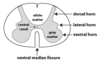
Where in the spinal chord cross section do motor neurons have their cell bodies?
Ventral horn (bottom)
Where in the spinal chord cross section do preganglionic neurons have their cell bodies?
Lateral horn (middle side bit)
What does it mean to say that sensory neurons are pseudounipolar?
It means that the cell body has moved so that the dendrite and the axon forms a single axon
Its called psuedounipolar because it starts off as bipolar
What kind of synapse connects an inhibitory neuron to a reflex arc
Axo-Axonic synapse
What is an axonal tracer?
A substance which can be carried along an axon in either direction
What is an anterograde tracer?
A tracer which is carried away from the cell body
What is a retrograde tracer?
A tracer which is carried towards the cell body
What does diffusion tensor imaging show?
The white matter tracts
Why can diffusion tensor imaging show the white matter tracts?
Water molecules are polarised
When they are constrained in very narrow structures such as myelinated axons the water molecules align along the axon tracts
Magnetic resonance signal at any given voxel has a vector
Vectors added together make fibres
What can diffusion tensor imaging not show about the white matter tracts?
The direction of axon travel
What information do the dorsal columns carry?
Information about touch and pressure
What information does the spinocerebrellar column carry?
Information about proprioception
What information does the spinothalamic column carry?
Information about pain and temperature
What information does the lateral column carry?
Movement information
Is white matter on the inside or the outside of the spinal chord?
on the outside
Is white matter on the inside or the outside of the brain?
On the inside
Name 3 types of glial cell
Macrophages
Astrocytes
Oligodendrocytes (schwann cels in PNS)
What is a brief role of macrophages?
Scavenging for dying cells, detritus
What are two brief functions of astrocytes?
- Regulate nutrient flow from blood
- Ionic balance and synapses
What is the function of oligodendrocytes?
They wrap axons in myelin
what does rostral mean in humans?
towards the top
what does caudal mean in humans?
towards the bottom
what does ventral mean in humans (and what is it also called)
towards the front / anterior
what does dorsal mean in humans and what is it also called
towards the back / posterior
where is the sub arachnoid space in the spinal chord and what are three things youll find in it?
between the arachnoid (middle) membrane and the pia(inner) membrane
its filled with cerebrospinal fluid
you’ll also find the nerve roots and blood vessels
visualise how to distinguish between lumber, thoracic and cervical spinal chord cross sections?
what is a feature of the thoracic spinal chord?
a small intermediate lateral horn sticking out
where is the ventral horn of the spinal chord?
where in the spinal chord are the motor neurons cell bodies?
in the ventral horn
where in the spinal chord are the preganglionic neurons cell body?
in the lateral horn
why are the ventral horns larger in the cervical levels and the lumbar levels
ventral horns contain motor neurons
the cervical level and lumbar levels are where most of the motor control of the upper limbs(cervical) and lower limbs (lumber) is situated
why is there only lateral horns in the thoracic segment
because sympathetic ganglia neurons are present in the lateral horns
and they are found only in the thoracic segment of the spinal chord
which roots do the preganglionic neurons and the motor neurons travel through?
the ventral root
is the following neurons myelinated or unmyelinated?
- preganglionic neuron
- postganglionic neuron
preganglionic neuron is myelinated
postganglionic neuron is unmyelinated
which neurons are pseudounipolar?
input sensory neurons to the spinal chord
their cell bodies sit in the dorsal root ganglia
how come our knee jerk reflexes dont happen when we are voluntarily moving our muscles
in the knee jerk reflex, the sensory neuron provides info about the muscle being stretched and the motor neuron responds by counteracting
there is an inhibitory neuron that is only activated during voluntary movement, so that this doesnt occur in voluntary movement
the inhibitory neuron is connected to the axon output of the sensory neuron with an axo-axonic synapse
