Week 3: Derm Flashcards

1
Q

What are the best practices for performing a skin exam? (8)

A
  1. Integrate skin exam in general exam
  2. Document what is present
  3. Ensure proper lighting
  4. Measuring tape/ruler
  5. Dermoscope
  6. Have patient in underwear in gown
  7. Ungloved hands if possible
  8. Scalp to toes
2
Q

What are the eight characteristics that the FNP should describe of every skin lesion?

A
  1. Number
  2. Size
  3. Color
  4. Shape
  5. Texture
  6. Primary Lesion
  7. Location
  8. Configuration
3
Q

define lesion

A

any single area of altered skin - may be singular or multiple

4
Q

How do you record the following characteristics: Number and size

A

Number: if multiple, record how many, estimate if numerous

Size: length and width in mm or cm

5
Q

What are some ways to describe color? (3)

A
  1. skin colored = same shade as patient’s skin
  2. blanching = red lesion that becomes white when pressure is applied, suggests inflammation
  3. non-blanching = bright red or violaceous that stays red when pressure is applied, suggests vascular involvement
6
Q

What are some ways to describe configuration? (4)

A
  1. unilateral
  2. dermatomal
  3. grouped
  4. linear
7
Q

What is this primary lesion?

A

Macule: flat and <1cm

Photo is an example of morbilliform drug eruption

8
Q

What is this primary lesion?

A

Patch: flat, >1cm

Photo is an example of seborrheic dermatitis

9
Q

What is this primary lesion?

A

Papule: raised <1cm

Photo is an example of basal cell carcinoma

10
Q

What is this primary lesion?

A

Plaques: raised >1cm, can be lichenified

Photo is an example of plaque psoriasis

11
Q

What is this primary lesion?

A

Vesicle: raised, clear fluid-filled, <1cm

Photo is an example of herpes zoster

12
Q

What is this primary lesion?

A

Bulla: raised, clear fluid-filled >1cm

Photo is an example of inherited skin fragility disorder

13
Q

What is this primary lesion?

A

Pusutle: small palpable collection of neutrophils or keratin that appears white

Photo is an example of bacterial folliculitis

14
Q

What is this primary lesion?

A

Furuncle: inflamed hair follicle, multiple furuncles combine to form a carbuncle

Photo is an example of both furuncles and a carbuncle

15
Q

What is this primary lesion?

A

Nodule: larger, deeper under the layer of the skin than a papule

Photo is an example of a keloid

16
Q

What is this primary lesion?

A

Subcutaneous mass vs. cyst: masses are typically a well defined area of abnormal growth, where cysts are a distinct collection of fluid

Photo on the left: excised cyst

Photo on the right: lipoma or subcutaneous mass

17
Q

What is this primary lesion?

A

Wheal: localized dermal edema

Photo is an example of urticaria

18
Q

What is this primary lesion?

A

Burrow: small linear pathways in the epidermis

Photo is an example of scabies

19
Q

What are some terms used to refer to the shape of a lesion? (5)

A
  1. Circular
  2. oval
  3. Annular: ring like with central clearing
  4. Nummular: ring like with no central clearing
  5. polygonal
20
Q

What are some terms used to refer to the texture of a lesion? (4)

A
  1. smooth
  2. fleshy
  3. verrucous/warty
  4. scaly (fine, keratotic, greasy)
21
Q

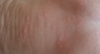
What is this condition? What are the classic characteristics?

A

Actinic Keratosis

  • Easier to feel than see
  • Superficial keratotic papules come and go on sun damaged skin
  • Precursor to SCC
22
Q

What is this condition and what does it mimic?

A

Superficial xerosis or sebhorreic dermatitis

  • Mimics actinic keratosis
23
Q

What is this condition and what are it’s characteristics? (hint there are 3 types)

A
  1. Superficial basal cell carcinoma: Pink patch that does not heal
  2. Nodular basal cell carcinoma: Pink papule often with translucent or pearly appearance and overlying telangiectasias, may have focal pigmentation
  3. Ulcerated basal cell carcinoma: nonhealing ulcer, resulting in rolled border
24
Q

What mimics basal cell carcinoma? (4)

A
  1. Actinic keratosis
  2. Sebaceous hyperplasia
  3. Fibrous papule
  4. Squamous cell carcinoma: smooth but firm border
25
What exam findings are concerning for malignant melanoma?
ABCDE(EFG): if two or more are present, consider biopsy and referral to dermatologist 1. Asymmetry 2. Irregular Border 3. Color - within the mole but also within surrounding moles 4. Diameter greater than 6mm 5. Evolving or changing 6. EFG: elevated, firm to palpation, growing rapidly over several weeks
26
What is this? What are it's characteristics (color and size, shape, pulsatility/effect of pressure, distribution, significance)
Spider Angioma * **Color and size:** firey red, from very small to 2cm * **Shape**: central body, sometimes raised, surrounded by erythema and radiating legs * **Pulsatility/effect of pressure**: often seen in the center of the spider when pressure with a glass slide is applied, pressure on the body causes blanching of the spider * **Distribution:** face, neck, arms, upper trunk * **Significance:** normal and common on the face and chest, seen in pregnancy and liver disease
27
What is this? What are it's characteristics? (color and size, shape, pulsatility/effect of pressure, distribution, significance)
Spider Vein * **Color and size:** bluish, size variable, from very small to several inches * **Shape**: variable, may resemble a spider or be linear, irregular, cascading * **Pulsatility and effect of pressure**: absent, pressure over the center does not cause blanching, but diffuse pressure blanches the veins * **Distribution**: most often on the legs, near veins, also on the anterior chest * **Significance**: often accompanies increased pressure in the superficial veins as in varicose veins
28
What is this? What are it's characteristics? (color and size, shape, pulsatility/effect of pressure, distribution, significance)
Cherry Angioma * **Color and size:** Bright or ruby red, may become purplish with age, 1-3mm * **Shape:** round, flat or sometimes raised, may be surrounded by a pale halo * **Pulsatility and effect of pressure**: absent, may show partial blanching especially if pressure applied with edge of a pinpoint * **Distribution**: trunk, also extremities * **Significance:** none, increases in size and numbers with aging
29
What are examples of sun damage? (6)
1. Solar lentigo: bilaterally symmetric brown macules located on sun-exposed skin, including the face, shoulder and arms and hands (left picture) 2. Actinic purpura: ecchymoses limited to the dorsal forearms and hands but not extending above the shirt sleeve line on the upper arm (right picture) 3. Wrinkles: increased sun damage and tanning leads to deeper wrinkles at an earlier age 4. Solar elastosis: yellowish white macules or papules in sun-exposed skin especially on forehead 5. Poikiloderma: red patches in sun-damaged areas, especially the V of the neck and lateral neck, with fine telangiectasias, both hyper and hypopigmentation 6. Cutis rhomboidalis nuchae: deep wrinkles on the posterior neck that crisscross
30
What are some abnormal nail findings? (7)
1. Onychomycosis: fungal infection 2. Onchyolysis: painless separation of the whitened opaque nail plate from the pinker translucent nail bed 3. habit tic deformity: pushing on nails/picking at cuticles, looks like a christmas tree 4. melanonychia : increased pigmentation in the nail matrix 5. Nail biting: can cause trauma to the nail 6. Paronychia: superficial nail infection 7. Clubbing: bulbous swelling of the soft tissue at the nail base
31
What skin findings would you expect in a patient with chronic renal disease? (5)
pallor, xerosis, uremic frost, half and half nails, calciphylaxis
32
What skin findings would you expect in a patient with Crohn disease? (4)
erythema nodosum, pyoderma gangrenosum, enterocutaneous fistulas, aphthlous ulcers
33
What skin findings would you expect in a patient with Cushing disease? (9)
striae, atrophy, purpura, ecchymoses, telangiectasaias, acne, moon facies, buffalo hump, hypertrichosis
34
What skin findings would you expect in a patient with diabetes? (7)
pruritis, diabetic dermopathy, acanthosis nigricans, candidiasis, neuropathic ulcers, necrobiosis lipoidica, eruptive xanthomas
35
What skin findings would you expect in a patient with dyslipidemias? (2)
xanthomas, xanthelasma
36
What skin findings would you expect in a patient with hypo (6) vs. hyperthyroidism (6)?
* Hypo: dry, rough and pale skin, coarse and brittle hair, myxedema, alopecia (eyebrows), skin cool to touch, thin and brittle nails * Hyper: warm, moist, soft and velvety skin, thin and fine hair, alopecia, vitiligo, pretibial myxedema, hyperpigmentation
37
What skin findings would you expect in a patient with Kawasaki disease? (5)
mucosal erythema, strawberry tongue, cherry red lips, polymorphous rash, erythema of the palms and soles with later desquamation of the fingertips
38
What skin findings would you expect in a patient with liver disease? (7)
jaundice, spider angiomas and other telangiectasias, palmar erythema, terry nails, pruritis, purpura, caput medusae
39
What skin findings would you expect in a patient with leukemia/lymphoma? (9)
pallor, exfoliative erythroderma, nodules, petechiae, ecchymoses, pruritis, vasculitis, pyoderma gangrenosum, bullous disease
40
What skin findings would you expect in a patient with meningococcemia?
angular or stellate purpuric patches and plaques with gunmetal gray center; progresses to ecchymoses, bullae, necrosis
41
What findings would you expect in a patient with neurofibromatosis? (4)
neurofibromas, café au lait spots, freckling in the axiallae (crowe sign), plexiform neurofibroma
42
What skin findings would you expect in a patient with lupus? (3)
malar erythema, periungual erythema, interphalangeal erythema
43
What are is the APGAR scoring for a newborn's skin?
* 0: blue, pale * 1: pink body, blue extremities * 2: pink all over
44
Where would you assess an infant’s skin for cyanosis? What would you consider on the DDx for an infant with cyanosis?
* Assess the mouth, tongue and conjunctivae as well as skin color * DDx: * Acrocyanosis: bluish discoloration in the palms and soles * Cyanotic congenital heart disease * Central cyanosis: hands and feet, lips, tongue, sublingual tissues * Harlequin dyschromia: transient cyanosis of one half of the body or extremity
45
What is this? What are it's characteristics?
Miliaria rubra: scattered vesicles on an erythematous based , result from obstruction of the sweat gland ducts
46
What is this and what are it's characteristics?
Erythema toxicum: erythematous macules with central pinpoint vesicles
47
What is this and what are its characteristics?
Pustular melanosis: small vesiculopustules over a brown macular base; seen more commonly in black infants
48
What is this and what are it's characteristics?
Milia: pinhead-size white, pearly papules, without surrounding erythema on the nose, chin and forehead result from retention of sebum in the openings of the sebaceous glands
49
What is this and what are its characteristics?
Cutis marmorata: vasomotor changes in the dermis and subcutaneous tissue, response to cooling or chronic exposure to radiant heat, bluish mottled & lattice-like appearance
50
What is this and what are it's characteristics?
physiologic jaundice: related to acclimation outside of the womb, if persists past 5 days consider abnormal
51
What is this and what are it's characteristics?
Midline hair tufts: over lumbosacral spine region suggests possible spinal cord defect
52
What are some benign birthmarks found in infants? (6)
1. eyelid patch 2. Salmon patch: splotchy pink mark 3. Café au lait spots: light brown pigmented lesions \<1 to 2 cm at birth 4. Multiple lesions might indicate neurofibromatosis 5. Congenital dermal melanocytosis 6. Mongolian patch/slate blue patches: dark or bluish pigmentation over the buttocks and lower lumbar regions
53
In the pediatric population, what are some wart-like lesions and how do they appear on exam?
1. Verruca vulgaris: dry, rough warts on hands 2. Verruca plana: small, flat warts 3. Plantar warts: tender warts on feet 4. Molluscum contagiosum: dome-shaped, fleshy lesions with central umbilication 5. Adolescent acne: open and closed comedones, inflamed pustules
54
What are some commons skin lesions during childhood and how do they appear on exam?
1. Insect bites: intensely pruritis, red, distinct papules characterize these lesions 2. Tinea capitis: scaling, crusting and hair loss along with painful plaque and occipital lymph node 3. Urticaria: pruritis, allergic sensitivity reaction 4. Scabies: intensely itchy papule and vesicles, sometimes burrows, most often on extremities 5. Tinea corporis: this annular lesion has central clearing and papules along the border 6. Pityriasis rosea: oval lesions on trunk, often in a christmas tree pattern, herald patch