Week 11: Pathophysiology of Congenital Heart Defects Flashcards
What are the 4 common congenital heart defects?
- Development of the atrial septum
- Development of the ventricular septum
- Division of the main outflow tract (from the truncus arteriosus into the pulmonary artery and aorta)
- The development of the valves
- It is very common for multiple defects to be present in one child
What is septation
- Septation is the division of parts by a septum, in the case of the heart, septation refers to the formation of the right and left hearts (both ventricular and atrial), via a septum
Describe the septation of the heart
- The atrial septum develops during 4th week of embryonic life as structure called septum primum. Descends from superior region of the primitive atrium
At same time, endocardial cushion appears.
Septum primum grows down towards the endocardial cushion.
Septum secondum begins to grow down from the superior region towards the endocardial cushion. –> leaves oval shaped opening int eh inferior region forming the foramen ovale.
Septum primum reaches endocardial cushion and regresses leaving only the lower poriton which forms a one way valve for foramen ovale.
Ventral septum achieved by endocardial cushion as well as muscular septum. Muscular septum grows upwards the endocardial cushion as it grows down to form intraventricular septum.
Endocardial cushion also helps form the tricuspid valve and the mitrall all on the left side.

Describe the development of the major arteries
6 -7 weeks there is one great vessel called truncus arteriosus
Divides into two

What congenital issues are associated with development of the major arteries
- Persistent truncus arteriosus
- Transposition of the great arteries
- Congenital aortic and pulmonic valve defects
what congenital issues are associated with septation
- Atrial septal defects
- Ventricular septal defects
- Atrioventricular valve defects
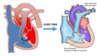
Describe the foetal circulation of Oxygen

- The oxygenated blood comes from the placenta, through the umbilical vein into the inferior vena cava (which also takes deoxygenated blood from the foetus)
- A large amount of oxygenated blood then goes through the foramen ovale, and into the left atrium, then to the left ventricle, then through the aorta
- the venous blood meanwhile flows through the superior vena cava and inferior vena cava and by some mechanism is mostly shifted into the right ventricle (along with some oxygenated blood)
- This then goes through the pulmonary arteries, however as the lungs are collapsed it can’t take it through its normal route
- Instead, there is a special connecting vessel between the pulmonary artery and the aortic arch known as the ductus arteriosus
- The ductus arteriosus is kept open as the pressure within the pulmonary artery is higher than in the aorta, and also through a chemical substance known as Prostaglandin E1 (PGE1)
- Then flows around the body, and back to the placenta for re-oxygenation

How does the formaen ovale naturally shut?
Pulmonary vessels open from childs first breath.
Causes pulmonary vessels to open. –> reduces pulmonary vascular resistance which causes the pulmonary pressure to fall while systemic circulation pressure rises.
- Systemic resistance starts to rise, increasing the pressure within the left atrium and aorta, whilst the pressure in the right atrium and ventricle starts to decrease causing the foramen ovale to naturally shut (6 months this will be completely closed)
How is the ductus arteriosus closed?
- This is facilitated by an increased pressure in the aorta which causes oxygenated blood to flow from the aorta to the pulmonary artery
- The exposure of high levels of oxygen to the ductus arteriosus results in the constriction of the structure closing it off by about 72 hours (as well as the decline of PGE1)
What are 5 symptoms of congenial heart defects?
- Poor growth (failure to thrive)
- Endocarditis
- Recurrent lower respiratory tract infections
- Dyspnea
- Exercise intolerance (easy fatigability)
- Poor feeding
- Arrythmia
- Tachycardia
- Reduced peripheral perfusion
- Pulmonary congestion
- Congestive heart failure
- Cyanosis
What are the 2 main factors for congenital heart disease?
- A genetic problem itself
- 30% of CHD have chromosomal abnormality - Environmental factors
- These are modifiable teratogens like;
- Alcohol, certain drugs
- Maternal rubella infection in the first trimester
- Maternal radiation
- Maternal diabetes
- Maternal age if greater than 40
Describe the pathology of obstructions in the heart in relation to congenital heart defects
- Is the pathology of abnormal narrowing’s in the hearts vessels or valves
- The obstruction may be with a valve not opening well causing an obstruction to the blood flow (from stenosis or coarctation of the aorta)
- It may even be because of the poor formation of valves or the failure to develop resulting in the path being completely blocked
- There may be a narrowed portion (particularly common in the aorta) of a vessel resulting in reduced blood flow causing increased resistance (increasing pressure)
- The workload of the heart is thus greatly increased possibly causing heart failure

Why can congenital heart defects go undiagnosed for so long?
Asymptomatic at first –> symptoms will worsen over time as the person grows as they need more cardiac output to supply there growth demands.
In ventricular septal defect, why would you have pale cold fingers, but they are not cyanosed?
Decrease Blood pressure will trigger baroreceptors –> activates sympathetic activity –> vasoconstriciton of peripherals
If a patient has coarctation of the aorta and patent ductus arteriosus, what kind of shunt would you expect?
From left to right
If symptoms become severe will eventually get left sided heart failure which will cause the shunt to shift to right to left.
In coartation of the aorta, why would a patient have reduced muscle development in their lower body compared with their upper body?
Less blood flow to lower limb due to narrowing of the aorta going to lower limbs.
Muscle development in upper limb due to increase blood flow to upper limb through subclavian artery etc.

why would you maintain the ductus arteriosus and formen ovale in a baby with TGA
so then blood can go from left to right and between ventricles and can mix oxygenated blood in the pulmonary artery so it can get sent around the body
One way to maintain the ductus arteriosus is to administer PGE to the baby. What is PGE and what its role in the management of a baby with TGA?
is a prostaglandin 1 –> keeps foramen and ductus arteriosus open
used in conditions with decrease blood flow to lungs
In tetralogy of Fallot, what is the mostl likely reason for cyanosis associated with crying/feeding?
crying –> increases pulmonary vascular resistance which increase pressure in the right side of heart which leads to a right to left shunt.
Feeding –> increases parasympathetic activity which therefore dilates vessels. This decreases pressure in left ventricle and therefore creates a right to left shunt.
both lead to cyanosis
why type of murmur would you expect to hear in a patient with tetralogy of fallot? what is the cause for the murmur?
Harsh systolic ejection murmur.
Because of teh stenosis –> during the systolic contraction the extra narrowing creates this murmur.
What is Eisenmengers syndrome?
A left to right shunt in ventral septum.
- The increased pulmonary blood flow will cause the gradual thickening of the pulmonary vessel wall (because of the exposure to oxygenated blood during early life)
- The tunica intima will proliferate and eventually fibrous, the tunica media will hypertrophy leading to reductions to the pulmonary lumen, increasing resistance
- Basically, increased pulmonary blood pressure in early life will lead to gradual pulmonary resistance increase, causing increased blood pressure, further causing hypertrophy to the right ventricle

What are murmurs due to?
- Murmurs are due to abnormal or turbulent blood flow that is occurring in the heart or vessels causing audible noise
IF murmurs occur after the 1st heart sound but before the 2nd they are known as ____
If murmurs occur after the 2nd heart sound they are known as ____
Systole
Diastole
What is cyanosis
- Is the bluish discolouration that occurs to the skin and mucous membranes of patients caused by an elevated arterial blood concentration of deoxygenated haemoglobin

What are 4 conditions that are acyanotic (those that do not experience cyanosis early in life, may develop it later on)
- Atrial and Ventricular septal defects
- Patent ductus arteriosus
- Congenital pulmonary and aortic valve stenosis
- Coarctation of the aorta
which are those conditions that are cyanotic (will see cyanosis develop very early on in life)
- Tetralogy of Fallot
- Transposition of the great arteries
- Truncus arteriosus
Explain what an atrial septal defect is and the complications it can lead to?
- This is characterised by a persistent opening in the interatrial septum after birth
- This means the blood can shunt from the left atrium to the right atrium (following the path of high to low pressure)
- This means there is mixing of blood
- The size of the defect determines the degree of shunting
- If it is mild to moderate, it will be asymptomatic
- Children may experience dyspnea on exertion, fatigue and recurrent lower respiratory tract infections (these infections are caused by the increase in pulmonary blood flow [as more blood is flowing into the right side from the left])
- Whilst these children may start out as asymptomatic, eventually the volume overload in the right atrium and right ventricle causes ventricular hypertrophy
- This will therefore increase the right ventricles pressure, and will eventually be greater then the left ventricles pressure causing blood to flow into the left ventricle (from high to low)
- The resulting right to left shunt causes cyanosis and eventually, heart failure (as the right ventricle has to generate far more pressure than it is used too)

What is a key clinical feature of atrial septal defect
systolic murmur
Explain what an ventricular septal defect is and the complications it can lead to?
- Ventricular septal defect has a very similar pathology to atrial septal defects, and is often associated with atrial septal defects, tetralogy of Fallot and transposition of the great arteries
- A small VSD is virtually asymptomatic, and even has the capacity to close spontaneously by the age of 2
- The hemodynamic changes are similar to atrial septal defect in that initially, the shunt is from left to right which increases the right hearts workload
- This then overloads the right ventricle, causing hypertrophy of the right ventricle
- As a result of this, slowly the pulmonary circulation increases (from RV hypertrophy), causing pulmonary hypertension
- The right ventricle pressure therefore eventually overtakes the left ventricle, reversing the shunt to right to left (causing the development of cyanosis) and eventually right sided heart failure

what is a key clinical feature of a ventricular septal defect
- A key clinical sign of a ventricular septal defect is a systolic thrill and systolic murmur
Explain what a patent ductus arteriosus is and what complications it leads to
- In foetal life, the ductus arteriosus is a structure which allows oxygenated blood to bypass the lungs by flowing straight from the pulmonary artery into the aorta
- Normally, this structure closes very rapidly after birth (hours, days after birth due to increased oxygen causing constriction and PGE1 also decrease after birth), if it doesn’t, it results in patent ductus arteriosus
- If the pressure remains higher in the aorta, it may have no clinical manifestations (as the shunt will be left to right)
- However, as the lungs will have more blood flowing to it, the will be increased load on the left ventricle eventually causing left heart failure
- Also, if there is more blood flowing into the pulmonary arteries, pulmonary hypertension may ensue
- If these things occur, a right to left shunt may develop (as the pressure within the pulmonary artery may be greater than the aorta) causing the development of cyanosis

Explain what is congenital pulmonary valve stenosis is and what complications it leads to
- Occurs when there is an obstruction to the right ventricle outflow, from thickened pulmonary valves (congenitally fused pulmonic valve cusps) which significantly reduces the valves opening, thus reducing the ejection of blood
- This makes it harder for the right ventricle to eject blood, or in other words means the afterload is increased
- This results in right ventricle hypertrophy and eventually, leads to right sided heart failure
- The severity depends on the degree of stenosis and if other congenital defects are also present
- Key clinical signs include a systolic ejection murmur (due to turbulence during systole) and dyspnea on exertion
- This can be corrected by a transcatheter balloon pulmonary valvuloplasty
- Surgery is also an option

Explain what is congenital aortic valve stenosis is and what complications it leads to
- Is virtually the same pathology as the congenital pulmonary valve stenosis, however involves the aortic valve instead
- This involves a narrowed aortic valve, and often is only bicuspid instead of tricuspid
- Blood will then have to be pushed through a narrowed opening, which results in LV afterload, causing LV hypertrophy and eventually, LV failure
- Key clinical signs include a prominent systolic murmur
- Can be corrected by a transcatheter balloon valvuloplasty or surgical replacement of the stenosed valve is it is severe

Explain what is coarctation of the aorta is and what complications it leads to
- Coarctation simply means the congenital narrowing of a short section of the aorta
- This can occur anywhere along the aorta, however commonly occurs next to the ductus arteriosus (meaning it is juxtaductal)
- This is often associated with a number of other anomalies including Turners syndrome and occurs more often in males than females
- The issue with this defect occurs as the narrowing means there is more workload to the left ventricle as the region proximal to the coarctation is going to be high, so the LV has to work hard to overcome it (as there is a greater afterload)
- This will eventually result in LV hypertrophy, and LV heart failure and in some cases aortic rupture

What are the clinical features of coarctation of the aorta and what type of murmur it would be?
- A key clinical feature of coarctation of the aorta is the tendency for a patient to have high BP in the upper extremities, but low BP in the lower extremities
- The femoral, popliteal and dorsalis pedis pulsations are weak
- The pulses in the arms and carotid vessels are bounding
- There may be a mid-systolic ejection murmur due to turbulence

Explain what is tetralogy of fallot is and what complications it leads to
- As the name suggests, tetralogy of Fallot is made up of 4 issues within the heart
- It is the most common form of cyanotic congenital heart defects after infancy
- The defects that will be evident include;
1. Pulmonary valve stenosis
2. Right ventricular hypertrophy (because of valve stenosis)
3. Ventricular septal defect
4. Overriding aorta (which is when the aorta is shifted towards the right, opening directly onto the intraventricular septum itself) - The severity of this condition is directly related to the degree of pulmonary valve stenosis
- That is, the greater the degree of stenosis, the earlier the manifestations of cyanosis
- This is because if worse the stenosis is, the greater the RV pressure will have to be
- The greater the RV pressure the more deoxygenated blood that will be shunted across to the left ventricle
- The direction of the shunt can also be affected by acute changes in systemic and pulmonary vascular resistance (which increases during exertion)
- In other words, during exertion (e.g. feeding, defecation, crying) the overriding aorta may receive more deoxygenated blood producing cyanosis
- This is because there is a change in the systemic and pulmonary vessels
- Upon exertion, systemic resistance will fall, as pulmonary resistance will increase causing RV pressure to increase, shifting the shunt from right to left

Explain what is transposition of the great arteries (TGA) is and what complications it leads to
- Is characterised by the two great vessels opening to the wrong chambers, that is the aorta opens to the right ventricle, the pulmonary artery opens to the left ventricle
- Babies with this heart defect can survive foetal life thanks to the foramen ovale
- However, once this closes, there is completely separate pulmonary and systemic circulation, they do not mix but rather run in parallel
- Deoxygenated blood just goes to the peripheral tissues, then back to the right heart then back around
- Oxygenated blood just keeps getting pumped through the pulmonary circulation then back and around through the left heart
- Survival of the child will only really occur if they have some other defect (such as atrial septal defect or ventricular septal defect) which allows some degree of mixing of oxygenated and deoxygenated blood

Explain what is persistant truncus arteriosus is and what complications it leads to
- During foetal development, the aorta and pulmonary artery form from one common primitive vessel, known as the truncus arteriosus
- At a point of foetal development this truncus arteriosus normally divides into the aorta and pulmonary artery, however in this disorder this division goes wrong causing persistent truncus arteriosus
- This usually will be accompanied by a ventricular septal defect
- As a significant amount of deoxygenated blood goes to the systemic circulation, there is early onset of cyanosis
- As the pulmonary circuit provides less resistance, more blood tends towards the lungs as opposed to the systemic circulation
- Because of this abnormally high blood flow to the lungs, pulmonary hypertension can often ensue which eventually shunts more deoxygenated blood into the aorta producing even more cyanosis
- This will lead to congestive heart failure

Name any substance that is used to measure in plasma oslmolarity clearance
PAH