W2: Organisation of the neck Flashcards
(30 cards)
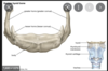
Important landmarks and levels
- Hyoid ( Body, greater horn) –C3
- Thyroid cartilage –C4 & C5
- Cricoid cartilage –C6
- External occipital protuberance
- Spine C7 ( vertebral prominence)

Describe hyoid bone
Has body and greater and lesser horns
Where does the thyroid gland lie in the neck?
The isthmus of the thyroid gland lies at the level of the 2nd and 3rd tracheal cartilage rings
Which triangles do we have?
The neck may be divided into two anatomical triangles, bordered by the sternocleidomastoid and trapezius muscles:
- Anterior triangle – area between the mid-line anteriorly and the anterior border of sternocleidomastoid posteriorly, bounded superiorly by the lower jaw. Subdivided into a further 3 triangles.
- Posterior triangle – area between sternocleidomastoid and the trapezius muscles, bounded inferiorly by the middle 1/3rd of the clavicle.
- Submandibular triangle
- Carotid triangle
- Muscular triangle
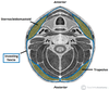
Discuss posterior triangle
Bounded by sternocleidomastoid (anterior) and trapezius (posterior) and the middle 1/3 of clavicle (inferior) forms the base
Contents:
Nerves:
- Spinal accessory nerve - sternocleidomastoid and trapezius - superficial and vulnerable to injury
- Cutaneous nerves (cervical plexus) (Lesser occipital nerve (C2) ,Great Auricular nerve(C2,C3),Transverse cervical cutaneous nerve (C2,C3),Supraclavicular nerve (C3,C4) - also phrenic
- Trunks of brachial plexus
Vasculature:
- Subclavian artery (3rd part)
- Branches of Subclavian artery
- Subclavian vein (central catheter)
- External jugular vein (fromed by retromandicular and posterior auricular) - superficially - emplies into subclavian vein
Muscles:
- omohyoid crosses
- Floor
- Lev scap
- Scalenes
What are the boundaries of anterior triangle?
Superiorly – inferior border of the mandible (jawbone).
Laterally – anterior border of the sternocleidomastoid.
Medially – sagittal line down the midline of the neck.

What can the anterior triangle be split into?
Carotid
Submental
Submandibular
Muscular
Discuss carotid triangle
The carotid triangle of the neck has the following boundaries:
Superior – posterior belly of the digastric muscle.
Lateral – medial border of the sternocleidomastoid muscle.
Inferior – superior belly of the omohyoid muscle.
The main contents of the carotid triangle are the common carotid artery (which bifurcates within the carotid triangle into the external and internal carotid arteries), the internal jugular vein, and the hypoglossal and vagus nerves.

Discuss submental triangle
The submental triangle in the neck is situated underneath the chin. It contains the submental lymph nodes, which filter lymph draining from the floor of the mouth and parts of the tongue. Also anterior jugular vein
It is bounded:
Inferiorly – hyoid bone.
Medially – midline of the neck.
Laterally – anterior belly of the digastric
The floor of the submental triangle is formed by the mylohyoid muscle, which runs from the mandible to the hyoid bone.

Discuss submandibular triangle
The submandibular triangle is located underneath the body of the mandible. It contains the submandibular gland (salivary), and lymph nodes. The facial artery and vein also pass through this area.
The boundaries of the submandibular triangle are:
Superiorly – body of the mandible.
Anteriorly – anterior belly of the digastric muscle.
Posteriorly – posterior belly of the digastric muscle.

Discuss muscular triangle
The muscular triangle is situated more inferiorly than the subdivisions. It is a slightly ‘dubious’ triangle, in reality having four boundaries. The muscular triangle contains some muscles and organs – the infrahyoid muscles, the pharynx, and the thyroid, parathyroid glands.
The boundaries of the muscular triangle are:
Superiorly – hyoid bone.
Medially – imaginary midline of the neck.
Supero-laterally – superior belly of the omohyoid muscle.
Infero-laterally – inferior portion of the sternocleidomastoid muscle.

What is the roof of these triangles made from?
deep (investing) fascia that surrounds the neck like a collar, enclos- ing the sternocleidomastoid anteriorly and the trapezius posteriorly.
Discuss floor of posterior triangle
The floor of the posterior triangle is formed by the pre-vertebral fascia, which surrounds the deep pre-verte- bral muscles of the neck.The vertebral vessels pass through a triangular gap in the fascia to reach the foramen transversarium of the cervical spine.
What can fascia be divided into?
Deep and superficial
What does the superficial fascia blend with?
Platysma muscles
What are the deep fascial layers? (superficial -> deep)
Investing layer
Pretracheal layer
Prevertebral layer
Carotid sheath
Discuss investing layer
The investing layer is the most superficial of the deep cervical fascia.
It surrounds all the structures in the neck. Where it meets the trapezius and sternocleidomastoid muscles, it splits into two, completely surrounding them.
The investing fascia can be thought of as a tube; with superior, inferior, anterior and posterior attachments:
Superior – attaches to the external occipital protuberance and the superior nuchal line of the skull.
Anteriorly – attaches to the hyoid bone.
Inferiorly – attaches to the spine and acromion of the scapula, the clavicle, and the manubrium of the sternum.
Posterior – attaches along the nuchal ligament of the vertebral column
Discuss pretracheal layer
The pretracheal layer of fascia is situated in the anterior neck. It spans between the hyoid bone superiorly and the thorax inferiorly (where it fuses with the pericardium).
The trachea, oesophagus, thyroid gland and infrahyoid muscles (sternothyroid, sternohyoid, omohyoid) are enclosed by the pretracheal fascia.
Anatomically, it can be divided into two parts:
Muscular part – encloses the infrahyoid muscles.
Visceral part – encloses the thyroid gland, trachea and oesophagus.
Discuss prevertebral layer
The prevertebral fascia surrounds the vertebral column and its associated muscles; scalene muscles, prevertebral muscles, and the deep muscles of the back.
It has attachments along the antero-posterior and supero-inferior axes:
Superior attachment – base of the skull.
Anterior attachment – transverse processes and vertebral bodies of the vertebral column.
Posterior attachment – along the nuchal ligament of the vertebral column
Inferior attachment – fusion with the endothoracic fascia of the ribcage.
The anterolateral portion of prevertebral fascia forms the floor of the posterior triangle of the neck. It also surrounds the brachial plexus as it leaves the neck and subclavian artery as it passes through the lower neck region – in doing so, it forms the axillary sheath.

Discuss carotid sheath
The carotid sheaths are paired structures on either side of the neck, which enclose an important neurovascular bundle of the neck.
The contents of the carotid sheath are:
- Common carotid artery
- Internal jugular vein.
- Vagus nerve.
- Accompanying cervical lymph nodes.
The fascia of the carotid sheath is formed by contributions from the pretracheal, prevertebral, and investing fascia layers. The carotid artery bifurcates within the sheath into the external and internal carotid arteries.
The carotid fascia is organised into a column, which runs between the base of the skull to the thoracic mediastinum. This is of clinical importance as a pathway for the spread of infection.

Discuss sternocleidomastoid
asses from the manubrium and clavicle to insert into the mastoid process of the temporal bone. When contracting unilaterally, tilts the head to the same side and rotates face, When contracting bilaterally, flexes cervical vertebrae so chin approaches manubrium. Sternocleidomastoid is
innervated by the spinal accessory nerve (CN XI).
Discuss trapezius
passes from the external occipital protuberance and spinous processes of C7-T12, to insert
into the clavicle, acromion and spine of the scapula.When contracted, elevates, retracts and rotates scapu-
lae.Trapezius is innervated by the spinal accessory nerve (CN XI).

Discuss scalene muscles
a group of three muscles within the lateral neck, innervated by C4-C7 spinal nerves, which
function to elevate the thoracic cage and bend the neck ipsilaterally.


