Vestibular System Flashcards

1
Q

What are the three main elements in signalling of the vestibular system?

A

Input - visual, pressure(proprioreceptors), vestibular organ (in the ear)

Central processing - mostly brainstem

Output - ocular reflex, spinal reflexes for postural control.

2
Q

What is the name of the balance organ in the ear? Where is it found?

A

labyrinth

Contained inside the temporal bone

3
Q

Describe the structure of the vestibular system in the ear?

A

Bony labyrinth covers the mebranous labyrinth.

5 organs:

  • Otolith organs - utricle and saccule
  • 3 semicircular canals all attached to utricle but different - horizontal, superior, inferior.

Fluid inside the membranous membrane = endolymph

Fluid outside membranous membrane = perilymph

4
Q

What are the static and kinetic parts of the labyrinth?

A

Static - otolith organs

Kinetic - semicircular canals

5
Q

Which two semicircular canals come together?

A

Superior(/anterior) and inferior(/posterior) - these have only one entrance into the utricle.

Horizontal canal has its won entrance.

6
Q

Why are the ends of the semicircular canals wider?

A

They contain the ampulla, cupula and kinocilia.

7
Q

What are the two branches of the vestibulocochlear nerve and where do they go?

A

NB: CNVIII

  • Vestibular part goes to the vestibular organ
  • Cochlear part goes to the cochlea
8
Q

What are the two types of cells involved in transduction in the vestibular organs?

A

Hair cells: type I and type II

9
Q

Which hair cells in the vestibular organ are more numerous? Describe their afferents and efferents.

A

Type I

  • are round and are more numerous.
  • have direct afferents around the cells and indirect efferents.
  • are the main cells responsible for the afferent signals of the vestibular system.

Type II

  • are long
  • have direct afferents and efferents but there are fewer of them.
10
Q

What do the otolith organs consist of?

A
  • Utricle
  • Saccule
  • Maculae (shown as the striped regions on picture below)
  • Hair cells
  • Gelatinous matrix
  • Otoliths = carbonate crystals
11
Q

What is the striola?

A

STRIOLA, a curved dividing ridge that runs through the middle of the MACULA

  • in the UTRICLE, the kinocilia are oriented TOWARD the striola,
  • in the SACCULE they are oriented AWAY from it
12
Q

What do the semicircular canals consist of?

A
  • Ampulla - dilated sac at the end of the semicircular canals
  • Crista ampullaris - contains the hair cells
  • Cupula - gelatinous projections which contain the crista which contain the hair cells
  • Kinociia - point in same direction on each side of the head

Semicircular canals are filled with endolymph.

13
Q

Describe the organisation of the macula.

A

Some type I and type II cells present. Hair bundles sit above and gel sits on top of this and the crystals sit on top of the gelatinous matrix.

14
Q

How are the semicircular canals layed out in relation to one another?

A

At 90 degrees to each other -

anterior canals are located at ~90o to each other; posterior canals are also located at ~90o to each other

15
Q

Describe the blood supply to the inner ear.

A

Via AICA - anterior inferior cerebellar artery (which also goes to the cerebellum so any problems e.g. stroke affecting it will also affect the inner ear)

This is a branch of the basilar artery.

16
Q

Name the two branches of the vestibular nerve. What do they supply? Where do they end?

A

Superior and inferior vestibular nerves

  • Superior sends sensory information from the UTRICLE and anterior and horizontal canals.
  • Inferior sends senory information from the SACCULE and posterior canals.

These end in the vestibular nuclei in the brainstem.

17
Q

What is the name of the ganglia of superior and inferior vestibular nerves?

A

Scarpi’s ganglia

18
Q

Name the different vestibular nuclei. Describe their organisation.

A
  • Superior
  • Lateral
  • Medial
  • Inferior

E.g. static labyrinth (otoliths) are connected mainly to the lateral and inferior nuclei.

E.g. kinetic labyrinth (semicircular canals) mainly send information to the superior and medial nuclei.

19
Q

What are the 5 areas tha vestibular nuclei project to?

A

Vestibular pathways can project to:

  1. Spinal cord – to be connected to the muscles to maintain posture/prevent falling
  2. Nuclei of the extraocular muscles – moving eyes when walking
  3. Cerebellum – Movement coordination, posture regulation, VOR(vestibulo-ocular reflex) modulation
  4. Centers for cardiovascular + respiratory control
  5. Thalamus
20
Q

Why do the bestibular nuclei project to the cerebellum?

A
  • Movement coordination
  • Posture regulation
  • VOR(vestibulo-ocular reflex) modulation
21
Q

Decribe the pathway of vestibular nuclei to the thalamus and cortex. What sensation do these projections account for?

A

Vestibular nuclei –> thalamus

–>thalamic nuclei –> the head region of the primary somatosensory cortex + to the superior parietal cortex(spacial orientation)

These cortical projections may account for feeling of dizziness during certain kinds of vestibular stimulation.

22
Q

What is the purpose of the vestibular system?

A

To detect and inform brain about head movements.

To keep images fixed in the retina during head movements.

To maintain postural control.

23
Q

Describe the firing of the nerve fibres of the vestibular system at rest.

A

Nerve fibres of the vestibular system are constantly firing – even when standing still.

  • Resting discharge – nerve fibres always sending signals at a certain rate
  • An increase in rate occurs when nerve cells are depolarised
  • Decrease in rate when nerve cells are hyperpolarised.
24
Q

What are the three types of responses of nerve fibres in the vestibular system? What is the mechanism?

A

So there are 3 types of responses:

  • resting,
  • excitation(hair bundles go in the direction of the kinocilium),
  • inhibition(hair fibres go against the direction of the kinocilium).

Process:

  • Kinocilia deflect, potassium and calcium enters. Neurotransmitter is released into the synapse and triggers a signal in the nerves.
25
Q

What types of movement are the otolith organs mainly involved in?

A
  • Utricule and Saccule – mostly involved in linear acceleration, tilt and gravity (since crystals have weight)
  • Otolith movement will cause depolarisation or hyperpolarisation
    • Utricule: horizontal movement, velocity and acceleration (NB: location)
    • Saccule: vertical movement, velocity and acceleration.
26
Q

Give examples of different types of motion and describe how the otolith organs ensures that the correct signal is sent to the brain.

A

Head tild backwards =/+acceleration backwards

Head tilt forward =/+acceleration forward.

  • Otolithic organs sense how quickly you are accelerating forward or backward, left or right, or up or down
  • Humans can sense head tilting and linear acceleration even in dark environments because of the orientation of two groups of hair cell bundles on either side of the striola.
  • Hair cells on opposite sides move with mirror symmetry, so when one side is moved, the other is inhibited.
  • The opposing effects caused by a tilt of the head cause differential sensory inputs from the hair cell bundles allow humans to tell which way the head is tilting
  • Semicircular canals are much more straight forward.
27
Q

What types of movement are the semicircular canals involved in?

A

Angular acceleration (not constant movement)

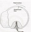
Work with the inertia of the endolymph (when the head moves in one direction the liquid will move in the other pushing the cupula and thus the hair bundles in that direction)

When the cupula moves it displaces the hair cells

28
Q

What direction does endolymph move in the SCC in relation to direction of acceleration?

A

In the opposite direction to acceleration

29
Q

What direction do kinocilia have to point in order to cause excitation in the semicircular canals?

A

Kinocilium is facing in opposite directions in each canal:

  • Superior and Inferior SCC: (+ve stimulation when) away from Utricule
  • Horizontal SCC: (+ve stimulation when) towards the Utricule
30
Q

What are the pairs in which SCC function?

A
  • Both horizontal (Lateral)
  • Left Anterior - Right Posterior (LARP)
  • Right Anterior – Left Posterior (RALP)
31
Q

Name 2 vestibular reflexes.

A

VSR – vestibulo-spinal reflex

VOR – vestibulo-ocular reflex

32
Q

What are the 2 VSR tracts?

A
  • Lateral vestibulospinal tract - ipsilateral
  • Medial vestibulospinal tract - bilateral
33
Q

Describe the lateral vestibulospinal tract. Where does it send its motor neurons?

A

Information travels from vestibular organ -> vestibular nerve -> lateral vestibular nuclei–> lumbar region of spinal cord –> ipsilateral limb muscles

  • Pathway – UNCROSSED (ipsilateral) and located in ventral funinculus
  • Motor neurons to proximal limb muscles
34
Q

Describe the medial vestibulospinal tract. Where does it send its motor neurons?

A

information travels from vestibular organ -> vestibular nerve -> medial vestibular nucelus –> upper thoracic spinal cord –> neck and back muscles

Pathway – bilateral but ipsilateral projection is more dense(as ipsilateral is most important).

Motor neurons to innervate mostly neck and back (axial) muscles.

35
Q

What is the function of the VOR? What connections are involved?

A
  • Function - keeps images fixed on the retina (when you turn your head)
  • Connections between the vestibular nuclei and ocular nuclei .
36
Q

What is the speed of the VOR?

A

This is the fastest reflex – around 8milliseconds

37
Q

Describe the VOR when you turn your head to the right.

A
  • Horizontal canals and horizontal extraocular muscles involved
  • žEye movement in opposite direction to head movement.
  • Moving head to the right activates the right horizontal canal and decreases the firing rate in the left horizontal canal.
  • This information goes to the ciliary nuclei and to the extraocular muscles.
  • To move the eyes to the left you need to contract the right medial rectus (in the right eye) and left lateral rectus (in the left eye)
  • Vice versa for the movement of the head to the other side.)
38
Q

What is the consequence of a lesion to one of the tracts of the VSR?

A

If patients have a lesion they will feel like they are sliding to one direction when trying to walk straight.

39
Q

What happens to the VOR when there is loss of one of the inner ear vestibular systems?

A

NYSTAGMUS occurs as result of the assymetry.

  • Information coming to vestibular nuclei from that side will decrease but the other side is still firing
  • This information is combined at the vestibular nuclei and interpreted as one side firing more i.e. like the head is moving in one direction
  • So signal is sent out to move the eyes in the opposite direction to head movement (even though actually still)
  • When the patient corrects this by looking straight again, the vestibular system takes over again and directs the eyes away etc
40
Q

Name 4 different vestibular tests.

A
  • Caloric test
  • vHIT
  • VEMP
  • Rotational test
41
Q

What are the 2 ways of classifying balance disorders?

A

In terms of location: peripheral vestibulat disorders and central vestibular disorders.

OR

Based on evolution: Acute, intermitent, recurrent, progressive.

42
Q

What parts of the vestibular system are involved in peripheral and central balance disorders? Give examples of conditions that can affect each.

A

Peripheral vestibular disorders: affect labyrinth and VIII nerve –eg, vestibular neuritis, BPPV, Meniere’s disease BVF, UVF.

Central vestibular disorders: CNS (brainstem/cerebellum) –eg, stroke, MS, tumours

43
Q

Give examples of the acute, intermittent, recurrent and progressive balance disorders.

A
  • Acute: can last for a few days e.g. vestibular neuritis (‘labyrinthitis’), labyrinthine concussion
  • Intermittent: can come and go e.g. benign paroxysmal positional vertigo (BPPV)
  • Recurrent: can come a few times a day in crises e.g. Meniere’s Disease – rare; Migraine – common.
  • Progressive: slow onset but builds up e.g. Neuroma (tumour of the CNVIII)