Vertebral column revision Flashcards
(44 cards)
how many pairs of spinal nerves?
where are they found?
what do they connect with?

where do spinal nerves emerge?
C1 will emerge from above C1 vertebrae
C8 will emerge below C7 vertebrae
this then continues all the way down

anterior and ventral are same thing
posterior and dorsal are same thing
sensory axon pathway spinal cord?
motor axon?
sympathetic?
spinal nerve composed of?
if thoracolumbar outflow will also have sympathetic outflow
spinal nerve = somatic sensory, motor + sympathetic
(visceral afferents travel alongside sympathetic nerves but are NOT part of the spinal nerve)


…
function of spinal nerve?
each spinal nerve pair supplies a body segment with:
- general sensory supply to all structures
- somatic motor supply to skeletal muscles
- sympathetic nerve supply to skin (sweat glands) and to smooth muscle of arterioles
dermatome vs myotome?
dermatome = area of skin supplied with sensory information from a single spinal nerve
(in reality, there is overlap)
myotome = skeletal muscles supplied with motor innervation from single spinal nerve
(again, most muscles not supplied by single spinal nerve)
landmarks dermatope map?
nipple = T4
umbilicus = T10
posterior scalp, neck and shoulder = C2-C4
uppe rlimb = C5-T1
lower limb, gluteal region + perineum = L2-Co1

B = T6
nerve plexuses?
examples?
intertwined anterior rami form a number of adjacent nerves
(NOT posterior rami)
cervical plexus (C1-C4) - posterior scalp, neck, diaphragm
brachial plexus (C5-T1) - upper limb
lumbar plexus (L1-L4) - lower limb
sacral plexus (L5-S4) - lower limb, gluteal region + perineum
(can be referred to toagether as lumbosacral plexus)

…
2 enlargements in spinal cord?
cervical (upper limb) + lumbar (lower limb)


…
spinal roots fuse to form?
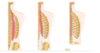
spinal cord terminates?
Roots fuse to form mixed spinal nerve which eventually produces posterior and anterior rami
Spinal cord terminated in cone shape called conus medullaris
Continues as thin connective tissue cordcalled filum terminale which is anchored at dorsum of the coccyx
(if you stand on your head your spinal cord doesn’t come crashing down)

spinal meninges continuous with?
Spinal meninges are continuous with cranial meninges via foramen magnum
meninges layers
Dura mater
Arachnoid mater
Pia mater
spinal cord suspended in canal vi?
formed from?
subarachnoid space?
Spinal cord is suspended in the canal by the denticulate ligament (found laterally)
The ligament is formed of pial and arachnoid tissue
Note - just like in brain, subarachnoid space filled with CSF
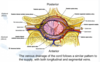
spinal cord made up of?

central canal spinal cord?
posterior vs anterior horn position?
opens into 4th ventricle superiorly
posterior = extends all way up to surface of spinal cord
anterior horn does not
(just helps with orientation)
what is present in spinal cord from T1-L2?
small lateral horn which contains the preganglionic sympathetic neurons

arterial supply to spinal cord?
3 major longitudinal arteries
- 1 anterior
- 2 posterior
- originate from vertebral arteries and run the entire length of spinal cord
Segmental arteries
- Derived from vertebral, intercostal and lumbar arteries
Radicular arteries
- Travel along dorsal and ventral roots

artery of adamkiewicz?
derived from?
also known as great anterior radiculomedullary artery
it is largest segmental artery of spinal cord
derived from 9-12th intercostal artery
venous drainage of spinal cord?
extrinsic muscles of the back?