Vertebral column Flashcards
how many vertebrae in adult vertebral column?

when do sacral verebrae fuse?
late puberty
curvatures of vertebral column?
primary curves (concave anteriorly) = thoracic + sacral/coccygeal (first to form in embryo)
secondary (convex anteriorly) = cervical + lumbarr

when do cuvratures of the vertebral column develop?
how do they develop?
thoacic and sacrococcygeal cuvratures are established in foetal development
cervical and lumbar secondary curves develop in infancy
- 2-3 months = independently hold head, compensatory secondary curvature develops in cervical region
- 6-8 months = further compensatory secondary curvature in lumbar region
1* are caused by the shape of the vertebrae
2* curves arise from changes to shape of intervertebral disc
(degeneration of discs in elderly results in more pronounced primary curvature)
some examples of pathological vertebral curvatures? (3)

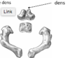
black line seperating?

vertebral body (top)
vertebral arch
juvenile vertebrae parts
never call centrum vertebral body - as vertebral body is more than just centrum (+ neural arch)

what is adult vertebral body derived from?
juvenile centrum plus and small portion of the neural (vertebral) arch
explain ossification of the centra (centrum)
where does it first appear?
progression?
begins in mesenchymal stage (undifferentiated mesoderm)
ossification begins dorsal to the notocord
true endochondral ossification (center -> periphery)
first appears in lower thoracic an upper lumbar regions (T10-L1) between 9-10 foetal weeks
bidirectional progression (reaches L5 by 3rd month and C2 by 4th foetal month)
(white is notocord)
(bottom area is centrum)
(black is ossification)

why is there a ring-shaped area of ossification of the centra?
notochord cells contain angiogenic inhibiting factor which delays vascular penetration to this region
so vertebral centra fro first trimester have an avascular region around notochord
this results in ring-shaped ossification
vascular supply centra?
avascular area around notochord
note that developing centrum has lots of grooves due to vascular supply

explain ossification of the neural arches
starts on inner surface of each hemi-arch
intramembranous ossification followed by endochonral

when does ossification of neural arches first occur and where?
progression?
initiation??
first appear in lower cervical and upper thoracic regions in 2nd foetal month
then spreads upwards and downwards
ossification in upper cervical/lower throacic initiated in response to gasp reflex (muscle contraction)
by 3rd foetal month ossification in lower throacic and upper lumbar regions
ossification here is initiated in response to lower limb movement (muscle contraction)
when and where does neural arch fusion occur?
when do cervical arches fuse?
lowest lubar arches?
therefore…..
1st year of life - neural arches fuse posteriorly at the spinous processes (at the posterior synchondrosis)
- occurs initially in lower throacic and upper lumbar regions and progresses upwards and downwards
cervical arches may not fuse until 2nd year
lowest lumbar arches may not fuse until end of the fifth year
therefore in any individual <6 y/o some degree of non-fusion of arches should be expected (not to be mistaken for fracture)
NB: a synchrondrosis is a primary cartilaginous joint (2 bones seperated by hyaline cartilage - hyaline cartilage can turn to bone with advancing age which is exactly what happens here)

neurocentral fusion?
when does it occur?
adult vertebral body formed from?
fusion between neural arches and centra (occurs ventral to pedicles at neurocentral junction)
occurs between 2-5 years
adult vertebral body formed from centrum and “boutons” of pedicles
- head of costal processes (ribs) only every articulates with these boutons and never with the centrum

where does neurocentral fusion occur first?
last?
1st = lumbar region
followed by cervical segment
last = thoracic vertebrae (possibly due to delay caused by ribs)
does neurocentral junction go away?
no it is maintained throughout life
it is an area of fracture weakness
many vertebral fractures occur at area of neurocentral fusion

…
C1 also called?
ossification?
what is different about C1?
incomplete ring?
atlas (top of the world)
C1 ossifies from 3 primary centres
still have neural arches, no centrum (anterior arch/bar instead)
incomplete ring up until about 2 years as anterior arch takes longer to form than neural arches
pic (A = appearance, F=fusion)

neurocentral ossification C1?
shouldn’t really be called this because there is no centrum
but occurs at 5-6 years

when should you expect a complete fused C1?
5-6 years
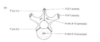
what will C1 initially appear as at birth?
appearance?
7 weeks?
2 neural arches (bony masses)
- larger concave articular facets anteriorly on upper surface
- smaller flatter articular facets on lower surface
at 7 weeks the centre for these lateral masses appears (posterior to articular pillar)
(articular pillar is part that articulates with occipital condyles)
(so its the little wedge shaped area in the first image)

how to differentiate between superior and inferior facets of atlas (C1) at birth?
superior facets are much larger (bear weight of skull, atlanto-occipital joint)
inferior facets and much smaller

what happens to atlas (C1) after birth?
morphology remains unchanged throughout 1st year after birth
but - increases in size (so looks the same just bigger)
in 1st/2nd year ossification occurs in anterior arch
(either as single/paired/multifocal nodules that extend directly from lateral masses)

what happens if there is an absence of the anterior ossification bar required to form anterior arch?
ossification bars will extend directly from lateral masses and form anterior arch
fusion of C1 (atlas)?
growth?
posterior arch is first to fuse (4-5 years) - however remains open in 1% of adults - called spina bifida atlantis
anterior neurocentral junctions may not fuse until 5-6 years
before fusion, endochondral growth occurs at all junctions to allow growth of vertebral canal
adult size reached by 4-6 years (early limitation of size of vertebral canal is clear indication of precocious maturation of CNS)

how to tell spina bifida atlantis apart from posterior arch that just hasn’t used yet?
if hasn’t fused yet - ends will flare towards eachother (pic 1)
in SBA - tips are pointed (pic 2)

line of union between between anterior arch of C1 and lateral masses?
passes through anterior portions of superior articular facet
(see picture)
so superior articular facet is combination of lateral arch and anterior arch
site of fracture weakness :))

C2 (axis) primary ossification?
5 primary centres for ossification
- one for the centrum
- one for each half of neural arch
- once for each half of the dens (odontoid process)
why are there 6 in the image?
final piece of bone is called ossiculum terminale (pointy bit)
generally considered to be a secondary centre of ossification

when do neural arches of C2 (axis) appear?
centrum?
odontoid process?
neural arches = 7-8 foetal weeks (in womb)
centrum = 4-5 foetal months
dens = 4-6 foetal months
why does juvenile dens have forked appearance?
2 ossification centres of odontoid process rapidly coalesce so that intradental synchrondorsis fuses by birth
(see previous cards)

fusion of axis (C2)?
posterior synchondrosis between neural arces fuses 3-4 years
dens fuses laterally to neural arches at dentoneural synchondrosis 3-4 years
- (fusion line passes across superior articular facet so medial 1/3 of facet is formed by dens and lateral 2/3 is formed by neural arch)
inferior articular facet froms entirely from neural arch
complete fusion of foramen transversarium 3-5 years
dentocentral junction (i.e. centrum and dens) + paired neurocentral junctions (centrum + neural arches) fuse 4-6 years
all lines of fusion disappear by 9-10 years

do all lines of fusion for C2 (axis) disappear by 9-10 years?
small horizontal crevice may remain in posterior dentocentral junction
(site of fracture weakness)
what happens to C2 (axis) at 2 years of age?
12 years?
ossiculum terminale appears at 2 years
os terminale fuses with dens at 12 years
2ry ossification centres C2 (axis)?
inferior annular ring?
5 (6 if include os terminale) secondary centres (epiphyses)
- 2 flake-like epiphyses for transverse processes (see picture)
- 2 plate-like epiphyses for bifid spinous process (bottom of pic)
- inferior annular ring
a 2ry centre appars in inferior anular ring and a small tongue of bone processes up to posterior surface of dens, filling the interdental groove

ossification of typical cervical vertebrae (i.e. not C1 or C2)?
develop with general ossification pattern of any typical vertebra
formed from 3 centres
- neural arches = 2-3 months
- centrum = 3-4 months

fusion of typical cervical vertebrae?
2 years = posterior synchondrosis
3-4 years = neurocentral fusion
- once neurocentral fusion complete, synovial uncovertebral joints (of Luschka) form on sides of neural element of vertebral body (red in pic)
single bony element close to adult morphology by 4 years

2ry ossification centres (epiphyses) of typical cervical vertebrae?
when do they appear?
fuse?
6 centres
- 2 x transverse processes
- 2 x spinous processes
- 1 x superior annular ring
- 1 x inferior annular ring
appear beginning of puberty
begin fusing end of puberty (18+), complete fusion by 24 y/o

typical thoracic vertebrae ossification?
general ossification pattern as previously outlined
3 primary centres present by 3 foetal months

typical lumbar vertebrae ossification?
again, general ossification pattern as previously outlined (2 neural arches + 1 centrum)
all 3 primary centres present by 3rd foetal month

thoracic and lumbar secondary ossification?
- superior annular ring
- inferior annular ring
- 2 x transverse processes
- 1 x spinous process
for thoracic - sometimes have centres on costal surfaces
for lumbar - sometimes on mamillary processes

sacrum ossification pattern?
complex - 21 seperate centres!!
each sacral vertebrae is represetned by typical 3 primary centres
- 1 x centrum
- 2 x neural arches
plus….
S1-S3 have paired lateral elements
these form ventral aspect of alae and are the site of sacroiliac articulation

describe pattern of sacral ossification
3rd foetal month - S1 + S2 centra appear
4th foetal month - S3 + S4 centra, S1-S3 neural arches
5th foetal month - S5 centra, S4-S5 neural arches
6th-8th foetal month - S1-S3 paired costal elements
birth - all centres present but tiny

sacral fusion?
remain seperate until?
neural arch unites with lateral elements = 2-5 years
- united component then fuses with central = 2-6 years
all primary centres fused except posterior synchrondrosis at spinous processes (interesting because in other vertebrae post. synchrondrosis is first to fuse - for sacrum it is last)
- fuses 7-15 years
individual sacral vertebrae remain eperate until puberty

2ry ossification centres (epiphyses) sacrum?
not consistent in number and varies between individuals
14 constnt centres:
- 10 x annular rings (2 per sacral centrum)
- 2 x auricular epiphyses (SI joint surface) - seen above lateral margin epihyses in image
- 2 x lateral margin epiphyses
multiple small variabel elements:
- flake-like epiphyses
- spinous process
- median sacral crest
- lateral sacral crest
- between sacral vertebrae

fusion of 2ry centres sacrum?
general rules?
12 years = lateral elements fuse, annular epiphyses commence fusion in caudocranial direction
- unusual - fusion is happening bottom to top so S5 fuses to S4, S4 to S3 and so on…
general rule
- if spaces can be identified between bodies <20 y/o
- if space only between S1 + S2 <27 y/o
- complete union not observed until >25 y/o
epiphysis for sacroiliac joint appear?
fuse?
what follows same pattern?
appear 15-16 years
fus eby 18 years
lateral margin epiphysis follows same pattern

when does sacroiliac joint form?
complete?
at birth appearance?
adult?
forms 2nd foetal month
complete by 7th foetal month
at birth - straight and parelell to vertebral colulm reflecting quadripedal form
mechanical forces associated with bipedalism cause joint to curve caudodorsally into adult morphology
features of sacroiliac joint?
non-weight bearing? and demonstrates no sexual dimoprhism
coccygeal ossification?
each coccygeal vertebrae forms from a single seperate centre
- Co1 may form from multiple - appears ~1 year
- Co2 = 3-6 yrs
- Co3 = 10 yrs
- Co4 = puberty
recognisable by puberty
