The Spinal Cord Flashcards
(34 cards)
Overview of the spinal cord
- Extends from the Atlas to L1
- spinal cord narrows at L1 to form conus medullaris
- the pia mater elongates to form the filum terminal which attaches to the coccyx
- there is Cervical and Lumbar enlargement of the spinal cord, where the cervical and lumbar plexus occurs
- Cervical: C3-T2
- Lumbar: L1-S3
- surrounded by the meninges (pia, arachnoid, dura)
Regions of the Spinal Cord
- Cervical: C1-C7
- the enlargement allows innervation to the upper limbs
- Thoracic: T1-T12
- Lumbar: L1-L5
- the enlargement allows innervation to the lower limbs
- Sacral: S1-S5
The spinal nerves of the spinal cord
- 31 pairs of spinal nerves attached to the anterior/motor roots/ efferent and the posterior/ sensory roots/ afferent
Grey Matter of the spinal cord
- H-shaped, with anterior and posterior grey horns
- horns are joined by a thin grey commissure which contains the central canal
- ventral,
- motor neurons
- lateral
- preganglionic synaptic neurons
- dorsal horns
- receiving sensory input
- amount of grey matter correlates with the amount of muscle innervated at that level (hence enlargements)
White Matter of the Spinal cord
- Surrounds grey matter, made up of myelinated axons
- made up of white columns/tracts/funiculi
- long ascending tracts carry afferent impulses to the brain
- long descending tracts carry efferent impulses from the brain
- Dorsal column contains ascending tracts
- Lateral column contains descending and ascending tracts
- Ventral column contains mainly descending tracts
What is the receptor type for touch, pressure and vibration sensations?
- describe the type of fibre present and the velocity of conduction
- Mechanoreceptors : they are A-Beta with a wide diameter, fast conduction velocity
- Merkel’s cells
- Ruffini end organs
- Pacinian corpuscles
- Bare nerve endings: they are A-Theta with a medium diameter and speed
What is the receptor type for pain and temperature sensations?
- describe the type of fibre present and the velocity of conduction
- Bare nerve endings: with A-theta fibres and a medium diameter and speed
- for fast ‘picking’ pain
- Bare nerve endings: with C-fibres thin diameter, and slow conduction
- for slow-burning pain, itch
- also for non-discriminative touch
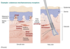
Explain the different types of receptors seen in this diagram
What factors allow somatosensory afferents to convey different functional information?
- Fibre type: axon diameter
- Their Receptive field
- for cutaneous afferents, this is the area of the skin surface over which stimulation results in a significant change in the action potential
- this along with innervation density allows for two-point discrimination
- Rapidly adapting afferents
- afferents that fall silent on continued stimulation
- good for conveying information about changes in ongoing stimulation
- Slowly adapting afferents
- better at conveying spatial information such as size and shape
- the type of stimuli
Explain lower motor neurons
- Lower motor neurons are collected in longitudinally organized columns
- Each column contains the larger, alpha (thick axon, high conductance velocity), and smaller, gamma (thin axon, low conductance velocity), motor neurons to one muscle (or a few functionally similar muscles).
- Each column extends through more than one segment of the cord.
- Each muscle receives motor fibres through more than one ventral root and spinal nerve
- Destruction of a single ventral root or a single spinal nerve will not produce paralysis, only weakness (paresis).
What are Proprioceptive sensory inputs?
- give examples of organs
Proprioceptive sensory inputs: is to give detailed and continuous information about the limbs and other body parts in space. Proprioceptive sensory organs include:
- Muscle spindles- negative feedback regulation of muscle length
- found in striated muscle
- made up of intrafusal muscle fibres surrounded by a capsule of connective tissue and distributed among extrafusal fibres (produce force)
- Innervation form group Ia afferent axons (rapidly adapting response) and group II afferents (sustained response to muscle length)
- Golgi tendon organs- negative feedback regulation of muscle tension
- formed by branches of group Ib afferents
Recreate this topographic map of the body musculature in the primary motor cortex

What are the effects of a spinal cord lesion: Anterior cord syndrome?

- Bilateral lower motor neuron paralysis and muscular atrophy in the segment of the lesion (due to damage to lower motor neurons).
- Bilateral spastic paralysis below the level of the lesion (due to loss of anterior descending tracts).
- Bilateral loss of pain, temperature and light touch sensations below the level of the lesion (due to loss of anterior and lateral spinothalamic tracts).
What are the effects of a spinal cord lesion: Brown-Séquard (cord hemisection) Syndrome

- Ipsilateral lower motor neuron paralysis and muscular atrophy in the segment of the lesion (due to damage to lower motor neurons).
- Ipsilateral spastic paralysis below the level of the lesion (due to loss of anterior descending tracts).
- Ipsilateral band of cutaneous anaesthesia in the segment of the lesion (due to loss of dorsal root).
- Ipsilateral loss of tactile discrimination and of vibratory and proprioceptive sensations below the level of the lesion (due to loss of ascending tracts in the dorsal white column on the side of the lesion).
- Contralateral loss of pain, temperature and light touch (due to loss of crossed lateral spinothalamic tracts on the side of the lesion).
What are the effects of spinal cord lesion: Complete cord transection syndrome?

- Complete loss of sensation and voluntary movement below the level of the lesion.
- Bilateral lower motor neuron paralysis and muscular atrophy in the segment of the lesion.
- Bilateral spastic paralysis below the level of the lesion (due to loss of descending tracts).
- Bilateral loss of all sensations below the level of the lesion (due to loss of ascending tracts).
- Bladder and bowel function no longer under voluntary control (due to loss of descending autonomic fibres)
Describe the circuit anatomy of the ascending tracts in the spinal cord
- First order (primary sensory) neuron
- Enters spinal cord via dorsal root
- Second order neuron
- Ascends spinal cord or brainstem
- Third order neuron
- Projects to the cerebral cortex
Give an overview of the Medial Lemniscus pathway (dorsal column)
- Fine touch: from cutaneous mechanoreceptors
- Proprioception: from muscle spindles, golgi tendon organs, joints
- Provides brain with positional information

Describe the pathway of the Dorsal columns Medial Lemniscus Pathway
- first-order
- second-order
- third-order
First-order
- Enter the spinal cord and ascend the dorsal column on the same side within the
-
Fasciculus gracilis (medial) –> Nucleus gracilis in the medulla
- information from lower limb
-
Fasciculus cuneatus (lateral) –> Nucleus cuneatus in the medulla
- information from upper limb
-
Fasciculus gracilis (medial) –> Nucleus gracilis in the medulla
- Fibres ascend dorsal column uncrossed and synapse on second-order neurons in the medulla
Second-orders neurons
- crosses in the medulla and ascends to the thalamus to form the medial lemniscus
Third-order neurons
- project from the thalamus to the somatosensory cortex (Brodmann areas 1, 2, 3)

Explain the cause and effect of damage to the dorsal column?
- Lesion on one side of the spinal cord results in loss of tactile discrimination & proprioception on the same side__
- Can be caused by multiple sclerosis
- Symptoms: Sensoy ataxia
- loss of coordination and balance without visual cues (no positional information)
- Clinical Test: Romberg’s sign
- sever swaying on standing with eyes closed/ feet together
What is the role of the Spinothalamic tract
Carry information about
- Pain (from nociceptors)
- Temperature
- Crude touch

Describe the pathway of the Spinothalamic tract
First-order
- Enters the dorsal horn and forms the tract of Lissauer
- collateral branches are given off at the tip of the dorsal horn
- runs up or down 1-2 spinal segments
- Synapses in the dorsal horn with second-order neurons
Second-order neurons
- Cross in the dorsal horn at each level
- Ascend in the anterolateral column to the thalamus
- fibres from lower limb: lateral in the tact
- fibres from the upper limb: medial in the tract
Third-order neurons
- project from the thalamus to the sensory cortex

Explain the effects and cause of damage tot he anterolateral column
- Lesion on one side of the spinal cord causes loss of pain, temperature and crude touch on the opposite side
- Outer tract injury causes loss of lower limb pain first as the fibres sit laterally
- cord compression due to herniated disk
- Inner tract injury causes loss of upper limb pain first as the fibres sit medially
- grey matter tumour
Give an overview of the Spinocerebellar tracts and its role?
- Unconscious muscle proprioception
- information from muscle spindles, golgi tendon organs
- for smooth motor coordination
- Two neurons in the pathway
- Comprise of three main tracts
- Anterior and posterior spinocerebellar tracts: carries info from trunk and lower limb
- tracts terminate in the cerebellum on the same side
- left cerebellum control left side of body
Describe the pathway of the posterior Spinocerebellar tract and the effects of damage.
First-order neurons
- synapse in the dorsal horn
Second-order neurons
- Ascend in lateral column to cerebellum (very fast axons)
- Lesion on one’s side of the spinal cord leads to uncoordinated lower limb muscular activity on the same side
- rarely damaged in isolation


