Superior Mediastinum Flashcards
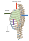
What is the sternal angle and why is it important?
- junction between manumbrium & sternal body
- important landmark
- inferior boundary superior mediastinum
- arch of aorta
- azygous vein drains into superior vena cava
- second costal cartilage
- bifurcation of trachea
What is the mediastinum & why is it clinically important?
- broad central partition that separates the two lateraly placed pleural sacs
- important
- organs
- large veins & arteries
- lymphatics
- nerves
- fascia
- dynamic nature
- breathing
- swallowing
- disease state
- body position

What are the boundaries of the mediastinum?
- superiorly
- the superior thoracic apeture
- inferior
- the diaphragm
- anteriorly
- sternum and costal cartilage
- posteriorly
- the thoracic vertebrae
What are the subdivisions of the mediastinum & what is the name of the line demarcating this boundary?
- transverse thoracic plane (red line)
- sternal angle & T4-T5
- Above this line = superior mediastinum
- Below this line = inferior mediastinum
- anterior
- small area (potential space)
- middle
- heart
- posterior
- the rest of the mediastinum
- anterior

What contents are located within the anterior mediastinum?
- Potential space
- Contents
- thymus gland
- fat
- lymph nodes
- branches of internal thoracic arteries & veins
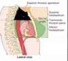
Describe the boundaries of the superior mediastinum
- Superiorly
- superior thoracic apeture
- inferiorly
- horizontal plan extending between sternal angle and IV disc between T4/T5 vertebra
- Anteriorly
- sternum
- posteriorly
- bodies of vertebrae T1-T4
- laterally
- mediastinal pleura
Identify the layers of from the superior view of the mediastinum


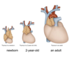
What is the thymus, where is is located & how does it change with age?
- diffuse lymphoid organ
- locatin
- posterior to manubrium
- occupies anterior part of superior mediastinum
- Age changes
- large in infants & young children
- replaced with fat in adults
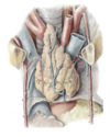
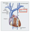
Identify the venous branches indicated provided image

- left & right brachiocephalic veins form from union of
- subclavian and internal jugular
- happening at the sternoclavicular joints
- left & right brachiocephalic veins unit to form the superior vena cava
- shut blood from the head, neck, and upper limbs to right atrium
- Azygous will drain into superior vena cava at the level of the sternal angel

Describe the difference between the left brachiocephalic vein & right brachiocephalic vein?
Tributaries?
- left
- left brachiocephalic is twice as long in length & more oblique trajectory b/c needs to cross the midline
- It is above the superior border of the manubrium in children
- anterior to the roots of the three major branches of the aorta
- tributaties
- vertebral
- first posterior intercostal
- left superior intercostal
- inferior thyroid
- internal thoracic
- Right
- shorter & more verticla trajectory
- tributaties
- vertebral
- first posterior intercostal
- inferior thyroid
- thymic
- internal thoracic
Describe the location of the superior vena cava with relation to its surroundings
For what clinical procedures is it important to understand the location of the superior vena cava?
- begins at the level of the inferior border of the first right costal cartilage
- lies in the right side of the superior mediastinum
- terminal half is in the middle mediastinum
- the azygous vein drains intot he SVC just above the pericardial sac
- Clinical correlation
- central lines are ususally passd through great veins to end in the superior vena cava or right atrium
- used to administer fluids, drugs, and blood
What disease can result from constriction of superior vena cava?
causes?
test you can perform?
Symptoms?
superior vena cava syndrome
- cause
- cancer
- benign tumor
- aneurysm
- problems with head, neck & upper limb draining
- lots of swelling
- associate with really large veins
- patient asked to raise hand up to the ears for a minute or so
- if face turns red with swelling = positive test

identify the branches & sections of the aorta
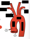
- 3 sections of the aorta
- ascending, arch (in superior mediastinum), descending
- branches of the arch
- brachiocephalic (largest)
- right subclavian
- right common carotid
- left common carotid
- left subclavian (posterior to LCC)
- brachiocephalic (largest)

The curved nature to the aorta creates what shape on an radiograph?

What is the name of the structure that attaches the aortic arch to the left pulmonary artery?
It is the remnant of what embryologicla structure?
Why is it clinically imporant?
- Ligamentum arteriosum
- remnant of fetal ductus artiosus
- Clinical importanc
- site where left recurrent laryngeal nerve can be damaged
- common site for aortic coartications
- abnormal narrowing (stenosis)
- blood flow to the inferior part of hte body is obstructed

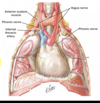
Describe the descent of the left & right phrenic nerves.
- phrenic (C3, 4, 5)
- anterior to root of the lungs
- travel with pericardiacophrenic vessels
- Right phrenic
- lays all on blue
- when enters mediastinum in contact with right brachiocephalic vein
- associated with superior vena cava
- in contact with right atrium
- in contact with inferior vena cava
- lays all on blue
- left phrenic
- posterior to left brachiocephalic vein
- anterior to arch of aorta
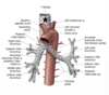
Describe the descent of the right and left vagus nerves
- Right
- descending from the neck
- anterior to right subclavian artery
- gives rise to right recurrent laryngeal nerve around here
- descends between right brachiocephalic vein & brachiocephalic trunk
- pass posterior to root of the lunk
- in contact with the trachea
- Left
- anterior to left subclavian artery
- posterior to left brachiocephalic vein
- anterior to the arch of the aorta
- will give off left recurrent laryngeal nerve around here
- close to ligamentum arteriosum
- posterior to rot of left lung
- never touches the trachea

What is the relationship between the vagus & phrenic nerve?
- Phrenic is lateral to vagus
- Phrenic : anterior to root of the lungs
- Vagus : posterior to root of lungs

Describe the trajectory of the right & left recurrent laryngeal nerve
- Right
- loops aroudn the right subclavian artery and ascends between the trachea 7 esophagus to the larynx
- NOT located in the superior mediastinum
- Left
- branch of left vagus that supplies many of the muscles of the larynx
- loops under arch of the aorta & ascends between teh trachea and esophagus to return to the larynx

What clinical problems can cause damage to the recurrent laryngeal nerve?
What is the result of damage to this nerve?
- any investigative (diagnostic) procedure or disease process in the superior mediastinum may injur these nerves & affect the voice
- bronchogenic or esophageal carcnoma, enlargemetn of the mediastinal lymph nodes, or an aneurysm of the arch of the aorta
- If the procedure is specifically to fix the ligamentum arteriosum, we are specifically talking about hte left recurrent laryngeal nerve
What is the relationsihp between the trachea & aortic arch?
Azygous vessels?
Where does it bifurcate into primary bronchi?
Which mediastinum compartments is it part of?
- descends posterior and to the right of the aortic arch
- divides into the right and left primary bronchi at the sternal angle (or Louis)
- azygous arches over the right primary bronchus while the aorta arches over the left primary bronchus
- part of superior mediastinum
- not a component of the posterior mediastinum
How does the esophagus enter the superior mediastinum?
What is its relationship with the azygous?
Left recurrent laryngeal nerve?
- Enters superior mediastinum between the trachea and vertebral column, wher eit lies anterior to the bodies of the T1-T4 vertebrae
- Azygoud crossed the esophagus on the right
- left side:
- left recurrent nerve is found between the trachea and esophagus

What is the position of the thoracic duct within the superior mediastinum?
What is its function?
How dos it enter the thorax and what is its trajectory?
- Most posterior structure in the superior mediastinum
- main lymphatic duct of the body
- begins in the abdomen and enters the thorax to the right of the aorta
- ascends to the base of the neck to drain into the venous system at the junction of the L. internal jugular and L. subclavian veins

