Posterior Mediastinum Flashcards
What are the boundaries for the posterior mediastinum?
- superiorly
- transverse thoracic plane
- from sternal angle to IVD between T4 &T5
- inferior
- diaphragm
- posteriorly
- vertebrae T5 - T12
- anteriorly
- pericardium
- laterally
- mediastinal pleura

What are the contents of the posterior mediastinum?
- Descendign aorta
- azygos system
- thoracic
- duct
- esophagus
- splanchnic & associated sympathetics

What vertebral level does the esophagus start & stop?
What is its spatial relationship with the root of the lung & the heart, the aorta & the azygous system?
- Esophagus starts at the level of C6
- at level of cricoid cartialge
- passes throug the diaphragm at T10
- Ends at the level of T11
- Runs posterior to root of lung
- behind the left atrium
- aorta will be to the left of esophagus
- however, aorta will become more midline as it descents & you will eventually have aortoesophageal decussation site
- azygous left & posterior to esophagus

What are natual areas of constriction within the eophagus?
Why are these important?
- Junction of esophagus w/ pharynx (C6)
- cricoid cartilage pressing back onto esophagus
- arch of aorta (TV4)
- left main bronchus (T5)
- aortoesophageal decussation site (T10)
They are important so that we are able to identify normal areas of constriction during barium studies compared to diverticula
Normal (left), divertcula (right)

What are the common diverticula that can form in the esophagus?
- Zenkers
- between cricopharyngeus & inferior pharyngeal constrictor
- parabronchial diverticula
- epiphrenic diverticula

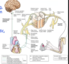
Describe the innervation of the esophagus
Covered in a plexus of nerves: esophageal plexus
- Sympathetic contribution
- sphanchnic
- parasympathetic contribution
- vagus
- As we come down further, will develop anterior & posterior vagal trunks, immediately superior to the diaphragm
- right vagus mainly contibutes to posterior vagal trunk
- left vagus mainly contributes to anterior vagal trunk
- Once through the esophagus, immediately start forming more plexus to innervate structures of the abdomen

What is the role of the sympathetic & parasympathetic innervation of the esophagus?
- Vagal control
- Proximal 1/3 is skeletal & under voluntary control
- Distal 2/3 more smooth muscle & under involuntary control
- parasympathetics in charge of reflexes such as modulating contractions
- sensory - monitoring, modulation
- Sympathetics
- basal motors & carry pain signals
- overlap for the levels of the heart & pain in the esophagus may be confused with cardiac pain (i.e. heartburn)
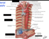
Identify the arterial branches that supply the esophagus
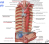
Identify the venous branches that are responsible for draining the esophagus


Identify the lymph nodes that drain the lymph of the esophagus

Eventually, most lymph will drain to the poserior mediastinal nodes
except most superior lymph on right side, which will got to the right lymphatic duct
everything else will make its way back through the thoracic duct
most inferior to left gastric nodes

Describe the start, stop & trajector of the thoracic aorta
- Starts at T4/T5 IVD
- left anterolateral position of our vertebral bodies
- as it descends it will take a more midline approach
- proximal position, esophagus will be to the right of the aorta
- Will end T12 as it passes through aortic hiatus of diaphragm
- by the time it reaches the abdomen, it will be in the midline
- Distal position, esophagus is anterior to aorta

Describe the spatial relationship between the thoracic aorta & the follow structures:
Root of the lung
pericardium
diaphragm
hemiazygous vein
thoracic duct
- posterior to root of the lung
- root of the lung:
- main bronchus
- pulmonary veins & arteries
- bronchial artery
- root of the lung:
- posterior to pericardium
- most inferior portion is posterior to diaphragm as well (before it crosses through)
- anterior to hemiazygous vein & accessory hemiazygous vein
- anterior to thoracic duct

Identify the branches of the thoracic aorta & what they supply

- bronchial
- supply bronchial walls
- nor responsible for taking deoxygenated blood away from the heart – supplying epithelium itself
- mediastinal
- posterior mediastinal stuctures like the lymph nodes
- not supplying the heart nor the pericardium
- esophageal
- middl portion of esophagus
- pericardial
- posterior pericardium of the heart
- superior phenic
- superior aspect of the diaphragm

What veins form the azygous sustem?
Describe the trajectory of the azygous vein
- form by lumbar veins joining with subcostal vein
- as they converge, we will form our azygous veing on the right side
- Travel on the right side of T12-T5, on right of thoracic duct & aorta; right & posterior to esophagus
- posterior intercostal veins on the right will be draining into the azygous system along its length
- At the level of T4 (transverse thoracic plane) will arch from posterior to anterior over the root lung, creating a depressing/fissure seen on radiographs
- drain into superior vena cava
- hemiazygous is on the left side & will drain into the azygous system on the right
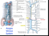
What is shown in the provided image?
the azygous arching over the lung & draining into the superior vena cava
What are the tributaries draining into the azygous system?
- Right broncial will drain directly into azygous
- Posterior intercostal
-
hemiazygous vein
- draining lower intercostal spaces as it ascends, gonna come over at the level of T9, passing posterior to the aorta, esophagus, thoracic duct as it crosses midline
- drain some of the mediastinal veins on their way to the azygous system
- left superior intercostal vein
-
accessory azygous vein
- Draining more superior 5-8 intercostal spaces
- as it descents, at level of T8, will cross midline & passs posterior to the aorta, esophagus, thoracic duct to drain into the azygous
- left bronchial vein

Describe the clinical problems that can arise due to the anastamotic nature of veins
- valvless system
- also a problem for easy spread of metastatic disease
- Superior vena cava
- if blood is unable to reach the heart through the superior vena cava do to a blockage (typically due to malignancy in apical portion of lung)
- present with distended jugular veins
- edema in face/upper extremities
- blood will find alternate route back to the heart
- Inferior vena cava syndrome
- similar to superior vena cava syndrome but will have edema in different areas
- elevated liver enzymes b/c blood pooling within hepatic veins
- portal hypertension
- blockage in portal vein
- travel back through gastric veins/esophageal veins
- & try to make is way back through the inferior vena cava
Describe the trajectory of lymphatic drainage from the cysterna chyli to the termination of the thoracic duct
- Cysterna chyli
- drain lymph from the abdomen
- dilated lymphatic vessel just inferior to the diaphragm at the level of L1/L2 at transpyloric plane
- will start to ascend through cysterna chyli & travel through thoracic duct as it enters the thorax
- posterior to aorta & esophagus
- to righ tof aorta & left of azygous vein
- At the level of T4, the thoracic duct will start arching to the left at junction of internal jugular & subclavian
- where the vast majority of the lymph will drain in the body

What Virchow’s node?
it is mainly associated with what diseases/
a lymph node in the left supraclavicular fossa (th area above the left clavicle) a group of supra clavicular lymp nodes
often see it with gastrointestinal malignancy
What spinal nerve roots are invloved in sympathetic innervation?
What routes can they take to innervate contents within the thorax?
Describe the position of the sympathetic trunk within the thorax
- T1-L2
- 12 bilateral sympathetic ganglio & 12 white rami
- enter trunk through white rami more lateral & longer
- exit trunk though grey
- “come in clean & go out dirty”
- visceral afferent fibers will enter through white rami to enter the spinal chord & transmit pain
- Can take
- spinal nerves
- visceral bb
- T. splanchnic nn
- Sympathetic trunk follows the neck of the ribs
- most lateal structure in the posterior mediastinum
- first thoracic ganglia may fuse to inferior cervical ganglia
- called a “stellate ganglion”

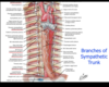
Identifiy the branches of the sympathetic trunk

Identify the vertebral level & conents for the following openings in the diaphragm
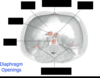
- Lumbocostal triangle (Bochdalek’s triangle)
- gaps in diaphram allowing parietal pleura & renal capsule to contact
- possible route for infection or hernia
- Caval opening T8
- IVC & right phrenic nerve
- Esophageal hiatus T10
- esophagus & anterior/posterior vagal trunks
- Aortic Hiatus T12
- descendign aorta
- thoracic duct
- medial crus T12-L1
- venous system & spanchnic nerves can travel through
- lateral crus T12-L1
- sympathetic trunk can travel through/least splanchnic
- Sternocostal triangle (Larrey’s cleft) T10
- passage for internal thoracic & superior epigstric

