SPR L8 Ankle Joint, Anatomy of the Foot Flashcards
Ankle Joint and Anatomy of the Foot
Learning Outcomes
- Describe the anatomy of the ankle joint
- Describe the anatomy of the tarsal tunnel and of the plantar aspect of the foot
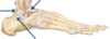
Identify the following bones of the foot
(see picture)
Tarsal Bones
Metatarsal Bones
Phalanges

Tarsal Bones (7)
- Calcaneus
- Talus
- Navicular
- Cuboid
- a) Cuneiform 1 b) Cuneiform 2 c) Cuneiform 3
Metatarsal Bones (5)
- a) MT 1 b) MT2 c) MT3 d) MT4 e) MT5
Phalanges (14)
- a-e) Proximal Phalanges
- a-e) Distal Phalanges
- a-d) Middle Phalanges (4)
Ankle Joint
During this lecture, the following will be considered…
(for general persual)
- Articular surfaces
- Classification
- Stability
- Arterial supply
- Nerve supply
- Movement
Ankle Joint: Articular Surfaces
- What are the articular surfaces of the ankle joint?
- What also contributes to the formation of the joint?
- the talus and the distal end of the tibia.
- The distal end of the fibula
The distal ends of the tibia and fibula (along with the inferior transverse part of the posterior tibiofibular ligament) form a malleolar mortise into which the pulley- shaped trochlea of the talus fits.

Ankle Joint: Ligaments
What are the ligaments of the ankle joint?

- Anterior talo-fibular ligament
- Posterior talo-fibular ligament
- Calcaneofibular ligament
- Deltoid Ligament

Ankle Joint: Ligaments
Name the ligaments identified in the image

Ankle Joint: Ligaments
Name the ligament identified in the image

Deltoid Ligament

Clinical Significance
What does this image show?

rupture of the anterior talo-fibular ligament.
Clinical Significance
What does this picture show?

rupture of the deltoid ligament.
Note the direction of force as indicated by the blue arrow
Movement
What movements take place at the ankle joint?
- dorsiflexion (extension of the foot at the anke joint)
- plantarflexion (flexion of the foot at the ankle joint).

Posterior Compartment
Superficial Muscles
What are these?
- Gastrocnemius m.
- Soleus m.

Posterior Compartment of the Leg
Deep Muscles
What are these?
- Tibialis posterior m.
- Flexor digitorum longus m.
- Flexor hallucis longus m.

Neurovascular Structures
What
- Nerve
- Artery
Is indicated by the arrows?

- Tibial Nerve (branch of the sciatic nerve)
- Posterior Tibial Artery
Neurovascular Structures
Where does the tibial nerve originate?
It is a branch of the sciatic nerve
Tarsal Tunnel
- Where is the tarsal tunnel?
- List the structures that run within it
1. located posterior to the medial malleolus
2.
- Tendon of tibialis posterior m.
- Tendon of flexor digitorum longus m.
- Posterior tibial artery and venae comitantes
- Tibial nerve
- Tendon of flexor hallucis longus m.

Plantar Aspect of the Foot
- How many layers of structures are within the plantar aspect of the foot?
- How many of these are muscular layers?
- What are the remaining 2 layers?
- What are the neurovascular structures?
- Why are there 2 neurovascular layers?
- 6
- 4
- Neurovascular
- the medial and lateral plantar arteries that run alongside the medial and lateral plantar nerves, respectively.
- these structures are initially located superficially and travel deep.
Arteries – Plantar Aspect of Foot
- What does the posterior tibial artery divide into?
- it’s two main terminal branches: the lateral plantar artery and the medial plantar artery

Nerves – Plantar Aspect of Foot
What does the tibial nerve divide into?
It’s two main terminal branches: Medial plantar nerve and Lateral plantar nerve

Plantar Aponeurosis
- When will the plantar aponeurosis be revealed?
- What is this fibrous structure analogous to?
- What is the function of this structure?
- What is the next stage of dissection, what lies underneath?
- Once you have made the relevant skin incisions and have removed subcutaneous tissue,
- the palmar aponeurosis in the hand and has the same function
- protection
- remove the plantar aponeurosis to reveal the 1st layer of muscles.

Muscle Layer 1
Which muscles are located in the first layer?

2. Flexor digitorum brevis m.
Two abductors:
1. Abductor hallucis m.
3. Abductor digiti minimi m.

Muscle Layer 2
Which muscles are located in the second layer?
Which tendons sweep into this muscle layer?
- Quadratus plantae m.
- Lumbrical mm. (attached to the flexor digitorum longus tendons)
tendons of flexor hallucis longus and tendons of flexor digitorum longus mm.

Muscle Layer 3
What muscles are in this layer?
- Adductor hallucis m. (oblique and transverse heads)
- Flexor hallucis brevis m.
- Flexor digiti minimi brevis m.

Muscle Layer 4
What muscle is in this layer?
the plantar interosseous muscles/plantar interossei

Cutaneous Innervation
Name the nerves that are involved with cutaneous innervation of the plantar aspect of the foot.

3 Medial plantar n.
- Lateral plantar n.
- Sural n.
- Saphenous n.
- Calcaneal nn. (these nn. are branches of the tibial nerve)


