Sports - Upper Extremity (Complete) Flashcards
What is the rotator interval of the shoulder?
[DeLee & Drez’s, 2015]
- Triangular space formed by:
- Supraspinatus
- Subscapularis
- Glenoid
- Contents
- Coracohumeral ligament
- Superior glenohumeral ligament
- Joint capsule

What is the critical shoulder angle (CSA)?
[JBJS REVIEWS 2018;6(8):e1]
- CSA is the angle between the plane of the glenoid fossa (the line from the inferior edge of the glenoid to the superior edge of the glenoid) and a line drawn from the inferior edge of the glenoid to the lateral edge of the acromion on a true anteroposterior (Grashey) shoulder radiograph
* Accounts for contributions from both glenoid inclination and lateral acromial length - Normal = 30-35°
- <30 = increased risk for GH arthritis
- Decreased CSA (<30°) increases compressive forces across the glenohumeral joint
- >35 = increased risk for rotator cuff tear
- Increased CSA (>35°) is thought to alter deltoid vectors, which results in increased superior shear forces on the rotator cuff muscles

What is the epidemiology of rotator cuff tears?
[Clin Sports Med 31 (2012) 589–604]
- Full thickness tear is present in 25% of patients in their 60s and 50% of patients in their 80s
- 50% of patients >65 with a symptomatic full thickness tear will have an asymptomatic full thickness tear on the contralateral side
- 50% of asymptomatic tears develop symptoms in 2-3 years
- 50% of symptomatic tears increase in size
Where is the ‘bare area’ located in the proximal humerus?
[J Am Acad Orthop Surg 2014;22:521-534]
- It is the triangular area between the humeral head articular surface and the medial margin of the posterior cuff insertion
- The superior apex of the triangle is where the supraspinatus and infraspinatus fibres converge

Where does the rotator cuff re-tear or failure of healing occur?
[JAAOS 2017;25:e261-e271]
Tendon-bone interface
Although adequate pain relief and patient satisfaction can be achieved in the absence of tendon healing following RTC repair, what are the benefits of tendon healing?
[JAAOS 2017;25:e261-e271]
- Higher strength
- Increased function
- Higher outcome scores
What risk factors are associated with lower tendon-bone RTC healing following repair?
[JAAOS 2017;25:e261-e271]
- Increased age
- Osteoporosis (independent of age)
- Chronic rotator cuff tear
- Muscle atrophy
- Fatty degeneration
- Larger size
- Tobacco use
- Low initial fixation strength
- Larger gap
- High tension repair
What is the DeOrio and Cofield classification for RTC tear size?
[J Am Acad Orthop Surg 2014;22:521-534]
Measurement based on “length of the greatest diameter of the tear” (ie. AP or ML)
- Small = 0-1cm
- Medium = 1-3cm
- Large = 3-5cm
- Massive = >5cm
What are the classification systems used to describe RTC tears?
[J Am Acad Orthop Surg 2014;22:521-534]
- Patte classification – D**egree of retraction
- Stage 1 = lateral margin of cuff close to footprint area
- Stage 2 = lateral margin of cuff at level of humeral head
- Stage 3 = lateral margin of cuff at level of glenoid
2. Goutallier Staging System – Fatty infiltration - Stage 0 - normal muscle
- Stage 1 - some fatty streaks
- Stage 2 - amount of muscle is greater than fatty streaks (<50% fat)
- Stage 3 - amount of muscle is equal to fatty streaks (50% fat)
- Stage 4 - amount of muscle is less than fatty streaks (>50% fat)
3. Thomazeau classification – Muscle atrophy - Stage 1 - normal or slight atrophy
- Occupation ratio = 0.6-1
- Stage 2 - moderate atrophy
- Occupation ratio = 0.4-0.6
- Stage 3 - severe atrophy
- Occupation ratio = <0.4
- Ellman classification – Degree of partial thickness tear
- Grade 1 - tear <3mm in depth
- Grade 2 - tear 3-6mm in depth
- Does not exceed 50% of tendon thickness
- Grade 3 - tear >6mm in depth
- Involves > 50% of tendon thickness
- Snyder classification – Tear type
- Involves > 50% of tendon thickness
- Type A - Articular sided partial tear
- Type B - Bursal sided partial tear
- Type C - Complete tear

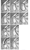
What is the classification of rotator cuff tear shape proposed by Davidson and Burkhart; Describe repair of each shape?
[J Am Acad Orthop Surg 2014;22:521-534]
- Crescent-shaped
- Most common
- Excellent medial-lateral mobility allowing tension-free repair back to GT
- U-shape and V-shape
- Apex of tear extends farther medial toward glenoid
- Medial-lateral mobility is limited, anterior-posterior mobility is adequate
- Repair by “margin convergence”
- Suture free margins together converting tear into a smaller crescent tear
- L-shape and reverse L-shape
- Have both a transverse and longitudinal component
- L-shape tears propagate along the interval between the supraspinatus and infraspinatus
- Reverse L-shape tears propagate through the rotator interval
- One edge is more mobile than the other
- Repair by technique similar to “margin convergence”
4. Massive, contracted, immobile - L-shaped or U-shaped
- Immobile in both AP and ML direction
- Interval slide technique to enhance mobility
- Anterior interval slide
- Incise the superior margin of the rotator interval and the CHL at the corocoid base
- Posterior interval slide
- Incise the interval between supraspinatus and infraspinatus towards the scapular spine
- ***Suprascapular nerve at risk
- Anterior interval slide
- Management options [JSES 2015; 24, 1493-1505]
- Nonoperative management
- Arthroscopic debridement with biceps tenotomy or tenodesis
- Complete repair
- Partial repair
- Patch augmentation
- Superior capsular reconstruction
- Tendon transfer
- Reverse total shoulder arthroplasty

What angle should a suture anchor be inserted to increase an anchors resistance to pullout?
45 degrees (the Deadman Angle)

What is the definition of a ‘massive’ RTC tear?
[International Orthopaedics (2015) 39:2403–2414]
Various definitions exist:
- >5cm tear in either the A-P or M-L direction (Cofield)
- Complete tears of at least 2 RTC tendons (Gerber)
- Coronal length and sagittal width ≥2cm on MRI (Donaldson)
What is the classification of massive rotator cuffs based on location?
[J Am Acad Orthop Surg 2013;21:492-501]
- Posterosuperior
* Involving the supraspinatus, infraspinatus, and possibly teres minor - Anterosuperior
* Involving the subscapularis and supraspinatus
What factors should be considered when determining if a RTC tear is repairable or irreparable?
[J Am Acad Orthop Surg 2013;21:492-501]
- Size
- Retraction
- Fatty infiltration and atrophy
* Goutallier stage 3-4 = generally considered irreparable - Acromiohumeral distance
* <7mm = generally considered irreparable - Static vs. dynamic superior migration
* Static migration = generally considered irreparable
What tendon transfers can be considered for irreparable RTC tears?
[J Am Acad Orthop Surg 2013;21:492-501]
- Latissimus dorsi for irreparable posterosuperior tears
- Pectoralis major for irreparable anterosuperior tears

What is the classification system for fatty infiltration on CT/MRI?
[J Am Acad Orthop Surg 2013;21:492-501]
Goutallier Staging System
- Stage 0 - normal muscle
- Stage 1 - some fatty streaks
- Stage 2 - amount of muscle is greater than fatty streaks (<50% fat)
- Stage 3 - amount of muscle is equal to fatty streaks (50% fat)
- Stage 4 - amount of muscle is less than fatty streaks (>50% fat)
***Note – fatty infiltration is not reversible

What factors contribute to retear rates after repair of massive RTC tears?
[J Shoulder Elbow Surg (2015) 24, 1493-1505]
- Increased fatty infiltration
- Decreased acromiohumeral space
- Smoking
- Size of the rotator cuff tear
- Increased tension on the repair
What are the indications for surgery for rotator cuff tears?
[Sports Med Arthrosc Rev 2018;26:129–133]
- Persistent pain despite nonoperative treatment (4-6 months)
- Options:
- Decompression with arthroscopic acromioplasty +/- debridement
- Indication
- Impingement
- Low grade partial articular sided tear
- Indication
- Rotator cuff repair
- Indication
- Symptomatic full-thickness tears
- Acute bursal-sided partial thickness tears that involve >25% of tendon thickness
- Partial articular-sided tears involving >50% of tendon thickness
- Indication
What patient factor predispose to developing calcific tendinitis of the RTC?
[J Am Acad Orthop Surg 2014;22:707-717]
- Female
- Age (30-60)
- Right shoulder > left shoulder
- Endocrine disorders
- Hypothyroidism
- Diabetes
- ?estrogen/menstrual disorders
- Tendon overuse
Where are the calcific deposits most commonly found in calcific tendonitis of the RTC?
[J Am Acad Orthop Surg 2014;22:707-717]
- 5-2 cm from the insertion in the hypovascular zone of the superior cuff
* Most common tendon involved is the supraspinatus
Describe the pathogenesis of calcific tendinitis of the RTC and the three main stages described by Uhthoff and Loehr
[J Am Acad Orthop Surg 2014;22:707-717]
- Calcific tendinitis of the RTC has a different pathogenesis than insertional RTC calcific tendinitis and calcific tendinitis at other sites (eg. Achilles, patellar tendon) which are degenerative
- Calcific tendinitis of the RTC is an active, cell-mediated process (rather than degenerative)
- Three main stages
- Precalcific stage
- Fibrocartilage metaplasia of the tendon in hypovascular zone
- Calcific stage
- Formative phase
- Calcific deposits form
- Resting phase
- Dormant
- Resorptive phase
- Calcific deposits replaced by fibroblasts and granulation tissue
- Most painful
- Postcalcific stage
- Formative phase
What are the two commonly used radiographic classification systems for calcific tendonitis of the RTC?
[J Am Acad Orthop Surg 2014;22:707-717]
- Gartner and Heyer
- Type I
- Well circumscribed, dense
- Type II
- Soft contour/dense or sharp/transparent
- Type III
- Translucent and cloudy appearance without clear circumscription
- Mole et al (French Society of Arthroscopy)
- Type A
- Dense, homogenous, sharp contours
- Type B
- Dense, segmented, sharp contours
- Type C
- Heterogeneous, soft contours
- Type D
- Dystrophic calcifications at the insertion of the rotator cuff tendons

What are the radiographic features of cuff tear arthropathy?
[AAOS comprehensive review 2, 2014]
- Superior humeral head migration
* Decreased acromiohumeral space - Acetabularization of the acromion
- Femoralization of the humeral head
* Rounding of the GT - Eccentric superior glenoid wear
- Osteopenia
- Snowcap sign
* Subarticular sclerosis - Absence of the typical peripheral osteophytes
* Lack inferior and medial humeral head osteophytes
Describe ‘pseudoparalysis’ of the shoulder
[J Bone Joint Surg Am. 2012;94:e34(1-11)]
- Defined as inability to actively elevate the arm in the presence of free PROM and in the absence of a neurologic lesion
- Occurs as a result of superior migration of the humeral head due to unopposed deltoid contraction in the presence of a rotator cuff tear (loss of the inferior directed force)