Sports - Lower Extremity (Complete) Flashcards
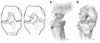
What are the arthroscopic hip portals and structures at risk?
[AAOS comprehensive review 2, 2014]
- Anterior
- Lateral femoral cutaneous nerve
- Femoral nerve
- Femoral artery
- Anterolateral
* Superior gluteal nerve - Posterolateral
* Sciatic nerve - Midanterior
* Lateral femoral cutaneous nerves

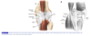
During hip arthroscopy, what nerve is at risk due to traction and the perineal post?
[AAOS comprehensive review 2, 2014]
Pudendal nerve
- Can result in:
- Hypoaesthesia of the perineum, scrotum and glans penis
- Erectile dysfunction
- Urinary incontinence
What are the indications for hip arthroscopy?
[Sports Health. 2017; 9(5): 402–413.]
- Central compartment
- Labral tears
- Chondral pathology
- Ligamentum teres pathology
- Septic arthritis
- Loose bodies
2. Peripheral compartment - Femoroacetabular impingement
- Subspine impingement
- Synovial disorders
- Capsular disorders
- Psoas tendon disorders
3. Peritrochanteric compartment - Greater trochanteric pain syndrome
- External snapping hip/iliotibial band disorder
- Deep gluteal space
- Ischiofemoral impingement
- Proximal hamstring disorders
- Sciatic nerve disorders
What are the contraindications to hip arthroscopy?
[Sports Health. 2017; 9(5): 402–413.]
- Advanced OA
- Ankylosis
- Acetabular and/or femoral dysplasia
- Severe deformity
* Retroversion, SCFE, Perthes - Obesity (relative)
- Neurological injuries/disorders (relative)
* Eg. pudendal neuralgia or peroneal or sciatic nerve palsy
What are the most common complications following hip arthroscopy?
[Bone Joint J. 2017 Dec;99-B(12):1577-1583]
- Nerve injury (0.9%)
- Pudendal > LFCN > sciatic > common peroneal > femoral
- Traction injuries include sciatic, common peroneal and femoral [Muscles Ligaments Tendons J. 2016 Jul-Sep; 6(3): 402–409.]
- Compression injuries include pudendal nerve
- Portal placement injuries include LFCN
2. Iatrogenic injury (0.7%) - Chondral > labral
3. HO (0.6%)
4. Adhesions (0.2%)
5. Infection (0.2%) - Superficial > deep
6. Other - DVT, perineal skin damage, hematoma, broken instrument, incomplete reshaping, femoral neck fracture, hip instability, iliopsoas tendinitis, AVN, ankle pain, arthrofibrosis, dislocation
What is the most common major complication following hip arthroscopy?
[Bone Joint J. 2017 Dec;99-B(12):1577-1583]
Intra-abdominal fluid extravasation
What is the innervation of the acetabular labrum?
[BMC Musculoskeletal Disorders 2014, 15:41]
Branch from nerve to quadratus femoris and obturator nerve
- Contains:
- Free nerve endings for nociception
- Nerve end organs (Pacini, Golgi, Ruffini corpuscles) for proprioception
- Higher concentration in:
- Anterosuperior and postersuperior labrum
- Articular side more so than the capsular side
What is the blood supply to the acetabular labrum?
[J Bone Joint Surg Am. 2010 Nov 3;92(15):2570-5]
Periacetabular vascular ring
- Originates from
- Superior and inferior gluteal vessels
- Medial and lateral femoral circumflex arteries
- Intrapelvic vascular system.

What is the function of the hip labrum?
[Journal of Biomechanics 33 (2000) 953-960]
- Deepens the acetabulum and extends the coverage of the femoral head
- Contributes to a negative pressure vacuum effect which adds stability to the hip joint
* Greater force required to distract joint - Provides a seal against fluid flow in and out of the intra-articular space enhancing lubrication mechanisms
* Encapsulates the fluid in the joint - Limits the rate of fluid expression from the cartilage during loading which enhances the cartilages ability to carry load and limit stresses on the cartilage
What is the Seldes classification of hip labral tears?
[Clin Orthop Relat Res. 2001 Jan;(382):232-40]
Type 1 – “Detachment”
- Detachment of the labrum from the articular hyaline cartilage at the transition zone
Type 2 – “Intrasubstance”
- One or more cleavage planes of variable depth within the substance of the labrum

What are the causes of hip labral tears?
[J Am Acad Orthop Surg 2017;25:e53-e62]
- Trauma
- FAI
- Dysplasia
- Hip hypermobility/capsular laxity
- Degeneration
How can you classify damage to the ligamentum teres, labrum and articular cartilage during hip arthroscopy?
[J Am Acad Orthop Surg 2017;25:e53-e62]
- Domb classification of ligamentum teres tears
- Grade 0 = No tear
- Grade 1 = <50% tear
- Grade 2 = >50% tear
- Grade 3 = 100% tear
- Seldes Classification of labral tears
- Grade 1 - chondrolabral junction tear
- Grade 2 - intrasubstance tear
- ALAD (acetabular labrum articular disruption) Classification
- Grade 1 - softening of the adjacent cartilage
- Grade 2 - early peel of cartilage
- Carpet delamination
- Grade 3 - large flap of cartilage
- Grade 4 - loss of cartilage
4. Outerbridge classification - Grade 0 - normal cartilage
- Grade 1 - cartilage with softening and swelling
- Grade 2 - partial thickness defect with fissures on the surface that do not reach subchondral bone or exceed 1.5cm in diameter
- Grade 3 - fissuring to the level of the subchondral bone in an area with a diameter larger than 1.5cm
- Grade 4 - exposed subchondral bone

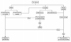
Describe the decision making algorithm when considering arthroscopic labral debridement vs. repair vs. reconstruction
[J Am Acad Orthop Surg 2017;25:e53-e62]
- Stable torn labrum
- Acetabuloplasty not needed = selective debridement
- Acetabuloplasty needed = repair
- Unstable torn labrum
- Viable tissue = repair
- Nonviable tissue, young patient = reconstruction
- Poor vascularity or advanced age = selective debridement
- Mostly calcified torn labrum
- Advanced age = selective debridement
- Young = reconstruction
What are the 3 main types of FAI?
- Cam impingement – femoral based abnormality
- Pincer impingement – acetabular based abnormality
- Combined/mixed-type
What are the features of a cam-lesion?
- Aspherical femoral head
- Reduced head-neck offset
- Characteristic ‘bump’ at the head-neck junction
- Pistol grip deformity
Where is the typical cam-lesion located?
[J Am Acad Orthop Surg 2013; 21(suppl 1):S20-S26]
Anterosuperior head-neck junction
What are the features of the pincer-lesion?
[J Bone Joint Surg Am. 2013;95:82-92]
- Global overcoverage
* Coxa profunda, coxa protrusio - Focal overcoverage
* Cephalad retroversion - Acetabular retroversion
What femur orientation contributes to FAI – anteversion or retroversion?
Femoral retroversion
What radiographs and radiographic findings are important in assessing FAI?
[DeLee & Drez’s, 2015]
- Radiographic views
- AP pelvis
- Lateral view
- Frog-leg lateral, Dunn views, cross-table lateral
- False profile view
- Signs of pincer-lesion
- Crossover sign [AAOS comprehensive review 2, 2014]
- Normally the anterior lip of the acetebulum lies medial to the posterior lip and converge at the superolateral aspect of the acetabulum
- With retroversion the anterior lip proximally lies lateral to the posterior lip and distally lies medially creating the crossover sign
- Prominent ischial spine sign
- Normally the ischial spine is hidden behind the acetabulum, if it appears more prominent it indicates acetabular retroversion
- Posterior wall sign
- Posterior rim of the acetabulum lies medial to the center of rotation of the femoral head indicating retroversion
- Lateral center edge angle
- Formed by a vertical line and a line connecting the femoral head center with the lateral edge of the acetabulum
- LCEA >40 suggests pincer-lesion
- Os acetabulum
3. Signs of cam-lesion [JAAOS 2013;21(suppl 1):S20-S26] - Alpha angle
- Formed by a line along the axis of the femoral neck and a line from the center of the femoral head to the point where the head diverges outside the circle
- >50 degrees is associated with FAI
- Head-neck offset and offset ratio
- Based on a lateral view, a line parallel to the long axis of the femoral neck is drawn along the anterior femoral neck and second line along the anterior aspect of the femoral head
- The distance between the two is the head neck offset
- <8mm likely represents cam-lesion
- Offset ratio is the distance between the two lines divided by the diameter of the femoral head
- <0.17 likely represents cam-lesion

What radiographic view best demonstrates the maximal CAM deformity?
[JAAOS 2013;21(suppl 1):S20-S26]
45° Dunn view
What radiographic view best demonstrates the anterior CAM deformity?
[JAAOS 2013;21(suppl 1):S20-S26]
Cross table lateral and frog leg lateral
What special tests should be performed during the physical exam for FAI?
[J Am Acad Orthop Surg 2013; 21(suppl 1):S16-S19]
- Impingement test (FADIR)
* With the hip at 90° of hip flexion the hip is internally rotated and adducted - Posterior impingement test
* Hip extension combined with external rotation - Log roll test
- Resisted hip flexion test
- FABER
Associated injuries in cam impingement include?
[Orthop Clin N Am 44 (2013) 575–589]
- Labral detachment from the acetabular rim
- Cartilage delamination (full and partial-thickness)
* Deeper compared to peripheral cartilage delamination in pincer impingement
Associated injuries in pincer impingement include?
[Orthop Clin N Am 44 (2013) 575–589]
- Labral pathology
- Peripheral cartilage delamination
- Contracoup chondrolabral lesions in the posterior acetabulum
* Due to anterior levering of the femur causing posterior shear