Soft Tissue and Joint Tumors Flashcards
Where is it thought that soft tissue tumors arise from?
• Thought that soft tissue tumors arise from pluripotent stem cells (we used to think they came from soft tissue and joints)
What should be the top three in your differential for a soft tissue tumor of the joint?
• Gangion Cyst
• Giant Cell Tumor of the Tendon Sheath
• Tenosynovial Giant Cell Tumor
What should be in your differential for a tumor of adipose tissue?
- Lipoma
- Liposarcoma
What should be in your differential for Fibrous tumors?
- Nodular Fasciitis
- Myositis Ossificans
- Fibromatosis (Superficial and Deep)
- Fibrosarcoma
What should be in your differential for Tumors of the Skeletal Muscle?
• smooth muscle?
Skeletal:
• Rhabdomyoma
• Rhabdomyosarcoma
Smooth:
• Leiomyoma
• Leiomyosarcoma
What are two soft tissue tumors of unknown origin?
- Synovial Sarcoma
- Undifferentiated Pleomorphic Sarcoma
How might someone present to you with a ganglion cyst?
• Pathogenesis?
• What histological features define this tumor?
Ganglion Cyst:
• This person might present with a fairly superficial mass overlying there tendon or other CT near a JOINT
Pathogensis:
• Thought to be degenerative process of the Tendons or CT
HISTO:
• Purple Myxoid degeneration inside of Cyst-like spaces with no real epithelial lining
What is typically the treatment for this disease?

This is a gangion cyst. Typically these are removed surgically.
Shown below is how this

How might someone with a giant cell tumor of the tendon sheath present to you?
• what will histology look like?
Presentation:
• Patient may present with a mass on a joint in the hand in either sex in their 20’s-40’s.
Histology:
• Multinucleated giant cells in a sea of histocytes and cells that resemble synoviocytes.
What is this?
• Key features?
• what might this tumor look like grossly (assuming it was found on the hand)

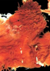
Giant Cell Tumor of Tendon Sheath (b/c near the hand)
KEYs on Histo:
• Giant cells surrounded by histocytes (eccentric nuclei with abundant cytoplasm) and cells that resemble synoviocytes
Gross:
• Is a WELL CIRCUMSCRIBED tumor that may appear yellow due to its contents being Lipid Laden Macrophage.

How might a patient with a tenosynovial giant cell tumor present?
• What are some key histological features to look for?
Presentation:
• A patient (male or female) that presents in their 20-40’s with a mass in their knee (should be worrying about osteosarcomas and giant cell bone tumors at this point)
Histology:
• SImilar to Giant cell tumor of the tendon sheath in that you see giant cells in a sea of histocytes and synviocytes BUT there will be a VILLONODULAR appearance and hemosiderin. EVIDENCE OF JOINT DESTRUCTION WILL ALSO BE PRESENT (not a feature of GST of Tendon Sheath)
What are the newer names for giant cell tumor of tendon sheath and tenosynovial giant cell tumor?
Giant Cell Tumor of Tendon Sheath = Localized Tensosynovial Giant Cell tumor (b/c it doesn’t invade the joint and is well circumscribed)
Tensosynovial Giant Cell Tumor = DIFFUSE TGCT now b/c it is nodular and has villi that show evidence of tissue invasion
What is shown here?
• Key features?
• How would you expect the tumor to present grossly?

(diffuse) Tenosynovial Giant Cell Tumor - you can tell by the nodular and villus appearance of the tissue on low power. On higher power expect a similar picture of giant cells in a background of histocytes.
Grossly you will see a similar Shaggy Picture of a Tumor that most commonly occurs in the knee
How might a patient with a Lipoma present to you?
• What would you expect to see histologically?
Presentation:
• This will likely present to you as an adult (male or female) with a large movable mass on there superficial extremitiy or trunk
Histologically:
Tissue resembles mature adipose tissue

What is this?
• key features?
• How common is this?
• Associated mutations?

Grossly this is well circumscribed tumor with the appearance of well differentiated fat cells on histology. = LIPOMA
55-70% of the time these will have HMGA2/HMGIC 12q13-15 fusion protein mutations
How would someone with a liposarcoma present?
• Key histiologic features? (3 subtypes)
Liposarcomas typically arise in people in their (40) 50’s and 60’s and involve DEEP (contrast to superficial in lipoma) soft tissues in the proximal extremities and the retroperitoneum.
3 Histologic Subtypes:
• Well Differentiated Liposarcoma
• Myxoid Liposarcoma
• Pleomorphic Liposarcoma
What tumor type is shown here?
• Key features?
• Mutations?
• Prognosis?

Liposarcoma is shown here with key feature being a Lipoblast with fat vaculoes that indent the nucleus (b/c the nucleus is too damn big for a fat cell)
Mutations associated with liposarcomas are MDM2 and CDK4 or 12q13-15 amplification
These tumors tend to recurr locally => prognosis may be okay or poor depending on subtype
What tumor types are shown here?
• Key features?
• how does prognosis vary on the basis of each of these subtypes?

Top Left:
• Well differentiated Liposarcoma - good prognosis these are pretty indolent
• These are adipocytes with scattered spindle cells
Top Right:
• Pleomorphic Liposarcoma - bad prognosis these tend to be very aggressive
• Looks angry AF
Lower Left:
• Myxoid Liposarcoma - intermediate prognosis
• Chicken Wire vessels propagating throughout with different levels of differentiated fat that looks like fetal fat.
In what region is the prognosis of a liposarcoma especially poor?
if its in the Retroperitoneum
How will someone with a nodular fasciitis present?
• What should you look for histologically?
Most likely this will be someone with previous trauma that may or may not recall experiencing the trauma (only 1/4 do). This tumor presents as a rapidly growing (occurs in wks - mos) mass that maxes out at about 5cm in the area of trauma.
Histology:
• Self limited CLONAL proliferation of cells in the SUBDERMAL fat
What is this?
• would you expect these cells to be monoclonal or polyclonal?

Nodular Fasciitis - you would expect these cells to be monoclonal

What soft tissue tumor is this?
• what are the key features?

This is a Nodular Fasciitis - you can see plump immature fibroblasts in short fasicles (said to look like tissue culture)
How do you expect someone with myositis ossificans to present?
• key features on histology?
Presentation:
Myositis Ossificans typically presents in a young athlete that has experienced trauma and will have a mass in their proximal extremities (need to distinguish from osteosarcoma)
Histology:
Mature bone is at the periphery with fibrous tissue resembling nodular fasciiitis at the center with osteoid between.
This happened as the result of traumatic injury to the femur.
• what is this likely to be?
• How long did it take to form?

Myositis Ossificans takes 3-6 weeks to form an ossification
What is this?
• Key features?
• How would you get rid of it?

Myositis Ossificans that could be removed by simple excision
Key Features:
• Mature bone at the periphery with fibrous tissue resembling nodular fasciitis at the center (see below for a picture of nodula fasciitis on high power)

How do you expect someone with superficial fibromatosis to present?
• Key histological features?
Superficial fibromatosis typically presents as a male patient that may have a thickening of the Plantar surface of the foot, dorsum of the penis, or palm of the hand that causes contracture of digits 4 and 5.
Histology:
• Spindle cells lesions with MATURE fibroblasts surrounded by dense dark pink collagen

This specimen was taken from the plantar surface of the foot where it was causing some of the digits to contract. What tumor type is this?

Superficial Fibromatosis
Who does a deep fibromatosis typically present in?
• Key histological features?
Deep fibromatosis typically arrises as a large infiltrative mass on either 1) shoulder, chest wall, or back or 2) intraabdominal in a female from her teens to 30s.
Histoogically:
• Appears as Broad Fascicles amid dense collagen
This tissue was biopsied from a 25 year old woman who has a thick, well circumscribed mass in her chest wall.
• what are some of the key histological features?
• what will the extracted tumor look like grossly?

Keys:
• Fasicles of fibrous tissue amid dense collagen on histology in combination with this histology is indicative of deep fibromatosis
Grossly this is a large, firm, white cute surface with INFILTRATIVE BORDERS

What immunohistochemical staining has been used on this deep fibromatosis?
• Why is this a useful stain?
***Deep fibromatosis located in the __________ is associated with _________.

This shows a Beta-Catenine stain on a deep fibromatosis. Is stains positive in these cells because they have increased ß-catenin/wnt/APC signaling taking place
***Deep fibromatosis located in the intraabdominal area is associated with Gardner’s Syndrome

Deep Fibromatosis

How does fibrosarcoma typically present?
• key histological features?
Fibrobrosarcoma typically presents in adults in the thigh or retroperitoneal area.
Histology:
• Variable and my resemble the spindle cells in fribromatosis or may loook like a herringbone pattern or be in architectural disarray
What causes this disease?
• How common is recurrence?
• How does it spread and where does it spread to?

Fibrosarcoma is shown here with herringbone patten on histology. Local recurrence is common and hematogenous spread to the lungs happens in more than 1/4 of cases.
How will this appear grossly?

• This a fibrosarcoma that will be UNencapsulated grossly and have necrosis and hemorrhage
How might a Rhabdomyoma present?
• what will histology look like?
Presentation:
Rhabdomyoma may present as a primary tumor in the heart (VENTRICLES) of a child
Histology:
• May appear as spider cells
What is this tumor?
• where was it likely found?
• Associated gene mutations?

Rhabdomyoma (not Rhabdomyosarcoma)
• This was likely found in the ventricles in a kid’s heart
• 1/2 of these are associated with TUBEROUS SCLEROSIS with mutations in TSC1 (hamartin) and TSC2 (Tuberin) tumor suppressor genes
How does rhabdomyosarcoma typically present?
There are 3 subtypes of this tumor that may present in different patients. The alverolar and embryonal subtypes present in children under 20 that have tumors arising in the head, neck, and genitourinary tract. The pleomorphic form typically presents in adults.
What is shown here?
• key feature?

These are strap cells in a rhabdomyosarcoma that are striated and trying to become muscle
***The are not always this obvious, a lower powered image is shown below***

What key feature of Rhabdomyosarcoma is shown here?

Eosinophilic inclusion bodies shown here are important features of Rhabdomyosarcoma histology in addition to the strap cells
What Histologic features distinguish the following Rhabdomyosarcomas?
• Embryonal
• Alveolar
• Pleomorphic
Embryonal:
• Spindled and round cells with a MYXOID stroma
Alveolar:
• Network of fibrous septae that divide the cells into clusters of aggregates (resembles alveoli). Cells appear as round cells with little cytoplasm
Pleomorphic:
• Numberous, large, sometimes multinucleated bizarre eosinophilic tumor cells
Where in does Sarcoma botyoides develop?
• how does it look grossly?
• what type of rhabdomyosarcoma is this a subtype of?
Forms a cambium layer in the wall of mucosal surfaces like the nasopharynx or vagina. Sarcoma botyoides is a subtype of embryonal rhabdomyosarcoma.
Gross appearance is a soft grey inflitrative mass
What translocations are associated with one type of rhabdomyosarcoma. What type?
• which translocation has the worst prognosis?
Alveolar Rhabdomyosarcomas are associated with t(2:13) and t(1:13) translocations. t(2:13) translocations have the worst px. (these lead to formation of a fusion gene of either FOX01-PAX3 or FOX01-PAX7)
What is often necessary to prove the pleomophic Rhabdomyosarcomas are what we think they are?
• Myogenin immunohistochemical stain is often necessary to prove a Rhabdomyosarcoma
What subtype of rhabdomyosarcoma is shown here?

Alveolar - typically arises in children and is associated with gene mutations invovling t(1:13) and t(2:13) with t(2;13) having the worst px.
Which of ALL of the rhabdomyosarcomas has the worst px?
• what patient is this likely to arise in?
Adult onset PLEOMORPHIC rhabdomyosarcomas have the worst px. (remember the other two typically arise in kids)
What is shown here?


How does a leiomyoma present?
• Histology?
A woman will present with various symptoms like infertility, pain, bleeding, or be completely asymptomatic. (found in 77% of women)
Histology:
• These appear as fasicles of densely eosinophilic spindle cells that often intersect at right angles
What is shown here?
• what would you see grossly?

- Fascicles of densely eosinophilic spindle cells that are intersecting at right angles indicative of leiomyoma
- Gross

Remember Leiomyomas most commonly present in the uterus but can present anywhere in the body due because they arise from smooth muscle
Who typically presents with Leiomyosarcoma?
• what do you see on histology?
Typically found in women, but is typically a painless tumor that may be large and bulky
• Histology shows Eosinophilic spindle cells with blunt-ended hyperchromatic nuclei in interweaving fascicles
Where are Leiomyosarcomas typically located?
• where does a particularly deadly form arise from?
Typically these are located in the deep soft tissues of the extremities or retroperitoneum
A particularly deadly form arists from the great vessels
What is this?

Leiomyosarcoma
Where do you most commonly see metastatis of sarcomas occur to? Be specific.
The Lungs, specifically the base of the lungs because this is were blood flow is the highest due to gravity
Who typically presents with a synovial sarcoma?
• what gene mutation is associated with this disease?
Typically this is a person in their 20’s-40’s. t(x; 18) mutations are common in these tumors
**Remember these are NOT limited to the synovium like originally thought**
What is shown on the left and right here?

BOTH are synovial Sarcomas:
Left: synovial sarcoma that is monophasic with uniform spindle cells and scant cytoplasm
Right: Biphasic has spindle cells but also gland-like structures
What is this?


