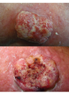
Skin cancer Flashcards

1
Q

Types of skin cancer

A

1. Non-Melanoma Skin Cancer

  • Basal Cell Carcinoma
  • Squamous Cell Carcinoma

2. Melanoma

3. Premalignant Conditions

  • Bowens Disease
  • Actinic Keratosis
2
Q

What’s the most common skin cancer?

A

Basal cell carcinomas are the commonest form of skin cancer making up 75% on the NMSC group

3
Q

Name (2) premalignant skin cancer conditions

A
  • Bowens Disease
  • Actinic Keratosis
4
Q

An 80 year old lady presents with a 2 year history of a pigmented lesion on the right cheek. Slowly increasing in size. On history alone what is the most likely diagnosis?

A: Melanoma

B: Lentigo maligna

C: Basal cell carcinoma

D: Squamous cell carcinoma

A

B. Lentigo maligna

5
Q

A 30 year old lady presents with a 5 year history of a lesion on the right upper back. Slowly increasing in size. History of excessive use of sun beds. 9mm pink nodule with small amount of central keratin. What is the most likely diagnosis?

A: Melanoma

B: Basal cell carcinoma

C: Squamous cell carcinoma

D: Seborrhoeic keratosis

A

B. Basal Cell Carcinoma

6
Q
A

D: Seborrhoeic keratosis

7
Q
A

C: Squamous cell carcinoma

8
Q
A

A. Melanoma

9
Q
A

B. Basal Cell Carcinoma

10
Q

Risk factors for skin cancer

A
  • Increasing age
  • Gender: M>F
  • Family history (PATCH gene: tumour suppressive)
  • Previous personal history of BCC
  • Photodamage
  • Fair skin, blue eyes, previous sunburn, sunbed use
  • Immunosuppressed
  • Diseases: e.g cutaneous lupus, naevus sebaceous, Gorlin syndrome
  • Previous ionizing radiation, arsenic exposure
11
Q

Features of Basal Cell carcinoma

A
  • Commonly affects head and neck
  • Slow growing papule, nodule
  • Skin coloured, pink or pigmented
  • Can bleed or ulcerate
  • Very rarely metastasise
12
Q

Different subtypes of Basal Cell Carcinoma

A
  • Nodular
  • Morphoeic
  • Infiltrative
  • Superficial
  • Basosquamous
13
Q

Spot diagnosis

A

Basal Cell Ca

14
Q

Spot diagnosis

A

Basal Cell Carcinoma (superficial type)

15
Q

Management of Basal Call Carcinoma

A

Surgery

  • Diagnostic biopsy initially
  • Excision
  • Moh’s micrographic surgery

Radiotherapy

16
Q

Management of superficial variant of Basal Cell Carcinoma

A

Superficial variant:

  • Cryotherapy
  • Curettage and cautery
  • Topical treatment
  • 5-FU; Imiquimod; Ingenol mebutate
  • Photodynamic therapy (PDT)
17
Q

Another name for Gorlin Syndrome

A

Basal cell naevus syndrome

18
Q

Features of Gorlin syndrome

A

Features:

  • Multiple BCC’s
  • Young age
  • Calcified falx cerebri
  • Broad forehead
  • Odontogenic cysts jaw
  • Abnormal ribs (bifid, fused, missing)
  • Palmar/plantar pitting
  • Other tumours – melanoma, breast carcinoma, NHL, ovarian fibroma
19
Q

Management of Gorlin Syndrome

A
  • Excision
  • Vismodegib binds to and inhibits the transmembrane protein Smoothened homologue (SMO) to inhibit the Hedgehog signalling pathway
20
Q

Pathogenesis of Gorlin syndrome

A

PATCH gene Chr 9; SUFU gene Chr 10

(defective tumour suppressor genes)

21
Q

Simple pathophysiology of Squamous cell carcinoma

A

Cancer arising from keratinocytes in the epidermis

22
Q

Risk factors for squamous cell carcinoma

A
  • excessive exposure to sunlight / psoralen UVA therapy
  • actinic keratoses and Bowen’s disease
  • immunosuppression e.g. following renal transplant, HIV
  • smoking
  • long-standing leg ulcers (Marjolin’s ulcer)
  • genetic conditions e.g. xeroderma pigmentosum, oculocutaneous albinism
23
Q

Features of squamous cell carcinoma

A
  • Short history – weeks to months
  • Rapidly enlarging papule/nodule
  • Tender
  • Can ulcerate
  • Commonly on sun-exposed sites
24
Q

Spot diagnosis

A

Squamous cell Ca

25
Q

Spot diagnosis

A

Squamous cell ca

26
Q

Spot diagnosis

A

Squamous cell Ca

27
Q

Subtypes of Squamous cell carcinoma

A

Many! E.g.

  • Keratoacanthoma
  • Acantholytic
  • Spindle
  • Clear cell
  • Basosquamous
28
Q

Management of Squamous cell ca

A

Surgery

  • Excision
  • Moh’s micrographic surgery
  • Shave, C&C
  • Radiotherapy

MDT

50% develop another SCC within 5 yeas (high risk group)

29
Q

Margins in surgery in squamous cell ca

A
  • Surgical excision with 4mm margins if lesion <20mm in diameter
  • If tumour >20mm then margins should be 6mm

*Mohs micrographic surgery may be used in high-risk patients and in cosmetically important sites

30
Q

Prognostic factors in squamous cell ca

A
31
Q

Pathophysiology of melanoma (simple)

A

form of skin cancer arising from melanocytes in the stratum basale

32
Q

Risk factors for melanoma

A
  • Increasing age
  • Family history
  • Previous personal history of melanoma/NMSC
  • Multiple naevi/multiple aytpical naevi
  • Photodamage
  • Fair skin, blue eyes, previous sunburn, sunbed use
  • Immunosuppressed
  • Association with pancreatic cancer
33
Q

Types of malignant melanoma

A
  • There are four main subtypes of melanoma.
  • Nodular melanoma is the most aggressive whilst the other forms spread more slowly
34
Q

Describe superficial spreading malignant melanoma

  • site
  • demographics
  • appearance
A
35
Q

Describe nodular malignant melanoma

  • site
  • demographics
  • appearance
A
36
Q

Describe lentigo maligna malignant melanoma

  • site
  • demographics
  • appearance
A
37
Q

Describe acral lentiginous malignant melanoma

  • site
  • demographics
  • appearance
A
38
Q

Diagnostic features of malignant melanoma

A

The main diagnostic features (major criteria)

  • Change in size
  • Change in shape
  • Change in colour

Secondary features (minor criteria)

  • Diameter >6mm
  • Inflammation
  • Oozing or bleeding
  • Altered sensation
39
Q

What’s that?

A
  • Hutchinson’s sign – pigment extending onto the proximal nail fold

!!!! Splitting/cracking of the nail + pigment = melanoma until proved otherwise

40
Q

Management of malignant melanoma

A

Surgery:

  • Excision
  • (punch, shave)
  • Wide local excision
  • Sentinel lymph node biopsy +/- LN resection

Oncology:

Immunotherapy

41
Q

Spot diagnosis

A

Nodular malignant melanoma

42
Q

Spot diagnosis

A

Lentigo maligna melanoma

43
Q

Spot diagnosis

A

Acral lentiginous