skin cancer Flashcards
describe the skin microanatomy
have the epidermis
dermis
hypodermis - has subcutaneous fat
bm
muscle
describe the structure of the epidermis `
have keratinocytes, melanocytes, DC - langerhans cells, merkle cells
keratinocytes start at the bm, mature and differentiate and move up through layers - get exposed to UV = mutatations
melanocytes are by the bm - can get exposed to UV = mutations
layers are:
- stratum corneum
- startum lucidum
- stratum granulosum
- stratum spinosum
- stratum basale
what are the different types of skin cancer *
keratinocyte derived - basal cell carcinoma, squamous cell carcinoma (aka non melanoma skin cancer)
melanocyte derived - malignant melanoma
vascular derived - kaposi sarcoma (common in AIDS), angiosarcoma
lymphocyte derived - mycosis fungiodes
general cause of skin cancer *
accumulation of mutations in key genes lead to uncontrolled cellular differentiation
causes of skin cancer *
genetic syndromes - give predisposition to cancer
- Gorlin’s syndrome - tendancy for basal cell carcinoma - defect in ORC1 gene
- xeroderma pigmentosum - defect in DNA repair by UV - develop multiple skin cancers
viral infections
- HHV8 in kaposi sarcoma
- HPV in SCC - particularly in the immunosuppressed
uv light - main cause
- BCC, SCC, malignant melanoma
immunosuppression
- drugs
- old age = immunosuppression
- HIV
- leukaemia
characteristics of malignant melanoma *
dark
irregular border
lumpy
incidence of malignant melanoma *
increasing in whit people
pale skin - suseptible to UV damage
increasing because people are living longer, more sun and behaviour change
higher incidence where higher exposure eg cornwall and norfolk
characteristics of basal cell carcinoma (BCC) *
pearly
grey/shiny
glistens
have telangiectasia - dilated small capillary blood vessels that look like they are branching
incidence of BCC *
increasing
describe how UV light contributes to skin cancer *
3 types UVA UVB UVC
UVC is blocked by the stratosphere
UVB and UVA hit the surface and contribute to mutations
UV damage to DNA leads to mutations in specific genes - cell division, DNA repair, cell cycle arrest - if these accumulate = cancer
why is sunlight essential
for photosynthesis
infrarred spectrum provides warmth
effect ion human mood
stimulates the production of vitamin D in the skin
describe how UVB causes skin cancer *
most important wavelength in carcinogenesis
directly induces mutations
induces photoproducts - these are cross links between bases - affects pyrimidines (C and T) - forming cyclobutane pyrimidine dimers eg T=T, T=C, C=C (ie thymine dimers etc)
this is usually repaired by nucelar exision repair
describe role of UVA in skin cancer *
100x more penetrates to surface than UVB
major cause of skin aging - penetrates deeper and effects collagen in dermis
contributes to carcinogenesis - causes cyclobutane pyrimidine dimers but less effectively than UVB
also forms free radicals whcih damage DNA and cell mmebrane
therapeutic use of UVA
in PUVA therapy for psoriasis
how is UV damage usually repaired *
DNA nucleotide excision
describe xeroderma pigmentosum *
genetic defect with nucleotide excision repair
means get skin cancer early with minimal exposure
ie <10yrs age, freckles and photosensitivity
sublings need to be tested
treated by removing the skin cancer and strict sun protection ie completely covered in sun including wearing a visor over face
what are the mutations that can cause cancer *
stimulate uncontrolled cell proliferation - mutations in p53 gene
alter responses to growth stimulating/repressive factors
inhibit programmed cell death - apoptosis
describe sunburn
the keratinocytes undergo apoptosis because of UV
this removes UV damaged cells that might otherwise form cancer cells
summarise photocarcinogenesis *
UV causes DNA damage
DNA can be repaired = normal cells
or if damage too severe - apoptosis
or if mutation accumulate = cell will transform = skin cancer
describe the immunomodulatory effects of UV *
UVA and B effect expression of genes in immunity - depletes the langerhans cells in the epidermis = immune suppression
there is reduced skin immunocompetence and immunosurveillance
further increases cancer causing potential of sun - normally langerhans cells (APC) cause cell death of cells exposed to UV
(basis for treatment of psoriasis - exposure to UV improves psoriasis)
what determines the host response to UV *
genetic influences - especially phototype
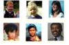
what are the fitzpatrick phototypes *
1 - always burns never tans
2 - usually burns, sometimes tans
3 - sometimes burns, usually tans
4 - never burns always tans
5 - moderate constitutive pigmentation - asian
6 - marked constituitive pigmentation - afro-carribean

describe melanin *
melanin pigmentation is responsible for skin colour
produced by melanocytes within the basal layer of the epidermis
skin colour depends on the amount of melanin produced as baseline, not the number of melanocytes (number of cells is constant)
describe melanocytes and production of melanin *
they are DC on the BM in epidermis that interdigitate and communicate with the keratinocytes - 1 melanocyte for 5 keratinocytes
they produce a baseline level of melanin
after UV exposure - keratinocytes send paracrine signal (MSH) that activates the melanocytes to make more melanin
melanin is taken up by keratinocytes and packaged around their nucleus to protect DNA
what are the types of melanin and what determines them *
eumelanin - brown/black - in later skin types
phaeomelanin - yellowish/reddish brown - red hair and pale skin
melanin is produced from tyrosine by a series of enzymes
MCR1 gene has >20 polymorphisms - the variation of melanin ratio produced depends on the polymorphism
this explains the different hair and skin coulour types
melanin detects skin sensitivity to UV damage
describe malignant melanoma *
tumour of the melanocytes - they become abnormal and have abnormal architecture
caused by UV exposure and genetic factors
there is a risk of metastasis leading to death
describe lentigo maligna *
it is melanoma in situ
there is proliferation of malignant (atypical) melanocytes in the epidermis
pagetoid spread - ie the melanocytes spread upwards, they should be at the bm
no risk of metastisis because they havent breached the epidermis
characteristics of lentigo maligna *
irregular shape
flaty
light and dark brown colours
>2cm usually
common in elderly at sites of skin exposure

treatment of lentigo maligna
excision
and skin graft
descrive lentigo maligna melanoma *
means there is invasion
dark patch
describe superficial spreading malignant melanoma *
lateal proliferation of malignant melanocytes
invade the bm
risk of metastasis
pagetoid spread - melanocytes in epidermis and dermis
also spread out and downwards - when head out = radial growth phase, when down = vertical growth phase
characteristics of superficial spreading MM *
dark - blue colour
irregular
how do you diagnose a superficial spreading MM *
ABCDE rule
asymettry
border irregular
colour variation - dark brown/black
diameter >7mm and increasing
erythma
what is the significance of a superficial spreading MM being pale in middle *
means middle has regressed - immune response against tumour
bad prognosis because means thicker and more invasive and perhaps already metastised
describe nodular malignant melanoma *
vertical proliferation of melanocytes - grows up and down
no previous horizontal growth
risk of metastasis
can arise from a mole/de novo
characteristics of nodular MM *
black and lumpy
describe nodular melanoma arising within a superficial spreading melanoma *
downward proliferation of malignant melanocytes
previous horizontal growth
nodule develops within an irregular plaque
prognosis is worse
can lose pigmentation
can have erythma - superficial reddening, these are amelanotic areas of the melanoma
describe acral lentiginous melanoma *
on soles or palms
lumps/flat pigmented plaques
common in darker skin types
describe amelanotic melanoma *
not pigmented
how do you determine the prognosis of melanoma
breslow thickness - depth of invasion of tumour cells in the skin
measurement from granular layer (just under the stratum corneum) to bottom of tumour
<1mm = thin
4= thick
1-4 = intermediate
what are the risk factors for the development of melanoma *
FH of dysplastic nevi or melanoma
UV irradiation
sunburns during childhood
intermittent burning exposure in unacclimitised fair skin - most important
atypical/dysplastic nevus syndrome
personal history of melanomas - if you have one you’re likely to have another
skin type 1/2
genetic markers - CDKN2A mutation
cingenital nevi
number of atypical nevi >5
high socioaconomic status
1equatoral latitudes
dna repair defects eg xeroderma pigmentosum
immunosuppression
describe kertaocanthoma *
grow rapidly then involutes
type of SCC
from keratinocyte - if well differentiated it produces keratin
describe SCC *
malignant tumour of kertainocytes
caused by UV, HPV, immunosuppression, may occur in scars/scarring process eg burns scar/disease with scar/long term ulcer
risk of metastasis if poorly differentaited is 10% - this is less than for malignant melanomas
well differentiated produces keratin - can see horns
can occur on lower lip from sun adn smoking
occur on ears of men and leg of women - exposed to sun
describe BCC *
malignant tumour from basal cell layer of epidermis
caused by sun exposure, genetics
slow growing - 3-4mm yr (can be faster)
invades tissue but doesnt metastise
common on face
common around eye - has telangiectasia - lost eyelash so destructive process
arborising telangiectasia - branching capillary bvs
describe nodular BCC *
glistening
telangiectasia
describe superficial BCC *
have rolled edge
glistening surface
crusty
describe mycosis fungoides *
look like psoriasis/eczema
pink scaly patch that progresses to plaque like lesion then tumour, then disseminates then die
it is a T cell lymphoma of skin
also called cutaneous T cell lymphoma
describe kaposi’s sarcoma *
HIV and HHV8 associated - HHV8 in AIDs and not
purplish nodules, plaques, flat legions
arise from the endothelial cells in lymphatics
orientation of plaque goes along the skin line
on palate, sole of foot, skin
treatment - excision, chemo/radio and treatment of HIV
describe epidermodysplasia veruciformis *
rare autosomal recessive condition
predisposition ot HPV induced warts and SCCs
role of immunomodulation in cancer
people with good immune systems resolve early cancers before they are detected
in immunocomprised people there is an increased occurrence of keratinocyte-derived skin tumours on sun-exposed sites.
role of p53 in skin cancer *
normally mutations in DNA = activation of p53 = expression of Bax gene = apoptosis
if you have p53 mutation - DNA with mutations might survive = more likely to get heratinocyte cancers