colorectal cancer Flashcards
epidemiology of colorectal cancer
major cancer in developed countries
4th most common cancer overall
2nd leading cause of cancer death overall
environmental (diet) factors and genetic factors are the aetiology
anatomy of the colon
caecum is hollow, large part of colon in R ileum
then have ascending colon
then hepatic flexure, transverse colon, splenic flexure, descending colon, sigmoid colon, rectum
in front of colon is the peritoneum
behind the colon is the mesentry where bv come in and out, it is fatty tissue
what is the function of the colon
extract water from faeces and therefore maintain electrolyte balance - therefore people with colorectomy become dehydrated - so gastroenterologists need to give them more water than normal and monitor electrolyte destrubances
is a faecal resevoir - this is an evolutionary advantage
bacterial digestion for vitamens especiall`y B and K
describe the structure of the colon
has mucosal surface folds
layers:
- area where the stool is
- epithelium (part of mucosa)
- lamina propria (part of mucosa)
- muscularis mucosa - thin layer of muscle
- muscularis externa
- submucosal
- muscularis propria - thick muscle
- fat and bv - this is the mesentry
- abdominal cavity
*

describe the colorectal crypt of lieberkuhn
have goblet cells that produce mucin - for immune function and lubrication
stem cells - constantly proliferating - move the cells up the crypt
mesenchymal cells are support cells - hold other cells in place

describe the colonic microanatomy
can see the lamina propria full of inflammatory cells
nucleus at the base of the cells and cytoplasm comes all the way up
below the nucleus have neuroendocrine cells

describe the turnover of colon cells *
a lot of turnover
proliferation renders cells vulnerable because that is the area where they are more likely to mutate
APC mutation prevents cell loss = mutation
normally there are protective mechanisms to eliminate genetically defective cells by natural loss of cells that are then replaced by normal cells, DNA monitors, repair enzymes that remove the damaged pieces of DNA and replace it
what is a polyp*
any projection from a mucosal surface into a hollow viscus and may be hyperplastic, neoplastic, inflammatory, harmartomatous etc
what is an adenoma *
a benign neoplasm of the mucosal epithelial cells - type of polyp
what are the colonic polyp types *
metaplastic/hyperplastic
adenomas
juvenille
peutz jeghers - familial disorder where get mucosal hyperpigmentation and polyps in GIT - can lead to higher risk of cancer
lipomas - benign growths of fatty tissue
others - any circumscribed intramucosal lesions
describe hyperplastic polyps *
very common
<0.5cm
90% of all lower intestinal polyps
often multiple
no malignant potential
15% have K-ras mutation
on histopath - still pink similar to normal, have serrated appearance

what are the different colonic adenoma types *
tubular when >75% adenoma is tubular - 90%
tubulovillous - 25-50% is villous - 10%
villous >50% is villous
flat - not polyp
serrated - similar to hyperplastic polyp but displastic
describe pedunculated and sessile adenomas *
pedunculated - means on stalk - A is intramucosal carcinoma that has not reached the musclaris propria; B has reached the muscularis propria it is invasive- but cutting stalk you can easily remove the adenoma
sessile adenoma - flat - more likely to spread into the muscle

what are the microscopic features of tubular adenomas *
columnar cells with nuclear enlargement, elongation, multilayering and loss of polarity (nucleus has moved to the lumen side of the epi)
increased proliferative activity - look at the mitotic count
reduced differentiation - how much look like normal colon
complexty/disorganisation of the architecture
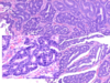
histopath of colonic adenoma *
more purple - increased nuclear material
nuclei look larger than hyperplastic
there is a loss of polarity

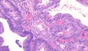
microscopic structure of villus adenomas *
mucinous cells with nucleus enlargement, elongation, multilayering and loss of polarity - these are signs of dysplasia
are exophytic frond-like extensions
may have hypersecretory function and result in excess mucus discharge and hypokalaemia - lose K into Gi tract because of mucin
small nucleoli
increased nucleocytoplasmic ratio

describe dysplasia *
means bad growth
abnormal growth of cells with some features of cancer
not invasive
there is subjective analysis - low, high or intermediate grade
describe the dysplasia

normal on R - relatively small nuclei on bottom and cells have plenty of mucin
low grade dysplasia on L - architecture still the same as normal
but nuclei are bigger, increased nucleocytoplasmic ratio, small nucleoli
describe this high grade dysplasia *

cells dont have normal architecture
high nucleocytoplasmic ratio
there is higher risk of it being carcinoma because of the high grade dysplasia
describe this high grade dysplasia *

nuclei becoming big
back to back glands
describe the polyp *

Large villous adenoma with an ulcerated adenocarcinoma in its center.
describe adenomatous polyposis coli *
it is a mutation on 5q21
the site of mutation determines clinical variants
- classic - 100% sure will get colon carcinoma
- attenuated - less adenomas
- gardner - tumour in bone and skin
- turcot - tumour in brain - glioblastoma and blastoma
develop 1000s of polyps
many pts have a profylactic colectomy <30yrs
describe the natural history of colonic adenoma *
25% of adults have adenomas at age 50
5% of these become cancers if left
large polyps have higher risk than small - because there are more cells
lead time 10yrs - time from development of adenoma to development of carcinoma
cancers stay at a curable stage for 2yrs
describe the progression from adenoma to carcinoma *
most colon carcinomas (CRC) arise from adenomas
there is residual adenoma in 10-30% of CRCs
adenomas and CRC have similar distribution within the kidney
adenomas usually precede carcinomas by 10-20yrs
endoscopic removal of polyps reduces chances of CRC
describe the genetic pathways that lead to CRC *
adenoma sequence - APC then K-ras then Smads then p53 then telomerase activation
microsatellite instability - microsatillites are repeat sequences prone to misalignment - some are in coding sequences of genes which inhibit growth or apoptosis or involved in surveillance eg TGFbR11
happens because you have mismatch repair genes repair mutations in the microsatellite regions - MSH2, MLH1 and 4 others - they are recessive genes - survive with 1, but if mutation in both = cancer
HNPCC is a germline mutation in these DNA repair genes
describe the adenoma-carcinoma sequence *
not when you have a problem with the microsatellite
- germline (inherited) mutations or somatic (acquired) mutation of cancer suppressor genes - 1st hit - APC, mismatch repair genes
- creates mucosa at risk - leads to methylation abnormalities = inactivation of normal alleles - 2nd hit - APC, B-catenin, MSH2
- protoonchogene mutation - K-ras - adenoma
- homozygoud loss of additional caner suppressor gene - p53 and LOH (p53 drives cell into apoptosis, stop mitosis and tell cell to repair DNA)
- additional mutations and gross chromosomal alterations - carcinoma
in same adenoma - see differnet stages ie some areas relatively normal - some are high grade

what are the 2 main pathways for genetic predisposition for CRC *
FAP - inactivation of APC tumour suppressor genes
HNPCC - microsatellite instability
why is APC particularly important in colon cancer *
APC holds B-catenin in the cytoplasm
if APC becomes mutated - b-catenin moves into nucleus = increase in cell proliferation by interferring with TFs
at bottom of crypt APC normally allows B-catenin into nucleus because want proliferation here
describe p53 in adenoma progression *
dark areas stain showing p53 - here p53 is overexpressed because there is a mutation in it so cells are making it more - this is block/diffuse positivity p53
normally p53 is presented at low and varying levels
describe the epidemiology of CRC *
35k per anum in UK
10% cancer related deaths
age range 50-80, sporadic rare <30
high in west, low in japan, mexico and africa - relate to the dietry factors - high fat, low fibre, high redmeat, refined carbohydrates
describe contribution of food to CRC *
contains carcinogens
also contains anti-cancer agents
heat modifies the chemicals further
bacteria modifies food residues
heterolytic amines are carcinogens found in red meat PhIP is formed when it is cooked
PhIP oxidation to N-OH-PhIP combined with deoxyguanosine leads to mutageneis
folate from green leafy veg is a co-enzyme for nucleotide synthesis and DNA methylation
MTHFR deficiency leads to disruption in DNA synthesis causing DNA instability - strands break and uracil incorporation = mutations, and decreased methionine synthesis leading to genomic hypomethylation and focal hypermethylation = gene activation and silencing
describe anti-cancer food elements *
Vit C and E - ROS scavenger - anti-oxidents - remove ROS that oxidise DNA which lead to mutations and cancer
isothiocyanites - cruciferous veg
polyphenols - green tea and fruit juice - activate MAPK - regulates phase 2 detoxifying enzymes as well as other genes (eg glut-s transferase) and reduce DNA oxidation
garlic associated apoptosis - ajoene, allicin
green tea - EGCG induced telomerase activity
usefulness of this unknown because difficult to isolate individual chemicals in food
describe the clinical presentation of CRC *
change in bowel habit
bleeding PR
unexplained iron def anaemia
mucus PR
bloating
cramps - colic
constitutional - weight loss, fatigue
people rationalise these symptoms with getting old, piles or irritable bowel - need to think about CRC
describe this barium enema *

apple core illusion
growth that is suppressing the lumen into the size of an apple core
describe macroscopic features of CRC *
small carcinomas may be present in larger polypoid adenomas, pedunculated or sessile
on top is polyp, then muscularis propria and then mesentry

describe the districution and presentation of CRC *
caecum/ascending colon- 22% - caecum is big so tumour not blocking the colon so present with IDA
transverse colon - 11%
descending colon - 6%
rectosigmoid - 55% - blood in rectum - close to anus - bleed PR
what are the microscopic structures of CRCs *
adenocarcinomas grade 1-3
mucinous carcinomas
singlet ring cell - worse prognosis
neuroendocrine - rarer
describe this micrograph of colonic carcinoma *

form glands embedded in desmoplastic/inflammatory reaction
describe this - colonic cancer *

nuclei wilder
various size nucleoli
large nucleus
increased nucleocytoplasmic ratio
describe this mucinous carcinoma *

worse prognosis
carcinoma in pool of mucin
describe this adenocarcinoma *

invaded through the muscularis propria = worse prognosis
describe this colon cancer *

breached the serosa - into the peritoneal cavity = worse prognosis
describe grading *
proportion of gland differentiation relative to solid areas or nests and cords of cells without lumina
can be well, moderately or poorly differentiated
describe Duke’s classification *
dukes A - growth limited to mucosa/submucosa - nodes negatibe
dukes B - growth into or beyond the muscularis propria - nodes -ve
dukes c1 - nodes +ve, apical LN -ve
dukes c2 - apical LN +VE
prognosis decreases down the levels
what are the clinical features that affect prognosis and how *
diagnosis in asymtomatic pts - improved
PR bleeding - improved
bowel obstruction/perforation - diminished prognosis
tumour location - colon better than rectum, L better than R (present earlier if close to the rectum),
age <30 diminished prognosis - more aggressive/not expecting to find CRC in this age gp
distant met - markedly worsened prog
pathological features that determine prognosis *
depth of bowel penetration - more = diminished prog
number of regional lymph nodes involved - more = worse
differentiation - well=better prog
mucinous (colloid) or signet ring - diminished prog
venous, lymphatic, peineural invasion - diminished prog
local inflamm and immunogenic reaction - if body is fighting the infection there is a better prognosis
why is it important to have an idea of prognosis *
because it determines treatment and how aggressive it should be - dont want to be too aggressive because treatment have SE
describe this tumour *

extramural vascular invasion
out of the bowel wall
adenocarcinoma in vessel
=bad prognosis
what are the treatment options for different stages of CRC*
1 - surgery
2 - sugery and 5FU
3 - surgery and 5FU/leucovorin (chemo)
4 - surgery and metastectomy, chemo, palliative RT
what makes people at high risk of colon cancer and so should be screened *
previous adenoma
1st degree relative affected by colorectal cancer before age 45 or >1 1st degree relative
evidence of dominant familial cancer trait including colorectal, uterine and other cancers
UC and CDs
heritable cancer families - including other sites
what is screening *
Screening is the practice of investigating apparently healthy individuals with the object of detecting unrecognised disease or people with an exceptionally high risk of developing disease, and of intervening in ways that will prevent the occurrence of disease or improve the prognosis when it develops.
what are the criteria for screening program *
importance of the disease - in respect to seriousness and frequency - in this case it is frequent and kills people
the natural history must be known to identify where screening can take place and to enable the effects of any intervention to be assessed
test characteristics - simple and acceptible to ptm sensitive and selective
the screening pop should have equal access to teh screening procedure
cost effectiveness
what is the NHS screening kit for colon cancer &
FOB - fecal occult blood test /FIT- fecal immunochemical blood test kit - find blood pt might have missed
FIT has replaced FOB
if positive send 60-75yrs for colonoscopy, 55-60 years to a sigmoidoscopy