SF3 1 EKG Flashcards

Second-Degree AV Block, Mobitz Type 1
* Progressive increase in PR Interval from beat to beat until single QRS absent
* PR interval goes back to initial length and cycles
* Usually benign
Stage of STEMI

Days Later
* ST Normalized
* T Wave inverted

Hypercalcemia
* Shortened QT Interval

Orthodromic Atrioventricular Reentrant Tachycardia
* Can be triggered by Atrial Premature beat in WPW
* No delta wave
* Conduction via AV node with reentry from accessory pathway
* QRS normal

Junctional Escape Rhythm
* No P Wave (impulse from below atria)
* Normal QRS
* Beat 40-60 bpm

Second-Degree AV Block, Mobitz Type II
* Sudden intermitten loss of AV conduction without gradual lengthening
* Block may persist two or more beats
* QRS often widened
* Conduction block beyond AV node
* Severe disease

Left Ventricular Hypertrophy
* Deep S in V1
* Elevated R in V5/V6

Sinus Bradycardia
* Normal P
* Normal QRS
* Slowed Heart Rate

Sinus Rhythm (WPW)
* Short PR Interval (<0.12 s)
* Slurred QRS “Delta Wave”
* “Fusion” (synced) AV and Accessory (Bundle of Kent) conduction
* QRS widened

Severe Hyperkalemia
* Flattened P
* Widened QRS

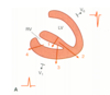
Third-Degree AV Block
* Complete heart block
* no relationship between P and QRS
* QRS width/rate dependent on whether AV node or His/Purkinje providing pacemaking
***In graph, second and fourth P wave superimposed on T wave

Right Bundle Branch Block
* Widened QRS
*RSR’ in V1 (Rabbit Ears)
* Prominent S in V6

Ventricular Premature Beat
* Ectopic ventricular focus fires AP
* Widened QRS (slow cell-to-cell conduction)
* Ectopic beat unrelated to preceding P wave
* T wave opposite polarity of QRS

First Degree AV Block
* PR Interval Lengthened (>0.2 s)
* Benign/Asymptomatic
Stage of STEMI

Acute
* ST Elevation
Stage of STEMI

Hours into it
* ST Elevation
* Depressed R Wave
* Q Wave Begins

Polymorphic Ventricular Tachycardia
* Example of “Torsades de Pointes” (waxing and waning pattern)
* QRS continually changes shape
* Rate varies
* Multiple ectopic foci or continually changing reentry circuit
* QT prolonged (LQTS)
* Abnormality of cardiac ion channel or calcium handling usually

Digoxin Therapy
* ST “Scooped” depression
* Mild PR Prolongation

Atrial Premature Beats
* Originate from Atrial focus outside of SA Node
* earlier-than-expected P Wave with abnormal shape
* QRS Normal
Stage of STEMI

Days 1-2
* T wave inversion
* Q wave deeper

Hyperkalemia
* Tall “peaked” T Wave

Sinus Tachycardia
Everything normal, SA node discharge > 100 bpm (typically 100-180 bpm)
Stage of STEMI

Weeks Later
* ST / T Normal
* Q Wave Persists

Atrial Flutter
* Rapid regular atrial activity at 180-350 bpm
* Many reach AV node during refractory period
* two or more beats of atria per ventricle
* usually caused by reentry over large Anatomically-Fixed Circuit

Monomorphic Ventricular Tachycardia
* QRS complex wide (>0.12 s)
* Rate of 100-200 bpm
* Rate regular
* QRS identical to one another
* Usually structural abnormality supporting reentry circuit (myocardial infarction or cardiomyopathy)
Right Ventricular Hypertrophy
* R > S in Lead V1
* Right Axis Deviation

Ventricular Escape Rhythms
* No P Wave
* Widened QRS Complex (distinguish from Junctional)
* Rate 15-40 bpm

Hypocalcemia
* Prolonged QT Interval

Hypokalemia
* ST Depression
* Flattened T
* Prominent U Wave

Atrioventricular Nodal Reentrant Tachycardia
* Normal QRS
* regular tachycardia
* P wave hidden/retrograde

Ventricular Fibrillation
* Disordered rapid stimulation of ventricles with no coordinated contraction
* chaotic irregular appearance, no QRS

Atrial Fibrillation
* chaotic rhythm with very high atrial rate (350-600 discharge/min)
* No P waves OR high frequency “noise”
* QRS-T normal but timing irregular
* Multiple Wandering Reentrant Circuits within Atria

Antidromic AVRT
* Wide QRS Complex
* Ventricles stimulated by anterograde conduction via accessory pathway
* reentry through AV node

Pathological Q wave. Typical of Myocardial infarction
(remember, must see grouping not single lead)

Left Bundle Branch Block
* Widened QRS
* Broad, notched R in V6
* Absent R and Prominent S in V1
Normal T Wave
Positive in all three bipolar limb leads
T Waves sensitive to…
1) Changes in electrolytes
2) Ischemia
3) drugs
(Normal ECG) Lead II
All complexes (P-QRS-T) normally positive
(Normal ECG) Lead aVR
All complexes (P-QRS-T) negative
(Normal ECG) Lead VI
* Small initial r wave (might not be able to see)
* Deeper S Wave
* T wave may be positive, biphasic, or negative
(Normal ECG) General T Waves
Normal T wave should begin with gradual rise with distal descent more abrupt. Sharp proximal rise in ST segment indicates something is wrong
(Normal ECG) V1-V6
Amplitude of R wave should be rising constantly from V1-V6
Equiphasic RS complex at V3
(Normal ECG) Lead V6
QRS complex typically begins with narrow Q Wave follow by large R wave
J Point
Point at which ST segment begins (end of S wave)
P Wave in Leads I, II, III
Should be upright in normal ECG
Septal Heart Leads
V1, V2
Anterior Heart Leads
V3, V4
Inferior Heart Leads
Leads II, III, and aVF
Low Lateral Heart Leads
V5, V6
High Lateral Heart Leads
Lead I and aVL
Inferior Myocardial Infarction (Leads and Artery)
Leads II, III, aVF
Right Coronary Artery
Anteroseptal Myocardial Infarction (Leads and Artery)
Leads V1-V2
Left Anterior Descending
Anteroapical Myocardial Infarction (Leads and Artery)
Leads V3-V4
Left Anterior Descending (distal)
Anterolateral Myocardial Infarction (Leads and Artery)
Leads V5-V6, I, and aVL
Left Circumflex Coronary Artery
Posterior Myocardial Infarction (Leads and Artery)
Leads V1, V2 (this is the weird one where it’s inverted because we dont have the right lead inplace)
Right Coronary Artery
