Repro Histo Flashcards
IgG transport to fetus
- IgG does get across during the 3rd trimester when the receptor for Ig is made by the syncitiotrophoblast
- Syncitiotrophoblasts grab the antibodies and transports them to the other side by trans-cytosis
- Child is born with the full complement of the mother’s antibodies
- Protects it for the first 2-3 months of life until it makes its own antibody


ejaculatory duct
epithelium of cervix
- Cervix has simple columnar epithelium with a lot of glands secreting mucus into the cervical canal
- Pap smears taking at the transition zone between the simple columnar epithelium of the cervix and the stratified squamous epithelium of the vagina

testes

Primary spermatocyte.
maternal side of the placenta
•Note the branching of the “bushy” villi (a lot of surface area) that are used for good exchange with the maternal circulation

testes

LABYRINTH-like, simple cuboidal epithelium
rete testis
testes

spermatozoa
3 layers of the uterus
endometrium
myometrium
perimetrium


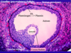
Secondary or antral follicle

efferent ductules
remnant of mesonephric duct
cellular change?

The simple columnar epithelium is changing via metaplasia to stratified squamous epithelium over a broad area. Will there be more of less mucus secretion in areas of metaplasia? Less!
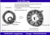
placenta:

maternal
placenta
Placenta
- Placenta will be divided into around 20 caudal regions that are separated by connective tissue that comes up from the maternal side
- Note the anchoring villus and branches
- Maternal blood (arrow) will come in on the outside of the villi and percolate around at a very sluggish pace (replenished every 2-3 minutes)
- Nutrients can then exchange with the fetal blood vessels that will go out into the umbilicus and to the fetus
- Maternal veins will drain the area
- Maternal and fetal circulations are generally pretty separated by the syncitiotrophoblasts
____________________

placenta:

fetal
mammry gland

Myoepithelial cell
stratum basalis
•Stratum basalis is basically staying the same and stains more darkly in H&E (also more cellular)


•corpus albicans
At 10 days after ovulation, LH levels will have fallen so much in the bloodstream that it can no longer maintain the corpus luteum
- No longer will make hormones
- Corpus luteum will degenerate completely, undergoing atresia
- Cells lose their nuclei
- Macrophages migrate in and start to digest them away
- Note the “acellular”-like and whitish appearance of the now called corpus albicans
- Note the one that has even degenerated further
ovary:

This is “rubber band” shape of a degenerating oocyte and its zona pellucida within an atretic follicle.

Myometrium: smooth muscle of the uterus.
uterus:

Endometrium: endometrial glands and stroma. Stratum functionalis. This layer will build up with Estrogen stimulation.
first meiotic division
- After the LH surge, roughly 36 hours after, ovulation occurs
- Ovulation is preceded by the first mitotic division, but not the second one
- Oocytes have been stuck in the first mitotic division since fetal life
- Factors produced by the near-by follicular granulosa cells are present that allow for only the first mitotic division to occur but prevent the second one from occurring
- After the first division, ovulation occurs
- Immediately following ovulation, there is dissolution of basement membrane and invasion of the theca interna and externa along with their vascular supply into the granulosa portion of the follicle themselves
- This is part of the process in the formation of the corpus luteum

vagina
stratified squamou epithelium
corpus luteum:

Theca lutein cells.
theca are dark stringy ones, granulosa are blobby ones
stratum functionalis in the proliferative phase
•endoetrium
In the proliferative phase (left images), there is an epithelium and long and straight glands that go down to the basalis layer