RBC disorder pt 1 Flashcards

1
Q

how do you classify anemias based on their mean corpuscular values (MCV)?

A
  1. Microcytic anemias: MCV less than 80
  2. Macrocytic anemias: MCV more than 100
  3. Normocytic anemias: MCV between 80 to 100
2
Q

What are the causes of Microcytic anemias?

A
  1. Iron deficiency anemia
  2. Anemia of Chronic Disease
  3. Thalassemias (β and α)
  4. Sideroblastic Anemias
3
Q

iron deficiency causing microcytic anemia occurs mostly on who in the population?

A

young children and women

4
Q

is iron actively secreted from the body?

A

no

5
Q

how do we eliminate iron from the body?

A
  • loss of epithelial cells from GIT,
  • epidermal cell loss from skin,
  • women in reproductive age
6
Q

how is iron balance achieved?

A

iron out of body must be equally intaked

7
Q

in what organ is iron absorbed?

what part of the organ?

what 2 pathways are used?

A

intestine

duodenum

Pathways:

1) uptake of heme iron
2) dietary non-heme

8
Q

uptake of heme iron pathway

heme iron is derived from what molecule?

what portion does the uptake of heme iron in intestine make?

what cells uptake the heme protein in the duodenum? what happens inside the cell?

A

Hb

2/3 of iron is uptaked though heme derived proteins

mucosal cells, heme oxygenase cleaves the porphyrin ring (heme is available for absorption htorugh non-heme pathway)

9
Q

dietary non-heme pathway

dietary non-heme is mostly present as what type of molecules?

what is an important factor in order to uptake this inorganic iron?

A

ferric hydroxide or bound to phytates, oxalates, sugars, citrate, lactate

low gastric pH

10
Q

Molecular mechanism of iron absorption

what enzyme do enterocytes use to import ferrous iron?

where do enterocytes express this enzyme?

can the enzyme be used to import ferric iron?

A

DMT1

expressed in apical surface

no

11
Q

Molecular mechanism of absorption of iron

non-heme is in ferric or ferrous state?

can this be transported by DMT1?

what has to be used? what does it do?

A

ferric

no

needs CYBRD1 to reduce ferric non-heme to ferrous so that DMT1 later can absorb it

12
Q

Molecular mechanisms of iron absorption

what happens to iron once it is absorbed by enterocytes?

what do you call iron in the plasma?

A

iron from enterocyte is transferred to plasma using FPN1 (ferroportin 1) to export it (outside)

transferrin

13
Q

Molecular mechanisms of iron absorption

where is transferrin made?

how many of the available transferrin binding sites are occupied by iron? why that amount?

A

liver

only one-third, in case there is excess iron

14
Q

Molecular mechanisms of iron absorption

whan happens to iron once it is in the plasma? (where does it go?)

once it is transferred to the other cell, what organelle uses up most of this iron 80-90%?

the other 10-20% of iron, where does it go?

A

iron is delivered to erythroid precursors (nomoblasts) by using transferrin receptors

mitochondria

rest of iron is stored as ferritin

15
Q

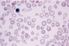
what are sideroblasts?

what stain can be used to view granules in sideroblasts?

A

normoblasts with granules

prussian blue

16
Q

how do sideroblast look in sideroblastic anemia?

A

ring-like

17
Q

Molecular mechanisms of iron absorption

what cell type can recycle iron?

A

macrophages

18
Q

Molecular mechanisms of iron absorption

how do alveolar macrophages recycle iron?

what is the exception after alveolar macrophages recycle iron?

A
  • they phagocytose erythrocytes & convert the iron to storage form
  • the iron cannot be returned to circulation
19
Q

what other type of macrophage can recycle iron?

what are the forms used to store iron in macrophages?

A

reticulo-endothelial macrophages

  1. ferritin
  2. hemosiderin
20
Q

what is ferritin?

in what cells is ferritin seen?

in what fluid compartment can ferritin be seen in small amounts?

A

protein that stores iron and releases it in controlled manner

seen in all cells

plasma

21
Q

what is the difference between ferritin and hemosiderin?

A

hemosiderin has:

  • higher amount of iron than protein
  • is less soluble in aqueous solution
  • is more stable and less available
22
Q

Iron deficiency anemia

when is iron deficiency anemia seen?

how is it corrected?

when are symptoms seen?

A
  • when iron balance tilts to negative
  • mobilizing the iron stores
  • when the tissue stores are depleted
23
Q

Iron deficiency anemia

the deficiency is seen due to what reasons?

A
  • dietary lack of iron
  • impaired absorption
  • there is increased use of it

(like in growth or menstruation, pregnancy)

  • chronic blood loss

(peptic ulcer disease, hemmorrhoids, carcinoma of stomach/colon, aspirin, ulcerative colitis, esophageal varices)

24
Q

when can you have unexplained iron deficiency anemia?

A

H. pylori infection

25
_iron deficiency anemia_ what will be seen in a lab report in the following values: Hb? heatocrit red cell indices (MCV, MHC, MCHC)
Hb= reduced hematorcrit = reduced MCV, MHC, MCHC = decreased
26
_iron deficiency anemia_ how will RBC look in a peripheral blood smear in iron deficiency anemia?
microcytic hypochromic with anisocytosis and poikilocytosis (pencil shape) anisocytosis = variation in size poikilocytosis = variation in shape
27
how do these red blood cells look? what anemia can this be?
microcytic and hypochromic iron deficiency anemia
28
what anemia is this? how do you know? where would you say this is? how is the bone marrow called during this anemia? how are the erythroid cells called in this anemia? how do you see the macrophages that have lost the iron?
iron deficiency anemia look at the prussian blue stain in the bone marrow hyperplastic marrow microcytic maturation gold standard stain
29
_Iron deficiency anemia_ what biochemical test can be used for this anemia?
1. serum ion concentration 2. TIBC 3. percentage saturation 4. serum ferritin
30
_Iron deficiency anemia_ how will serum iron concentration appear in this anemia? how will TIBC show in this anemia? What is TIBC? what relationship does ferritin have with TIBC? how will % saturation? (% of transferrin bound to iron) serum ferritin will appear how?
low increased total iron binding capacity when ferritin is low, TIBC is high decreased low
31
_Iron deficiency anemia_ what are the clinical features of iron deficiency anemia?
fatigue angina pectoris koilonychia low immunity (spoon shaped nails) Pallor angular chelitis (tongue burning sensation) **Plummer-Vinson Syndrome**
32
_Iron deficiency anemia_ what is Plummer-Vinson Syndrome?
triad of findings: 1) microcytic hypochromic anemia 2) atrophic glossitis 3) esophageal webs: can cause dysphagia
33
what is being shown here?
esophageal webs
34
_Iron deficiency anemia_ how do you treat iron deficiency anemia? how do you know the treatment works?
supply iron (ferrous sulfate) iron-carbohydrate complex blood tranfusion you will see reticulocyte count increase to max in 7-12 days
35
_Anemia of chronic disease (ACD)_ who gets this disease? what are the categories for ppl who get the disease?
ppl with chronic disease infections or non-infectious inflammatory disorders 1. Chronic microbial infections 2. Chronic immune disorders 3. Neoplasms 4. Alcoholism
36
_Anemia of chronic disease (ACD)_ what are examples of chronic microbial infections that lead to this anemia?
1. TB 2. osteomyelitis 3. pelvic inflammatory disease 4. bacterial endocarditis 5. lung abcess
37
_Anemia of chronic disease (ACD)_ what are examples of chronic immune disorders that lead to this anemia?
1. RA 2. SLE 3. vasculitis syndromes
38
Anemia of chronic disease (ACD) what is a neoplasia that may lead to this anemia?
hodgkins lymphoma
39
_Anemia of chronic disease (ACD)_ what is the pathogenesis of this anemia?
1) infection causes release of IL-1, TNF, IFN. 2) these cause liver to make hepcidin 3) hepcidin inhibits release of iron (iron cant go to transferrin) 4) ferritin & iron stores increase in **Marrow and macrophages** 5) IL-1 and TNF block iron absorption in intestine
40
_Anemia of chronic disease (ACD)_ Lab report: how do RBC's look under microscope? MCV? serum iron? serum ferritin? TIBC? bone marrow hemosiderin?
normocytic normochromic MCV = normal serum iron = reduced serum ferritin = increased or normal TIBC = decreased or normal bone marrow hemosiderin = low or absent
41
_Hemolytic anemia_ why does it happen? what reasons lead to it?
because there is an increase in the rate of red cell destruction (premature destruction of RBC) ## Footnote 1) intrinsic abnormality of RBC 2) extrinsic abnormality of RBC
42
_Hemolytic Anemia_ what are the intrinsic causes of this anemia? (membrane defect, Hb defect, Enzyme defect, acquired defect)
1. membrane defect: hereditary spherocytosis 2. Hb defect: sickle cell disease and thalassemias 3. Enzyme defect: G6PD deficiency 4. Acquired: paroxysmal nocturnal hemoglobinuria
43
_Hemolytic anemia_ Whata rethe extrinsic causes leading to this anemia?
1. immune mechanisms: hemolytic disease of newborn, incompatible blood transfusion 2. mechanical: microangiopathic hemolytic anemia, cardiac prosthetic valve
44
_Hemolytic Anemia_ how do you tell the site of RBC destruction? how do you tell the site of intravascular hemolysis? how do you tell the site of extravascular hemolysis?
look for free Hb and Hb products destruction of RBC within circulation, will see free Hb its exaggerated response of removing old RBC by the reticuloendothelial system (they get phagocytosed)
45
_Hemolytic Anemia_ what is an example of extravascular hemolysis?
hereditary spherocytosis
46
_Hemolytic Anemia_ What effect will this anemia cause on the bone marrow? when is it called anemia?
it will make it produce more RBC so bone marrow hyperplasia when the bone marrow cant compensate anymore for the RBC destruction
47
_Evidence of increased Hb breakdown_ is hyperbilirubinemia and jaundice a reliable guide of the rate of hemolysis? what determines the amount of bilirubin made? if there is no jaundice..can there still be anemia?
no the amount of Hb broken down and the ability of liver to excrete it yes
48
_Evidence of increased Hb breakdown_ where is haptoglobin made? what does haptoglobin do? can Hb pass through glomerulus? when can it not? what happens if there is persistent hemolysis? in what type hemolytic anemia is haptoglobin reduced? what is the normal haptoglobin levels?
liver bind to free Hb in plasma yes when it is bound to haptoglobin plasma haptoglobin is low both types 0.3-2.0
49
_Evidence of increased Hb breakdown_ what does plasma hemopexin do? where is plasma hemopexin made? does plasma hemopexin bind to Hb if Hb levels are very high? what happens if Hb levels are high? what is bound to ferriheme? why? when do we see hemopexin low?
it binds to free heme in a 1:1 ratio liver no Hb can convert to methemoglobin and then turn into ferriheme and globin hemopexin, so that it doesnt get lost in glomerular filtration in intravascular hemolysis
50
_Evidence of intravascular hemolysis_ what happens to Hb from destroyed RBC's? what happens if the Hb exceeds the haptoglobin capacity? what happens if the binding capacity of hemopexin is exceeded? what will it form? presence of this indicates what?
Hb gets released into the plasma unbound Hb turns into methemoglobin and then dissociates into ferriheme and globin ferriheme binds to albumin, forms methemalbumin methemalbumin tells us there is severe hemolysis
51
_Evidence of intravascular hemolysis_ what is the normal level of free Hb in plasma? what is the level of free Hb in plasma for intravascular hemolysis? what color will plasma be when Hb is high? how is it refered to? what happens when renal threshold for Hb reabsorption is exceeded? what will be the color of urine ?
low or 0.6 100-200 pink/red (hemoglobinemia) hemoglobinuria is noted pink or black (oxyhemoglobin/methemoglobin)
52
_Features of accelerated hematopoiesis_ what are the features of accelerated hematopoiesis?
1) reticulocytosis 2) Presence of nucleated red cells in peripheral blood smear 3) Bone marrow hyperplasia (erythroid) 4) LDH = increased 5) Extra medullary hematopoiesis (liver, spleen, LN) 6) Cholelithiasis (gallstones) 7) skeletal abnormalities
53
what is this?
reticulocytes (polychromatophelia)
54
what are these? what stain?
reticulocytes supravital stain
55
_Thalassemia syndromes_ what are the 2 types of thalassemia syndromes? what is deficient in each?
Alpha thalassemia: no alpha chain Beta thalassemia: deficient synthesis partial or complete
56
_Thalassemia Syndromes_ what is the difference between ß+-thalassemia and ß0-thalassemia?
ß+-thalassemia = partial absence of ß-globin chain ß0-thalassemia = total absence of ß-globin chain
57
_Beta thalassemia: Pathogenesis_ what is the Hb that is inadequately produced? what imbalance will be produced? what will the imbalance cause? how will RBC's look? what happens inside the bone marrow? what symptoms will be seen then?
HbA (a2ß2) more alpha chains available will clump and cause RBC to precipitate more alpha chains will cause damage to RBC membrane leading to extra-vascular hemolysis RBC's look microcytic hypochromic inside bone marrow: more alpha chains clump, form inclusion bodies in erythroblasts which get destroyed symptoms: ineffective erythropoiesis, increased iron absorption, iron overload (2ry hemochromatosis)
58
_Beta thalassemia major (lab)_ what can we expect about the following lab values in this condition: Hb? MCV? Peripheral Blood smear? Reticulocyte count? Hb Electrophoresis? bone marrow?
Hb = severe anemia MCV = low MCV Peripheral Blood smear = ansiopoikilocytosis, microcytic hypochromic, fragmented cells Reticulocyte count = increased Hb Electrophoresis = HbA2, HbF increased / absent HbA bone marrow = expanded and changed
59
what is this?
ß-thalassemia cells are fragmented, hypochromic
60
what is this?
ß-Thalassemia
61
_Beta thalassemia major_ What are clinical features? how do you treat this?
pallor jaundice growth retardation splenomegaly by 3 years gall stones skeletal abnormality give blood transfusion and splenectomy
62
what disease is this?
ß-thalassemia major
63
_Beta Thalassemia Minor_ what is another name for this? how will the anemia be for this? when does the anemia become evident? what type of anemia will be seen? how will ferritin levels appear? how will the MCV appear? what will a Hb electrophoresis show?
ß-thalassemia trait mild anemia during pregnancy microcytic anemia normal low mild deacrease in Hb A, increase in Hb F and Hb A2
64
_Beta Thalassemia Minor_ how do you differentiate this from iron deficiency anemia?
iron deficiency anemia improves with iron therapy, ß-thalassemia trait or minor is worsened by iron therapy
65
_Alpha Thalassemias_ what happens in it? what Hb are affected? what determines the severity? What are the 3 clinical types of alpha thalassemias? what happens in each of these 3 types?
alpha chains absent Hb A, Hb F, Hb A2 number of alpha chains missing 1) silent carrier: 1 alpha chain deleted 2) alpha thalassemia trait: 2 alpha chains deleted 3) HbH: 3 alpha chains deleted
66
_Alpha Thalassemia_ What happens in hydrops fetalis? in what part of the world is it common? What is so bad about hydrops fetalis? what can we expect if we look at the MCV? what can we expect if we look at the RBC count? what other Hb will a kid use to survive? what can happen in the 3rd trimester as a fetus? can lead to what?
all 4 alpha chains are deleted and gamma chains form tetramer south east asia binds O2 strongly but it wont let it go, tissue suffers hypoxia decreased MCV increased Hb portland 1/2 will see fetal distress, can lead to death in utero
67
_Alpha Thalassemia_ what symptoms can be detected on the fetus during hydrops fetalis? what symptoms can be detected on the mother during hydrops fetalis?
fetus: severe anemia (Hb 3 to 8 gm%), generalized edema, massive hepatosplenomegaly , signs of cardiac failure if alive mother: toxemia of pregnancy (hypertension, fluid retention with or without proteinuria), polyhydroamnios or oligohydroamnios
68
what condition is this?
hydrops fetalis
69
_Sideroblastic Anemia_ what characterizes this anemia? how can this characteristic be spotted? what 2 reasons can be given as to why we get this anemia?
iron deposits in the mitochondria of erythoblasts the mitochondria of erythroblasts will look as ring 1) insufficient generation of heme due to problem in synthesis pathway 2) mitochondrial defect involving iron pathway
70
_Sideroblastic Anemia_ what are the 2 types of Sideroblastic anemia? what is the defect we find in each? What causes them?
1) Hereditary: ALA synthetase 2 2) Acquired: * can be: - primary: myelodysplastic syndrome - secondary: chronic alcoholism, drugs (isoniazid), pyridoxine deficiency (vit b6), lead poisoning
71
what is this?
sideroblastic anemia (see the ring)
72
what is this?
sideroblastic anemia (see the ring)
73
_Sideroblastic Anemia_ what is the pathogenesis? Lab findings: peripheral blood smear? MCV? Bone Marrow? TIBC? serum iron? serum ferritin levels? urine porphyrin profile?
* iron is brought into mitochondria to form heme * if **protoporphyrin** is absent, iron would remain within mitochondria of developing red cells Lab findings Peripheral blood smear = normocytic, macrocytic, * pappenheimer bodies present MCV = low Bone Marrow = erythroid hyperplasia, will see ring sideroblasts TIBC = decreased serum iron = increased serum ferritin levels = increased erythropoietic porphyria
74
what can be viewed in this picture?
sideroblastic anemia with pappenheimer bodies
75
_Sideroblastic Anemia_ how do you treat withdrawal of drug here?
with pyridoxine