Radiology of Lung Cancer and Staging Flashcards
What percentage of patients with lung cancer present with advanced disease?
66%
What do you need to check when looking at a chest X-ray?
Name/marker/rotation/penetration
Lines/metal work
Heart
Mediastinum
Lung (zones - upper, middle, lower)
Bones
Diaphragm
Soft tissues
What are the first 4 things you should look at in a chest X-ray?
Name
Marker
Rotation
Penetration
What are the zones of the lungs in a chest X-ray?
Upper
Middle
Lower
What is A?

Mediastinum
What are you looking for in the mediastinum?
Hilar vascular structures crisply defined
No widening of mediastinum
Trachea should be central

What are you looking for in the lungs?
Compare upper, middle and lower zones
Look between ribs for lung detail
Remember to look ‘behind’ the heart

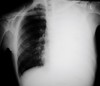
What is this?

Peripheral lung carcinoma
What is this?
Central lung carcinoma
How should we identify lung cancers on X-rays?
Compare with previous films
Always look at review areas
Remember lesions are often more subtle
What are the review areas of a chest X-ray?
Hila
Lung apices
Behind the heart
Behind the diaphragm

What is this?

Left hilar mass
What is this?

Right hilar mass
What is this?

Mass behind the heart
What is this?
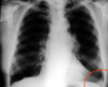
Mass left costophrenic angle
What is this?

Right apex tumour
What could the clinical history for lung cancer include?
Increasing shortness of breath in smoker
History of pulmonary fibrosis
Recent haemoptysis
What follows taking a history and examining the patient?
CT
What should be evaluated using a CT scan?
Size
Shape
Atelectasis
Border
Density
Solid or non-solid
Dynamic contrast enhancement >25HU
Growth
What is atelectasis?
Collapse of lung resulting in reduced gas exchange
What is the collapse of the lung resulting in reduced gas exchange called?
Atelectasis
What is a pulmonary mass?
Opacity in the lung over 3cm with no medistinal adenopathy or atelectasis
What is an opacity in the lung over 3cm with no mediastinal adenopathy or atelectasis called?
Pulmonary mass
What is a pulmonary nodule?
Opacity in the lung up to 3cm with no mediastinal adenopathy or atelectasis
What is an opacity in the lung up to 3cm with no mediastinal adenopathy or atelectasis called?
Pulmonary nodule
What is the difference between a pulmonary nodule and a pulmonary mass?
Pulmonary mass is over 3cm and pulmonary nodule is up to 3cm
What could a solitary pulmonary nodule or mass be?
Lung cancer
Metastasis
Benign lung neoplasm
Infection
Vascular haemotoma
What could suggest a solitary pulmonary nodule or mass is a metastasis?
Previous history of breast. renal, seminoma or sarcoma cancer
What are examples of benign lung neoplasms?
Carcinoid
Hamartoma
What does the staging of lung cancer take into account?
Clinical history/examination
Performance status
Pulmonary function
What system does the staging of lung cancer use?
TNM international system for staging lung cancer
What does the TNM international staging of lung cancer consider?
Size and position of tumour (T)
Whether cancer cells have spread into the lymph nodes (N)
Whether the tumour has spread anywhere else in the body, metastasis (M)
What is T?
Size and position of tumour
What is N?
Whether tumour has spread to lymph nodes
What is M?
Whether the tumour has spread into other parts of the body, metastasis
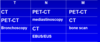
What investigations can be done to determine T?
CT
PET-CT
Bronchoscopy

What investigations can be done to determine N?
PET-CT
Mediastinoscopy
CT
EBUS/EUS (endobronchial ultrasound)
What does EBUS stand up for?
Endobronchial ultrasound
What investigations can be done to determine M?
PET-CT
CT
Bone scan

What is the most common tracer used?
FDG (flourodeoxyglucose)
What does FDG stand for?
Flourodeoxyglucose
What is often used for the staging of lung cancer?
Flourodeoxyglucose PET
What can be said about the availability and cost of FDG PET?
Expensive
Limited availability in the UK
What is the labelled glucose analogue used in FDG-PET?
18F-FDG
What is the half body time of 18F-FDG?
60 minutes
What does TX mean?
Primary tumour cannot be assessed
What does T0 mean?
No evidence of primary tumour
What does Tis mean?
Carcinoma in situ (has not spread to surrounding tissue, group of abnormal cells in the place where they formed)
What is carcinoma in situ?
Group of abnormal cells which are still where they were formed, have not spread to nearby tissue
What is T1?
Less than or equal to 3cm in diameter
Surrounded by lung or visceral pleura
Without bronchoscopic evidence of involvement of the main bronchus
What is T1a?
Less than or equal to 1cm
What is T1b?
Less than or equal to 2cm
What is T1c?
Less than or equal to 3cm
What are the sub classes of T1?
T1a
T1b
T1c
What is T2?
More than 3cm but less than 5cm
What are the different classes of T2?
T2a
T2b
What is T2a?
More than 3cm but less than 4cm
What is T2b?
More than 4cm but less than 5cm
When are tumours classified as T2a although they are less than 3cm?
Invades main bronchus
Invades visceral pleura
Associated with atelectasis or obstructive pneumonitis that extends to the hilar region involving part or all of the lung
What is T3?
More than 5cm but less than 7cm
When are tumours classified as T3 althouh they are less than 5cm?
Invades any of:
Chest wall
Phrenic nerve
Parietal pericardium
or has seperate tumour nodules in the same lobe as primary
What is T4?
More than 7cm
When is a tumour T4 although it is less than 7cm?
Invades any of:
Diaphragm
Mediastinum
Heart
Great vessels
Trachea
Recurrent laryngeal nerve
Oesophagus
Vertebral body
Carina
or seperate tumour nodules in a different ipsilateral lobe
What does N staging range from?
N0 to N3
What is N0?
No regional lymph node involvement
What is N1?
Ipsilateral peribronchial, hilar or intrapulmonary nodes including by direct extension
What is N2?
Ipsilateral mediastinal, subcarinal
What is N3?
Contralateral mediastinal, contralateral hilar, scalene or supraclavicular
How does the number of lymph nodes change with size?
There are many small lymph nodes and few large ones

How does the prevalence of metastasis change with the size of lymph nodes?
Large lymph nodes are more likely to have metastasis

What percentage of patients present with metastasis?
33%
What are common metastasis?
Cerebral
Skeletal
Adrenal
Liver
What does M staging range from?
M0 to M1
What is M0?
No distant metastasis
What is M1?
Distant metastasis
What are the different classes of M1?
M1a
M1b
M1c
What is M1a?
Seperate tumour nodes in a contralateral lobe, tumour with pleural or pericardial nodules or malignant pleural or pericardial effusion
What is M1b?
Single distant metastasis
What is M1c?
Multiple distant metastasis
What are some of the advantages of PET/CT scanning in staging?
Performs whole body staging in single study excluding cerebral disease
Discloses metastasis and other pathology no detected by other means
Excludes metastasis where structural imaging abnormal
Non invasive
What are some limitations of CT/PET?
All tests have false positives and false negatives
Cost
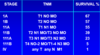
How does 5 year survival change with staging?
As staging increases survival decreases
What are some examples of tissue diagnosis methods?
Bronchoscopy with endobronchial ultrasound
Percutaneous image guided biopsy, flouroscopy/CT/US guided
Mediastinoscopy (sample mediastinal nodes)
Mediastinotomy (anterior mediastinal nodes)
Video assisted thoracoscopic surgery (VATS)
Explorative thoracotomy
