Radiology Flashcards
CXR
most important radiology-related nobel prize
Roentgen (1901, physics) : discovery of X-radiation
what is electromagnetic radiation ?
- X-rays belong to this group of radioation
- it’s the transport of energy through space as a combination of electric and magnetic fields
what’s the radiation we use to expose x-ray ?
gamma radiation (member of the electromagnetic radiation family)
What is PACS ? What are the advantages and problem with it ?
- Picture, Archiving and Communication Systems) : a technology supplant analog film/screen technology
- Advantages :
- more technical flexibility
- magnification
- brightness
- accessible to multiple sites
- easier to teach with
- no lost films
- more technical flexibility
- problems :
- serious confidentiality issues,
- Advantages :
Define Fluoroscopy
A medical imaging that shows a continuous X-ray image on a monitor, much like an X-ray movie.
Why would a PA view be prefered from an AP view ?
because with a PA view, there’s less magnification of the hearth on the final image (since it’s closer to the screen)
What is computed tomography ?
CT : the basic principle behind it is that the internal structure of an object can be reconstructed from multiple projections of the object.
Explain de basic principles of ultrasound
- sounds travel faster through dense objects
- when sound reaches a different density than the one it was travelling in, a number of sound waves will be reflected while others will continue
- A probe transmits sound waves and receives the reflected echos
- the image is formed by calculating the distance of the reflection based on tje time of travel of the sound waves
define MRI
- Magnetic resonnance imaging
- How it works
- certain nuclei in the body naturally react to a magnetic field… they become excited (when you put in a magnetic field, the hydrogen molecules that where running randomly in the body reorient from random)
- A radio pulse creates a second magnetic field at a right angle (90o) to the first, forcing the nucleo to make a quarter turn
- When the radio pulse is then turned off, the nuclei return to their original state, emitting their own caracteristic radio signal which can be converted into 2-dimentional images (they will return at their position after the first magnetic field at different rate depending of the organ they are in or wether there’s a tumor in that organ. It’s the energy that’s release during their return to the normal direction that an image can be made from)
Principe advantages and disadvantages of MRI
- Advantages
- no gamma radiation
- Disadvantages
- cost a lot
- the test take a ong time to take (you can only do about a patient an hour while with CT scan you can do about 5 patients/hour).
Why do we say that all ionizing radiation is harmful ?
Because the data are not available to indicate if there’s a thereshold below which no harmful effect will occur.
What radiation unit do we use today ?
- Sievert (Sv) or Rem
- 1 Sv = 100 Rem
Name natural sources of background radiation
- Cosmic rays - radiation that reaches earth from space
- Rocks and soil - some rocks are radioactive and give off radioactive radon gas (over 50% of our exposure)
- Living things - plants absorb radioactive materials from the soil and these pass up the food chain
What is a BED ?
- Banana equivalent dose (BED)
- potassium 40 (source of natural radiation from living things)
- correlated to 0,1 micro Sv
- about 1% of the average daily exposure to radiation
- CT scan = 70 000 BED
What’s the procedure giving the most radiation ?
- CT of the abdomen or pelvis
- equivalent to 500 chest X-ray
All ionizing radiation is harmful. How can we manage that?
- Do the BEST test first (if you can get an MRI, don’t do a CT scan)
- Do what is clinically appropriate
- think about what is the risk of not doing the test
- 10-20% medical imaging studies are unnecessary or appropriate
- Follow acceptable guidelines
What is the lowest level of cumulative exposure at which any increase in cancer risk is evident ?
100 milliSv
True or false. Artificial Intelligence (AI) will replace radiologists one day.
FALSE. AI will make us better radiologists, but not replace radiologists.
Basic principle of how X-rays are obtained ?
- To obtain a CXR, the x-ray beam travels through the patient to a film or detector
- Different tissues in the body will stop or “attenuate” the photons of the x-ray beam to different amounts, depending on their density
- a structure which is not very dense (ex: air filled lungs) will not stop amuch of the x-rays beam and will appear darker or more black on the x-ray film
- a structure which is very dense (ex: bone) will stop most of the x-ray beam and appear lighter or more white structure on the film
Number of density visible on CXR and what are they ?
5 densities
what is an interface ?
- when you have 2 structures of different densities in contact with each other, it creates an interface.
- You must have an interface to see things as 2 separate structures
- no interface = cannot be resolved as separate structures (ex : heart and aorta (both soft tissue))
What are the different CXR possible positions ?
-
PA (posterior-anterior) and lateral upright
- best option if possible
-
AP (anterior - posterior)
- done portable, can be upright or supine
-
Decubitus
- to evaluate effusion
-
Lordotic
- to see lung apex
-
expiration
- to evaluate pneumothorax
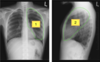
Why is a lateral CXR useful ? How is it done ?
- it’s a view taken with the xray beam passing from right (x-ray source) to left (detector) in the patient
- it helps localize disease in the chest and provides a better view of certain structures than with an PA view
- often a PA CXR will be accompanied by a lateral CXR
Usually, are PA and lateral view taken at the end of an expiration or an inspiration ? What’s the exception ?
- Usually obtained at the end of the inspiration
- at end of an expiration if we are specifically looking for a pneumothorax
When is an AP CXR obtained ? Do we get a lateral view with it ?
- An AP CXR is obtained in a patient who is unable to have a PA and lateral CXR
- traume patient in ER
- intubated patient
- No lateral view is obtained
- usually done with a portable x-ray machine that is not as good quality as dedicated equipment
- usually referred as «portable» for this reason
What is a decubitus CXR ? Why do we use it ?
- Obtained with the patient lying on their side
- Occasional done to evaluate pleural effusions
- see if there’s fluid in the pleural space (you get a better idea of how much fluid)
What is a lordotic CXR ? Why is it useful ?
- Patient leans back at 30 degree angle
- Demonstrates areas of the lungs apices that appear obscured on the PA CXR
- see apex of the lungs that are usually hidden by clavicule, soft tissue, upper ribs, etc.
What will markers on the film tell you ?
How it was obtained
- upright, erect, sitting, supine, semi-supine
- inspiration, expiration
Before interpreting CXR, what 2 questions should you ask ?
- How was the CXR obtained ?
- Is it a good quality CXR ?
What can affect the quality of a CXR ?
- rotation (patient no facing the detector)
- inspiration (patient doesn’t take deep breath)
- motion artifact (patient moves during CXR)
- not all areas of the chest are included on the film
- overlying structures obscuring areas of interest
How do you assess the patient’s rotation ?
Observing the clavicular heads and determining whether they are equal distance from the spinous process of the thoracic vertebral bodies
How do you assess inspiration ?
You count the ribs down to the diaphragm
- the diaphragm should be intersected by the 6th anterior rib or the 10th posterior rib in the mid-clavicular line
On which kind of CXR can you identify the horizontal fissure ?
most lateral and PA CXR
On which kind of CXR can you identify the oblique fissure ?
most lateral CXR but no PA CXR
identify those structures
both are right upper lobe
identify those structures
both are right lower lobe
identify those structures
both are right middle lobe
what lung structure is against the right heart border ?
right middle lobe
which lung strucutre will go behind the right upper lobe ?
right lower lobe (also a lot of this structure goes behind the diaphragm
identify those structures
both are left upper lobe
identify those structures
both are left lower lobe
what lung strucutre is on the left border of the heart ?
lingula (replacing the right middle lobe)
Why is the localization of the carina on CXR important ?
- it’s an important landmark to identify, especially in intubated patient to determine correct placement of the endotracheal tube (ETT)
- should be 2-6 cm above the carina
- too low = go into one of the mainstream bronchi and block ventilation to the other lung
- too high = patient may spontaneously dislodge the tube
- should be 2-6 cm above the carina
which of the hemidiaphragm is usually a little higher than the other ?
The right
what happens with the heart, left and right hemidiaphragm on a lateral view ?
The left and right hemi are almost superimposed. Anteriorly, the left hemi blends with the heart and becomes indistinct (since you loose the diffenrentiation because they are both muscles)
What are the costophrenic angles ? How could they indicate a disease ?
- The costophrenic angle is the point where the diaphragm and chest wall meet
- They are seen laterally on the PA CXR and posteriorly on the lateral as inverted “v” shaped structures
- They should normally appear as a sharp angle
- Rounding of the angle is termed “blunting” and is most commonly due to pleural effusions
define airspace disease. How does it present itself on a CXR ?
- A descriptive term for a pattern of disease where the airspaces (alveoli) are filled with material (fluid, pus, blood, tumor)
- On CXR
- It is seen as an area of increased density (more white) compared to the normal dark lung
- It causes loss of the normal lung markings (the vessels)
- It does not usually result in volume loss of the affected lung
what are the 2 CXR signs of airspace disease ?
- air bronchogram
- silhouette sign
what is an air bronchogram ?
- in a normal lung, the air-filled alveoli and air-filled airways are adjacent to each other and have the same density
- when the alveoli are filled with fluid or tissue from various causes, they become more white, or more dense
- however the airways do not fill with material and remain air-filled
- we then see the contrast between the fluid filled airspaces and the air filled airways
what is a silouhette sign ?
- when you develop an area of airspace disease in part of the lung, it now becomes soft tissue density instead of air density
- we then lose the normal interface between the air filled lungs and the adjacent soft tissue structure (heart, chest wall, diaphragm)
- when this occurs, we say the structure is «silhouetted» by the abnormal lung.
The silhouette sign can not only confirm the presence of airspace disease, but also can be helpful to identify the location of the disease based on which structure is silhouetted. In those cases of silhouette, where wuld the disease be ?
- right heart border
- left heart border
- right/left hemi-diaphragm
- right mediastinum
- left mediastinium/aortic arch
- right middle lobe
- lingula
- right or left lower lobe
- right upper lobe
- left upper lobe
define interstitial disease ? What are the most common causes ?
- descriptive term for lung disease that involves the interstitum of the lung
- it’s most commonly appears as increased linear opacities throughout the lungs
- it’s different from airspace disease (consolidation) in that it doesn’t obscure the pulmonary vascular markings (you can still see little white-ish vessels at the border of the lungs)
- Causes
- pulmonary edema
- pulmonary fibrosis
- infection (viral, mycoplasma pneumonia)
- malignancy (limphatic carcinomatosis)
What is a pneumothorax ? How can it be identified on a CXR ?
- The presence of air in the pleural space
- spontaneous
- traumatic
- Iatrogenic (caused by medical treatment)
- CXR findings
- identify the pleural line
- Sharp white line running parallel to the chest wall
- Represents the visceral pleura which has separated from the chest wall
- Between the pleural line and the chest wall, there is a lucent space without lung markings
- Expiratory view makes the pneumothorax more visible
- identify the pleural line
what is the type of pneumothorax that requires urgent treatment ? What are the signs ?
-
tension pneumothorax
- progressive build up of air in the pleural space
- a one-way valve mechanism of injury allows air to enter, but not exit the pleural space
- the increased pressure obstruct venous return to the heart and leads to circulatory compromise
- signs
- deviation of the trachea away from side of pneumothorax
- shift of the mediastinum away from the side of pneumothorax
- depression of the hemidiaphram on the side of the pneumothorax
What is a pleural effusion ? what are the main causes ?
- collection of fluid in the pleural space
- fluid
- pus
- blood
- many causes
- pulmonary edema
- infection
- malignancy
How can you see pleural effusions on CXR ? supine ? decubitus ? upright ?
- Fluid is heavier than the lung, therefore the effusion will collect in the most dependent part of the chest
- Upright CXR
- Blunting of the lateral and posterior costophrenic angles ○ Meniscus sign
- Decubitus CXR (patient lying on their side)
- Fluid will layer on dependent side
- Supine CXR
- Fluid collects posteriorly and is difficult to see ○ Overall increased density of the hemithorax
- Upright CXR
