Radiology Flashcards
- Which of the above x-ray tubes contains rotating anode?
A
B
both
none of them
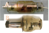
B
- Which part of the x-ray machine is marked in the picture?
generator
collimator
x-ray tube
control panel/computer

generator
- What kind of radiopacity has the marked organ in the picture?
gas
bone
soft tissue
fat

soft tissue
- What kind of radiopacity has the marked structure in the picture?
gas
bone
soft tissue
metal

metal
- What conclusion can be drawn when comparing the opacity of the structure in the circle and the rib?
The structure is a malignant tumour .
The structure and the rib have the same diameter
The structure lies further from the cassette than the rib .
The structure probably contains calcium .

The structure probably contains calcium .
- Why can’t we see the stifle in this image ?
It is amputated .
It is too far from the cassette .
It is relatively overexposed .
It is relatively underexposed .

It is relatively overexposed .
- What type of error is visible in the picture?
overexposed
under exposed
blurry
low contrast

blurry
- The structure of which tool is demonstrated in the picture?
intensifying screen
grid
collimator
cassette

grid
- Was this dog in sternal or dorsal recumbency?
sternal
dorsal
cannot be told
only the professor knows that

cannot be told
- What opacity does the marked area have?
fat
fluid
soft tissue
bone

fat
- What opacity does the marked area have?
fat
metal
soft tissue
bone

soft tissue
- Where is the bullet?
in the iliac bone
under the iliac bone
above the iliac bone
cannot be told

cannot be told
- Using the usual recumbency, what is the name of the projection?
a. laterolateral
b. lateromedial
c. mediolateral
d. mediomedial

mediolateral
- Is the positioning correct in this picture?
Yes.
No, the chest is rotated.
No, the forelimbs are not pulled forward.
No, the entire lung is not visible.

Yes
- The position of which image is correct for the interpretation?
A
B
Both
None of them.

none of them
Which statement is false regarding the image?
the bladder is full
the small intestines are gas filled
there is faces in the colon
the caecum is not visible

the small intestines are gas filled
Which statement is true regarding the image?
the contrast is only in the colon
complete obstruction cannot be ruled out
the contrast medium is surely barium sulfate
d. there is some contrast in the stomach too .

d. there is some contrast in the stomach too .
Which organ is marked bye the X?
a. stomach
b. kidney
c. spleen
d. intestine

intestine
Which statement is false regarding the image?
the animal is lying on its right side
it is a growing animal
the gastric fundus is filled with fluid
the bladder is small

it is a growing animal
Which statement is true regarding the image?
the marked organ is on the right side of the animal
the marked organ is not the gallbladder
the animal was lying on it’s sternum because the spinous processes are well seen
it is a suspected pneumoperitoneum

the marked organ is not the gallbladder
Which statement is false regarding the image ?
it is a suspected gastric volvulus
the liver is enlarged
the heart is enlarged
the chest is slightly rotated

the chest is slightly rotated
Which statement is true regarding the image?
it is a pneumothorax
it is an abdominal effusion
intestinal obstruction cannot be ruled out
the stomach is filled with gas

the stomach is filled with gas
Which one is correct?
x-soft tissue opacity, y-fluid opacity, z-gas opacity
x-fluid opacity, y-gas opacity, z-fat opacity
x-soft tissue opacity, y-fat opacity, z-fluid opacity
x-soft tissue opacity, y-fat opacity, z-gas opacity

x-soft tissue opacity, y-fat opacity, z-gas opacity
Which statement is true regarding the measurement ?
a cardiac disease can be diagnosed with it
the name of the method is HVS (horizontal vertical scale)
9.5 value is in the normal range
it cannot be used in old animals

a cardiac disease can be diagnosed with it


