Radiographic Teeth Flashcards
14#19
Left Bitewing

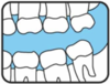
Right Bitewing - #2/#31, #3/#30, #4/#29, #5/#29

Mandibular Right - #32, #31, #30

Mandibular Right - #30, #29, #28

Maxillary Right - #3, #4, #5

Mandibular Left - #19, #18, #17

Maxillary - #8, #9

Maxillary Right - #6, #7

Maxillary Left - #12, #13

12/#21
Left Bitewing

Mandibular Left - #23, #22, #21

Mandibular Right - #28, #27, #26

Maxillary Left - #14, #15, #16

Right Bitewing - #1/#32, #2/#31, #3/#30

Mandibular Left - #21, #20, #19

Maxillary Right - #1, #2, #3

Mandibular - #26, #25, #24, #23

Maxillary - #10, #11
1.

Dentin
2.

Enamel
3.

Pulp Chamber
4.

PDL Space
5.

Lamina Dura
6.

Root Canal
7.

Cancellous bone

Nutrient canal in Maxillary Sinus Wall
1.

Primary Canine
2.

Primary first molar with partially resorbed roots.
3.

Permanent Canine
4.

Permanent first premolar
1.

Outline of nose
2.

Incisive foramen
3.

Lateral fossa
4.

Nasal fossa
5.

Nasal Septum
6.

Border of nasal fossa
7.

Anterior nasal spine
8.

Median palatine suture
1.

Incisive foramen
2.

Outline of the nose
3.

Lateral fossa
4.

Nasal fossa
5.

Nasal Septum
6.

Border of nasal fossa
7.

Anterior nasal spine
8.

Median palatine suture
1.

Lateral fossa
2.

Nasal fossa
3.

Inverted Y landmark
4.

Maxillary sinus
5.

Superimposition of first premolar over canine
1.

Lateral fossa
2.

Nasal fossa
3.

inverted Y landmark
4.

Maxillary sinus
5.

superimposition of premolar over canine

Magnified soft tissue outline of nose
1.

Border of maxillary sinus
2.

Maxillary sinus
3.

Zygomatic process of maxilla
4.

Septum in maxillary sinus
5.

Zygoma
6.

Border of zygomatic arch
1.

Border of maxillary sinus
2.

Maxillary sinus
3.

Zygomatic process of maxilla
4.

Zygoma
5.

Lateral pterygoid
6.

Border of zygomatic arch
7.

Maxillary tuberosity
8.

Coronoid process of mandible
1.

Mental ridge
2.

Nutrient canal
3.

Nutrient foramen
4.

Genial tubercles
5.

lingual foramen
6.

Inferior border of mandible
1.

Nutrient canal
2.

Torus mandibularis
1.

PDL space
2.

Lamina dura
3.

Mental foramen
4.

Submandibular fossa

Turus mandibularis
1.

Oblique Ridge
2.

Mylohyoid ridge
3.

Mandibular canal
4.

Submandibular fossa
1.

Radioluscent composite resin
2.

Radioluscent dental base
3.

Radioluscent glass ionomer
4.

Radiopaque cement under crown
5.

Porcelain crown
6.

PFM crown
7.

Silver point endodontic filler
1.

Dental base
2.

Amalgam
3.

Retention pin
1.

Irregular margins of amalgam
2.

Smooth edges of full metal crown
3.

Broken dental bur
1.

Amalgam
2.

Overhang
1.

Composite resin
(Appears slightly more radiopaque than dentin)
2.

Amalgam
1.

Glass ionomer bonding
2.

Orthodontic wire
1.

Full metal crown
2.

PFM
(Porceline Full Metal Crown)
1.

Radiopaque metal shell
2.

Less radiopaque ceramic porcelain crown
1.

Stainless steel crown
(Notice the see through appearance)
1.

Full metal crowns form bridge abutments
2.

Metal pontic
3.

amalgam
4.

Composite resin
5.

Gutta-percha
6.

Post and core
7.

PFM crown
8.

Base material
9.

Retention pin
1.

Radiopaque pins
2.

Radiopaque amalgam restorations

PFM
(Implant replaced tooth)

External Resorbtion

Surgical wire

Amalgam tattoo

Odontoma
1.

Second premolar did not develop under primary tooth
2.

Severe caries

Supernumerary tooth

Distomolar
1.

Supernumeray tooth with dilacerated root
2.

Periapical radiolucency

Dens in dente

Dilaceration

Fusion of mandibular lateral and central incisors
1.

Caries
2.

Radioluscent lesion
(Abscess, granuloma, or cyst)
1.

Dentigerous cyst involving
2.

Impacted third molar

Follicular cyst

Incisive canal cyst

Globulomaxillary cyst

Radiopaque lesion

Radiopaque lesion

Hypercementosis

Retained root fragment

External resorption

Internal resorption
(Widening of the pulp chamber)

Periapical cemental dysplasio (PCD)

Calcified styloid ligament
