Pulmonary embolism Flashcards
What are 6 clinical features of pulmonary embolism?
- Chest pain - typically pleuritic
- Dyspnoea
- Haemoptysis
- Tachycardia
- Tachypnoea
- Respiratory examination
- classicaly chest will be clear
- in real-world practice findings are often found
What are 4 frequently clinical findings in pulmonary embolism and what are their respective frequencies?
- Tachynpnoea (resp rate >16): 96%
- Crackles: 58%
- Tachycardia (HR>100): 44%
- Fever (temp> 37.8): 43%
What proportion of patients with PE present with the triad of symptoms: dyspnoea, pleuritic chest pain and haemoptysis?
few - 10%
What do the cardiorespiratory signs/ symptoms of PE depend on?
location and size
What 3 basic investigations should be performed initially in patients with symptoms or signs suggestive of PE?
history, examination, CXR to exclude other pathology
What is the tool introduced for the assessment of likelihood of venous thromboembolism by NICE in 2020?
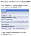
PERC rule: pulmonary embolism rule-out criteria
How does the PERC rule work when assessing likelihood of PE?
all criteria must be absent to have negative PERC result, i.e. you can rule out PE (probably <2%)

When should the PERC rule be used when assessing the likelihood of PE?
if there is a low pre-test probability of PE, but want more reassurance that it isn’t the diagnosis
(low = <15% probability)
If your suspicion of PE is greater than ‘low’ that would make you consider using the PERC tool, which tool should be used instead?
2-level PE Wells score
What does the 2-level PE Wells score consist of? 6 aspects

How is the result of the 2-level PE Wells score interpreted?
clinical probability simplified scores:
- >4 points: PE likely
- 4 or less: PE unlikely

What is the next step in investigation if PE is considered likely from the Wells score?
arrange immediate CTPA (computed tomography pulmonary angiogram)
if delay in getting CTPA, interim therapeutic anticoagulation should be given until scan is performed
What anticoagulation should be given in the interim if waiting for CTPA to be performed and there is a delay?
DOAC: apixaban or rivaroxaban
What is the next step in management/investigations based on the result of CTPA for a Wells score >4?
- positive: PE diagnosed
- negative: consider proximal leg vein ultrasound scan if DVT suspected
What is the next step in investigation for PE if Wells score is 4 points or less?
D-dimer test
Based on the results of the D-dimer test following a Wells score of 4 points or less what are the next steps in management?
- d-dimer positive: arrange immediate CTPA, if delay consider interim therapeutic anticoagulation
- d-dimer negative: PE unlikely, stop anticoagulation and consider alternative diagnosis
What are 4 advantages of CTPA over V/Q scans to diagnose PE?
- CTPA faster
- CTPA easier to perform out of hours
- Reduced need for further imaging following CTPA
- Possibility of CTPA providing alternative diagnosis if PE excluded
What are 3 situations in which a V/Q scan may be performed initially rather than CTPA?
- If appropriate facilities exist
- If CXR normal
- If no significant symptomatic concurrent cardiopulmonary disease
When is V/Q scanning the investigation of choice for suspected PE (over CTPA)?
if there is renal impairment - no requirement of contrast
When should age-adjusted d-dimer levels be considered?
patients >50 years
What are the classic ECG changes in PE?
- S1Q3T3
- prominent S waves in lead I
- large Q wave in lead III
- inverted T wave in lead III

In addition to S1Q3T3 what are 3 further ECG changes which are seen in PE?
- Right bundle branch block
- Right axis deviation
- Sinus tachycardia - commonest finding
Which patients with suspected PE are recommended to have a chest x-ray?
all patients to exclude other pathology
What findings may be seen in PE on a chest x-ray?
typically normal, may see wedge-shaped opacification
focal oligaemia
What are the sensitivity and specificity of a V/Q scan for PE?
sensitivity 75%, specificity 97%
What are 4 other causes of a mismatch in V/Q in addition to PE?
- Old pulmonary emboli
- AV malformations
- Vasculitis
- Previous radiotherapy
What change will be seen in COPD on a V/Q scan?
matched defects
What type of PE may be missed on a CTPA?
peripheral emboli affecting subsegmental arteries
What type of PE is shown in the image?
saddle embolus

What are 4 recent key changes to the guidelines for managing VTE in 2020?
- Use of DOACs as first line treatment for most peple with VTE
- Use of DOACs in patients with active cancer, as opposed to LMWH as per previous recommendation
- Outpatient treatment in low risk PE patients
- Routine cancer screening no longer recommended following VTE diagnosis
How should you determine which patients with PE should be admitted vs which shouldn’t?
those deemed low risk are able to be managed as outpatients - British Thoracic Society supports use of Pulmonary Embolism Severity Index (PESI) score
What are 3 key requirements for a patient with PE to be managed as an outpatient?
- Haemodynamic stability
- Lack of comorbidities
- Support at home
Which anticoagulants should be offered first-line following the diagnosis of PE?
apixaban or rivaroxaban
How have the guidelines changed regarding which anticoagulants to use for suspected PE?
previously NIC said use LMWH until diagnosis confirmed, but now adovcate using a DOAC once diagnosis suspected, and continue if diagnosis confirmed
If neither apixaban or rivaroxaban are suitable for use in a patient with confirmed/ suspected PE, what should eb used instead?
LMWG followed by dabigatran or edoxaban OR LMWH followed by vitamin K antagonist (i.e. warfarin)
How have the guidelines changed for management of PE in patients with active cancer?
previously LMWH recommended, now guidelines recommend using DOAC unless contraindicated
In patients with renal impairment with eGFR <15, what is the recommended management of PE?
LMWH or unfractionated heparin or LMWH followed by warfarin
What is the management of a PE in a patient with antiphospholipid syndrome?
LMWH followed by vitamin K antagonist (warfarin)
What is the minimum length of time that patients with PE should have anticoagulation for and what causes the total length to differ?
- 3 months
- whether provoked or unprovoked:
- provoked - if obvious precipitating event, can stop after initial 3 months (3-6 months if active cancer)
- unprovoked - typically continued for 6 months in total
What is the guidance for the length of treatment for a provoked VTE i.e. obvious cause?
typically stopped after initial 3 months
How long is anticoagulation recommended to be continued for VTE caused by active cancer?
3 to 6 months
How long is anticoagulation recommended to be continued for patients with unprovoked VTE?
6 months total
What should the length of time which anticoagulation for VTE is continued for be based on?
provoked vs unprovoked, but also balance risk of recurrence with risk of bleeding
What score can be used to determine risk of bleeding with anticoagulation treatment when managing VTE?
HAS-BLED score
What is the first line treatment for massive PE where there is circulatory failure (i.e. haemodynamic instability e.g. hypotension)?
thrombolysis (or can perform embolectomy)
What is a management option for patients who have repeat pulmonary embolisms despite adequate anticoagulation?
consider inferior vena cava (IVC) filters
How do inferior vena cava filters work to prevent PE?
stop clots formed in the deep veins of the leg from moving to the pulmonary arteries
What is the definition of massive PE?
acute PE with obstructive shock or SBP <90 mmHg
What is the definition of submassive PE?
acute PE without systemic hypotension (SBP >90) but either RV dysfunction or myocardial necrosis
What are 3 pathophysiological effects of PE?
- Increased pulmonary vascular resistance leading to right ventriular failure and hence obstructive shock
- Increased alveolar dead space can cause V/Q mismatch so pulmonary vasoconstriction occurs to optimise gas exchange
- Pulmonary infarction
What are 5 cardiovascular signs on examination of PE?
- Tachycardia
- Hypotension
- Elevated JVP
- Parasternal heave
- Loud P2 (closure of pulmonary valve, forms S2 with A2 usually being main component)
What are 5 thrombophilias which can predispose to PE?
- Factor V Leiden mutation
- Prothrombin gene mutation
- Hyperhomocytsteinaemia
- Antiphospholipid antibody syndrome
- Deficiency of antithrombin III, protein C or protein S
What can elevated JVP indicate in PE?
underlying right heart strain i.e. massive PE
What are 6 blood tests that you should request in suspected PE, in addition to d-dimer if Wells score supports this?
- FBC
- U+Es
- LFTs
- Coagulation
- CRP
- Troponin (?)
What should you do in the initial management of PE if the patient is hypovolaemic?
administer 500ml bolus Hartmann’s or 0.9% NaCl over 15 min (250ml if patient at increased risk of fluid overload)
make sure to reassess BP/ HR after. give up to 4x 500ml boluses


