Protozoal parasites Flashcards
How are protozoal parasites classified?
Presence or not of inactive cyst stage (usually extracellular)
lost motility
reduced metabolism
no growth of and multiplication.
Usually modified outer wall.
The cyst/ ”spore” resistant and often the infective stage
What are the four types of method of motion for protozoal parasites?
Amoeboid (Sarcomastogophora)
Flagellate (Sarcomastogophora)
Ciliate
Sporozoan- bad ones, filamentous complex at one end. can forms spores and malaria is usually under this group (Apicomplexa)
How do protozoal parasites reproduce?
Homologous recombination machinery is present in most (??all) species- expressed in certain points in the cycle-Host enzymes not required
Suggests meiosis/ sexual division may occur however appears rarer than binary fission
Single haploid cells only observed in some protozoa forms e.g., plasmodia (?? Other forms of recombination without haploid fusion… amoeboid parasites, conjugation (sexual material transfer without reproduction- ciliates)
What are the mitotic forms of division seen in protozoal parasites?
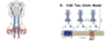
Binary fission: replication of DNA –> cytokinesis –> two identical daughter cells (get image)
Schizogony: cell –> reproduce multiple times to form schizont –> cytokinesis –> merozoites formed
Tell me about Flagellates
How do they reproduce?
What are the most sever flagellates
Mastigophora (flagella)
One or more flagella - sometimes with an undulating membrane (e.g., Trypanosomes)
Often two stages mastigote (flagellate)- (which has 3 sub -forms pro-, epi-, and trypo-) extracellular and amastigote, intracellular stages
Predominantly reproduce by asexual binary fission (do show sexual reproduction, in sense that you see DNA transfer)
Anterior is at flagellar tip end, which acts as a propeller to drag parasite along
No cyst stages (most severe)
- intestinal and urogenital flagellates e.g., Giardia, Trichomonas,
- Blood and tissue flagellates e.g., (haemoflagellates- kinetoplastids) Trypanosoma, Leishmania,

What are Amoeboid’s and whats are their two stages?
Sarcodina (flesh like)
Two stages
- Trophozoite (trope, nourishment + zoon, animal) from moves and capture food via pseudopods, temporary extensions of the cell into which the endoplasm then flow- nutritional stage. Usually non-ciliated (not always)
- Resistant long-lived cysts some multinucleate (Entamoeba histolytica)
How do Amoeboids reproduce?
Asexually via binary fission
What may some amoeboids show, provide an example for this?
Some may show third transient (1-2 hrs) flagellate state!! E.g., Naegleria Fowleri
Naegleria Fowleri brain eating amoeba - primary amoebic meningoencephalitis - ~ 100% lethal – found in tepid stagnant water - used as a model to study changes from amoeboid to flagellated stages- flagellate state for movement and reverts to amoeba
What are Ciliate’s and what are their two stages?
Ciliated protozoa- cilia distributed in rows or patches
Two stages: Cyst and Trophozoite
Tell me about the reproduction of ciliates
They have macronucleus-regulates metabolic activities and micronucleus -storage site for the germline genetic material
Show both sexual conjugation and binary fission
micronucleus gives rise to the macronucleus and is responsible for the genetic reorganization during conjugation
macronucleus organises protein production
diploid nucleus is the store of genetic material that converts to macronucleus
division: miotic stage–> two organisms attach via conjugation –> before that’s finished the micronucleus go through meiosis –> end up with 4 haploid micronuclei –> one of those divides again –> 5 haploid nuclei–> transfer across of the two fused conjugated cells –> 2 haploid micronuclei come together to form diploid micronucleus –> division of cell, reproduction –> old macronucleus is destroyed and replaced by micronuclei that becomes macronucleus

Give an example of a ciliate that is the only human parasite?
Balantidium coli, a giant intestinal parasite of humans and pigs (only human parasite)
Tell me about Sporozoa or Apicomplexa
What stages do they have?
What do they lack?
Tell me about their life cycle
Spore like stages
Lack flagella, cilia, or pseudopodia but capable of gliding movements.
Specialised apical complex secretes enzymes which allow the sporozoite (main infective from) to enter a host cell.
Have complex life cycle with alternating sexual and asexual reproductive phases involving two different hosts (digenetic).
Tell me about the complex reproduction of sporozoa or apicomplexa
asexual schizogony forming many offspring (merogony forming merizoites, and gametogany forming gamete)- which fuse for
sexual sporogony forming sporozoites – infective form for transfer
What are the two classes of sporozoa or apicomplexa and give examples of whats included in the classes?
Coccidian class – Coccidia, Neospora, Toxoplasma
Haematozoan class includes -Plasmodium species
Define Schizogony
Asexual multiple fission comes in a number of forms
Define Sporogony
Sexual and asexual phase in definitive host e.g., mosquito, male and female gametes fuse in the enteric area of the mosquito forming oocyst- undergoes Meiosis now
Tell me about the Schizogony of sporozoa or apicomplexa
Oocyst multiple asexual divisions Schizogony- forming multinucleate cells which split to single nucleate sporozoites- enter salivary glands and passed to secondary host
Define asexual schizogony
Asexual phase in secondary host e.g., mammal utilitising a schizont large multinucleate cell
Define Pre-erythrocytic schizogony (merogony)
Pre-erythrocytic Schizogony (merogony) – sporozoites enter cells (eg liver) and show nuclear replication form a Schizont –– this divides to single nucleate merozoites which are released
What are the two outcomes of erythrocytic schizogony (merogony or gametogany)
- Merozoites enter RBCs and form trophozoites – these show nuclear replication form Schizont– this divide to merozoites – released and repeats (merogony)
OR some merozoites instead undergo meiosis and form gametocytes which are released (gametogany)
- Gametocyte (in some sporozoa these differ in size eg Plasmodia)
Male - microgamete (can be flagellated!)
Female – macrogamete
In some cases with erythrocytic schizogony, there is no schizont, instead what conversion is there?
Trophozoites –> Merozoites directly
What parasites are included under the term flagellates?
Trypanosoma and Leishmania
Tell me about the flagellates main differentiation stages
? primarily insects’ parasites, vertebrates a minor part of biological cycle
Main differentiation stages- different structure, behaviour, and metabolism
- Flagellated Trypomastigote- in mammal, insect gut and transfer organism
Extracellular (some subversions- metacyclic (usually infectious to us), bloodstream, stumpy, procyclic (dividing form in insect))
- Flagellated epimastigote- found in insect tissue, extracellular
- Non-flagellated amastigote- present inside mammalian cells, intracellular- doesn’t need flagella because it is inside the cell
There is a promastigote in Leishmania…
Give an example for a Hemoflagellate
Trypanosoma (for Leishmania- see last year)
Tell me the main differentiation stages of Haemoflagellates
Body Borer
Believed to initially been a parasite of insects (may still be and mammals a minor part of biological cycle)
3 main differentiation stages
Promastigote
Trypomastigote - flagellated, in mammal and transfer organism to and from insect. Extracellular
Epimastigote – flagellated form found in insect tissue
Amstigote
Amastigote – non -flagellated form present inside mammalian cells (show binary fission). Intracellular
Tell me the morphology of the trypanosome…
Variety between species?
Adaptation?
characteristics/ features?
Different species have different morphological forms
Trypanosomes adopt different morphological forms in different hosts
Flagella appears at side in tyropmastigote and epimastigote
At and in promastigote
What are the different forms of the Trypanosome that appear in the life cycle?
Trypomastigote-posterior localization of mitochondrial DNA aggregate, (kinetoplast K), to the nucleus (N). An attached flagellum exits from the flagellar pocket (FP) near the cell posterior.
Epimastigote-kinetoplast is anterior to the nucleus. While the flagellum is still attached to the cell surface, it also has a long, cell-free overhang.
Promastigote-similar arrangement of DNA-containing organelles as epimastigote, but the flagellum is unattached after exiting the FP pocket at the cell anterior. (Seen in Leishmannia – not Trypanosomes)
Amastigote – smaller, spherical, kinetoplast anterior to the nucleus. The flagellum very short and nonmotile, - INTRACELLULAR FORM

Tell me about the Microtubule quartet (MtQ) in trypanosomes
The microtubule quartet (MtQ) has opposite polarity to the other array microtubules- (nucleated near the basal body and grows towards the cell anterior may act as a signal for the formation of the flagella).
The flagellum attachment zone (FAZ) filament interrupts the array and attaches the flagellum to the cell surface.
PFR, para-flagellar rod.
FP- flagellar pocket has unique composition

Classification of trypanosome species: Morphology and genetics show marked difference

Flagellate morphology
Different species have different morphological forms
Trypanosomes adopt different morphological forms in different hosts
Promastigote- Leissmania
Trypomastigote-?
General structure of the trypanosome
Position of flagella changes with nuclei position
Plasma membrane covered in protein coat made of VSG proteins

Give the structure of the trypanosomes
Unicellular eukaryotes
The cell membrane consists of at least three discrete domains: the cell membrane, the flagellar membrane, and the flagellar pocket
Possess a microtubule corset closely opposed to the cell membrane – define the shape of the cell
Have a kinetoplast – a mass of cytoplasmic DNA found at the base of the flagellum and continuous with the (single) mitochondrion.
Possess a paraflagellar rod – only partially characterised with unknown function (attachment to insect host? Aids motility?)

Tell me about the Kinetoplast
Mass of mitochondrial DNA which sits where the flagellar pocket will be at the end of the mitochondria
Kinetoplast splits first, then flagellar then nucleus during division
The kinetoplast is a specialized region of the mitochondria of trypanosomatids that harbors the most complex and unusual mitochondrial DNA found in nature. Kinetoplast DNA (kDNA) is composed of thousands of circular molecules topologically interlocked to form a single network.
What does the paraflagellar rod help with?
Possess a paraflagellar rod – only partially characterised with unknown function (attachment to insect host? Aids motility?)
- Attachment to insect host? Dense hemi-desmosome- like plaques associated with proximal flagellum and unsect cells
- Aids motility?
- Stabilises flagellum
Tell me about Kinetoplasts and their genetic information?
Unique to kinetoplastids (flagellates inc. Tryp and Leish)
Mitochondrial DNA
Mass of circular maxi- and mini- kDNA near flagella polar body
Present at one end of the branched single continuous mitochondria
10-40 copies of the mitochondrial genome (maxicircles)
1000 +copies of guide DNA (minicircles- forms gRNA) – unique correction system of mitochondrial genes akin to CRISPR

Name some significant flagellates (non-human)
- Crithidia
- Phytomonas
Tell me about Crithidia and provide an example
Crithidia – mongenic insect parasites (possible direct ancestor of group)
Only found in choanomastigote form
e.g., C. bombi parasite of bumblebees implicated in decline of wild bee populations

Tell me about Phytomonas and provide examples
Phytomonas (phyto= plant)
- digenic insect
- plant parasites.
P. staheli causes wilt in coconut /oil palms Latin America (>$1Billion lost/ yr).
P. leptovasorum coffee phloem necrosis in Liberica and Arabica ($8 billion / yr).
Other ssp infect tomatoes
No treatment, try to prevent spread
Probably initiated as insect monogenic parasite –maybe initiator of all parasitic flagellates and used as models for human flagellate trypanosomal diseases.
Digenic parasites:
Digenetic parasites are those that need more than one host (usually two) to complete their life cycles.

Species of Trypanosoma
T. ambystomae. in amphibians
T. antiquus, extinct (Fossil in Miocene amber)
T. avium, which causes trypanosomiasis in birds
T. boissoni, in elasmobranch
T. brucei, which causes sleeping sickness in humans and nagana in cattle
T. cruzi, which causes Chagas disease in humans
T. congolense, which causes nagana in ruminant livestock, horses and a wide range of wildlife
T. equinum, in South American horses, transmitted via Tabanidae,
T. equiperdum, which causes dourine or covering sickness in horses and other Equidae, it can be spread through coitus.
T. evansi, which causes one form of the disease surra in certain animals
T. everetti, in birds
T. hosei, in amphibians
T. irwini, in koalas
T. lewisi, in rats
T. melophagium, in sheep, transmitted via Melophagus ovinus
T. paddae, in birds
T. parroti, in amphibians
T. percae, in the species Perca fluviatilis
T. rangeli, believed to be non-pathogenic to humans
T. rotatorium, in amphibians
T. rugosae, in amphibians
T. sergenti, in amphibians
T. simiae, which causes nagana in pigs. Its main reservoirs are warthogs and bush pigs
T. sinipercae, in fishes (other host Leaches)
T. suis, which causes a different form of surra
T. theileri, a large trypanosome infecting ruminants
T. triglae, in marine teleosts
T. vivax, which causes the disease nagana, mainly in West Africa, although it has spread to South America
T grayi – crocodiles spread by Tsetse flies
T. clandestinus crocodiles spread by Leaches
Gicve examples of Trypanosomas found in america
America- some esp. rodent ones widespread
T. lewisi, in rats T musculi in mice…initially Americas (flea)-pathogenic to man too
T cruzi in man – Chagas disease – infectious to wide spectrum of mammals
Give examples of Trypasoma in Africa
T. vivax
T. Congolese
T. brucei
T. equiperdum
T. evansi
How many subspecies of T.brucei are there
What does it cause and in what mammals?
T brucei – 5 subspecies (flies /bats!)
Cause Nagana in ruminants
Sleeping sickness in man
Dourine in horses
Surra numerous animals including dogs/ cats ruminants (Africa/ middle east /south America
What does T.vivax cause?
Ruminants- Nagana- West Africe (Tsetse fly)
What does T. Congolese cause?
T. Congolese, causes nagana in ruminant livestock, horses, and a wide range of wildlife
What does T.equiperdum cause?
T. equiperdum, which causes dourine or covering sickness in horses and other Equidae (also mating)
What does T. evansi cause?
T. evansi, surra in certain animals
Mutant forms of T.brucei can’t grow in insects so what do they act as?
A vector
What is a salivarian trypanosome?
What hosts is it found in?
Salivarian trypanosomes - passed to the recipient in saliva
- insects normally tsetse flies (Glossina spp.)
- also, tabanid (horse) flies, vampire bats South America
- tsetse fly is an amplification step
African trypanosomiasis
What is a Stercocarian trypanosome?
What host is it found in?
Stercocarian trypanosomes – passed to the recipient in faeces of insects e.g., triatomid (assassin) bugs
also, tabanid flies, fleas
South American trypanosomiasis
With the African Trypanosome, what are the four main diseases caused by the T. brucei sub forms?
Sleeping sickness in man
Nagana in cattle and horses
Surra in many animals
Dourne in equids
What does the disease form of the African Trypanosomiasis depend on?
The trypanosome and species infected
Name the subspecies in the African Trypanosomaiasis which are the human diseases and those which are the animal diseases
Human Disease
Trypanosoma brucei rhodesiense (2%) - more acute disease, Zoonotic
Trypanosomoa brucei gambiense (98%).
Animal Disease
Trypanosoma. brucei. bruceii in cattle causes Nagana
Trypanosoma. brucei equiperdum, which causes dourine or covering sickness in Equidae (also spread at mating)
Trypanosoma. brucei evansi, surra in many species’ animals
Last 2 cannot reproduce in insects so only use them a mechanical vector
Morphologically identical – genetically different
What is the life cycle of the African Trypanosomiasis?
overtime will end up in CSF in CNS
When present in blood in high amount it goes to Stumpy form
Can show sexual production as evidence for DNA transfer occurring
Cattle can also be infected by T.b.rhodesiense (this is not seen with gambiense form)

Define procyclic form (TM)
Procyclic form (TM) – the dividing form of the parasite found in the insect host. Mostly binary fission but are capable of recombination (? sexual reproduction)
Define Epimastigote
Epimastigote – smaller form invasive form can leave gut of fly and enter its salivary gland has different flagella attachment to TM forms
Define Metacyclic form (TM)
Metacyclic form (TM) – the human infective form of the parasite found in the insect host- in salvarian forms it can resist salivary enzymes and reduces their production (see below for Chagas disease)
Define bloodstream form (TM)
Bloodstream form (TM) – the dividing form found in the bloodstream of the mammalian host
Where is the Tsetse fly found?
What is its life length?
How many species are there?
What are the hosts?
Tropical Africa (grassland and forest)
Long lived obligate parasites- female to 4 months as adult
At least 23 species in tropical Africa
Different subspecies are infected with different trypanosomes
Primary host: insect
Secondary host: mammals
Tell me the number of eggs that a female Tsetse fly will produce and how many across its life time?
Females feed on vertebrate blood through biting
Multiple egg broods >6 but only requires mating once (may mate more often)
Has a uterus and releases larva at 3rd Instar stage (9d)- hatches after 4 days, then stays in for a further 9 days as continues to develop
Produces one larva at a time released from uterus.
Larvae and burrows into sand or soil to pupate ie depended on mother for nutrition till adult
hence female must feed often-exclusively on blood
different species have different chosen hosts
can take >100% body weight in blood
Dynamic populations – drop by >90% in dry season
Tell me some general features of Tsetse flies
Day feeding
Attracted to smell, movement, and colour (specifically blue) – large animals
Often quite host specific
Very good transmission system – both Mechanical and Biological Transmission, must feed regularly
(Horse flies can act as Mechanical vector for trypanosomes)
The presence of these flies / their parasites is a direct cause of sub-Saharan poverty
– and failure in agriculture

What are some other ways of being infected with T.brucei species?
Other biting flies (e.g., Horse) – mechanical transfer only
Vertical trans-placental
Blood transfusion/ organ transplantation
Needle (rare must be when highly infectious)
Sexual contact
All these are relatively rare – the fly primary host is a very efficient amplification step
What are the two sub-strains of human T.bucei (that cause sleeping sickness in man)
Tell me some info about each strain
T. b. gambiense is found in Central and West Africa
- More Chronic - ~98% of all ASS
- Limited reservoirs
- Easier to control
T. b. rhodesiense is found primarily in East Africa (Botswana, Ethiopia, Kenya, Malawi, Tanzania, Uganda, Zaire, and Zimbabwe)
- More Acute - ~2% of all ASS
- Wildlife and Livestock infected too
- Harder to control

What are the different stages of human disease?
Tell me about each
Three (? two) defined stages – 100% fatal if not treated
Acute means speed of infection, severity is whether it will kill someone or not
Stage 0/ Initial infection – localised Trypanosomal chancre (response to localised presence of trypanosome)
at the site of the infectious fly bite ~ 2 days of infection. More common in T. b. rhodesiense infection,
Parasites multiply here – pathology due both to trypanosome and type IV hypersensitivity response to fly salivary proteins
Stage 1/ Haemolymphatic stage – parasite found in the blood/ Lymph
Nonspecific, generalized symptoms malaise, weakness, anaemia fatigue, pruritis, hepato-splenomegaly, weight loss and intermittent fevers in waves
(Immune evasion strategies here- parasite is highly antigenic)
~ 1–3 weeks after the tsetse fly bite T. b. rhodesiense
~ weeks to months after the tsetse fly bite T. b. rhodesiense
V common lymphadenopathy
Stage 2/ CNS stage (often overlaps with stage 1)
T. b. rhodesiense ~ 21–60 days.
T. b. gambiense ~ 300–500 days,
invasion of the CNS (CSF contains trypomastigotes) causes neuropsychiatric disease, (fever occurring less frequently)
sleep/wake cycle reversed, daytime somnolence, nocturnal insomnia, hence the common name
Behavioural -hallucinations, delirium, apathy, aggression, mania
Motor - weakness, ataxia, tremor,
Sensory - hyperaesthesia, anaesthesia, pruritis, visual hallucinations
-> resulting in seizures, coma, DEATH (inevitable if untreated)
Tell me about the Variant specific glycoprotein (VSG) involved in immune evation (present up to 500 days and strong immune response)
Variant specific glycoprotein– VSG – surface glycoprotein ~ 90% of outer coat and close to 100% external surface
Many (2000 VSAs (Variant specific antigen) genes or parts of genes in genome –one expressed at one time)
~65kD, 500 amino acids, 3 domains.
N terminal 360 amino acids, most superficial and seen by immune system poor sequence conservation but similar secondary/ tertiary structure
The 120 C-terminal amino acids highly conserved but masked lying next to the plasma membrane.
The C terminal pro protein has typical transmembrane hydrophobic sequence attaching the protein to the plasma but this cleaved and the mature protein is linked to the membrane cell surface by a phosphatidylinositol glycolipid link. This allows tighter packing and masks other more conserved membrane proteins from antibodies.
What is the VSG part of?
What happens as the parasitemia rises?
VSG part of 12-15nm thick outer layer on membrane
As the parasitemia rises, the host raises antibodies to the coat killing most of the parasites
Some of the remaining parasites switch their coat protein and therefore evade the immune response
TEMs of TSs of trypanosomes
Why is the VSG varied?
VSG is varied because have genetic recombination and VSG expression site switching in respect to the promotor and position relative the telomere (the most telemetric version is expressed) causes changes in VSG expression
What does the VSG do during immune evasion?
VSG recombination to novel forms
Stripping and replacement of VSG also occurs
?? role of flagellar groove
VSG shedding and attachment on to RBC causes
RBC lysis and anaemia
Recruitment of complement and antibodies away from the parasite
Hypocomplementemia- lower complement levels as they are being used to lyse RBC

What is the infective form of the Trypanosoma?
Tell me about this
The infective form is the stumpy trypomastigotes
Transition from slender (blood) to stumpy (infectious non proliferating) forms caused by a quorum sensing mechanism, stumpy induction factor (SIF) accumulates as parasite numbers increase during parasitaemia. I.e., so non-dividing infectious form only produced when high levels (consider the opposite)
Where the long form is ingested, they die in the fly

How do you diagnose infection by tryptosoma?
Clinical diagnosis initially hard – non-specific signs
Haematology –parasite only observed at time of high parasitaemia in early infection
Lymph node biopsy
PCR on blood/ CSF
CSF smears
Serology not good – main antibodies against VSGs …. which one?
- There are some screening Ab tests.
What are the two treatments for infection by tryptosoma?
Those drugs which enter the CNS and those which don’t
Tell me about the treatment for tryptosoma that does not enter the CNS
Haemolymphatic stages (these drugs don’t enter CNS)
- Pentamidine, for T.b gambianse - binds AT rich regions of parasite DNA -> crosslinking (resistance observed) – discovered 1937 Resistance to Pentanidine caused by mutations,
TbAT1/P2 transporter
High Affinity Pentamidine Transporter
Resistant trypanosomes were selected for in the field by farmers under-dosing
- Suramin, for T.b rhodesiense – binds trypanosomal glycolytic enzymes, inhibiting energy metabolism - discovered 1916
Tell me the treatment for tryptosoma that enters the CNS
What are some side effects?
*Nausea, vomiting, diarrhoea, headache, skin tingling, and weakness. Sore palms of the hands and soles of the feet, trouble seeing, fever, and abdominal pain may also occur
**headache, conjunctive injection, fever, tachycardia, hiccup, diarrhoea, vomiting, tremor, slurring of speech, usually followed by convulsions and ultimately coma, death
CNS stages- these enter CNS, but toxicity rules out use for earlier stages
Nifurtimox (a nitrofuran) undergoes reduction and releases oxygen radicals and H2O2 which destroys parasite DNA. Mammalian cells are protected by peroxidases and superoxide dismutase
Melarsoprol** (arsenical) irreversibly binds sulfhydryl groups on pyruvate kinase reduces energy production Limited selective toxicity (1949)
Eflornithine** (polyamine biosynthesis inhibitor) irreversibly inhibits ornithine decarboxylase which makes polyamines e.g., putrescine needed in cell division, 1970
** Severe side effects - ~95%+ clearance of parasite
What are some new treatments for infection by tryptosoma?
Fexinidazole - 2018, first drug for advanced-stage sleeping sickness in thirty years- Nitroimidazole compound
Acts against both stages if not severe taken orally- taken orally and enters CNS
Parasite nitro reductases reduce fexinidazole to reactive intermediates free radical and superoxide release which are capable of damaging DNA and proteins

What are some preventions put into place for infection by tryptosomas?
WHO has targeted West African trypanosomiasis for elimination as a public health problem?
Strategies for elimination of West African trypanosomiasis
: active and passive case - screening
: treatment of the confirmed cases,
: vector control to reduce the tsetse population.
East African trypanosomiasis far greater challenge due to reservoir infection in Wildlife and Livestock. Vector control is the primary strategy along with animal health.
There is no vaccine or prophylactic drug
What are some preventions that can be done in order to reduce Tsetse fly attack?
Environmental
Map highly effected sites – localised sequential aerosol spaying with plant spread insecticides in severe outbreaks
(Ground treatments not used – residues)
Bush Clearance – ecological issues
Pour on systemic insecticide treatment of animals (East African trypanosomiasis)
Baits and coloured traps (pheromone and insecticide traps)
Human
Long-sleeved shirts / pants of medium-weight material in neutral colours
Inspect vehicles - flies are attracted to the motion and dust from moving vehicles.
Avoid bushes.
Minimise activity during the hottest part of the day.
Together can reduce Tsetse fly attack by >99%

Tell me about the disease Nagana
Nagana- Trypanosoma brucei brucei and friends
Similar disease in cattle to sleeping sickness in humans
Caused by T.b. brucei, T.b. rhodesiense (T. vivax and T. congolense)
More widespread and commoner than sleeping sickness – more vectors and quicker reproduction in fly
Large areas of sub-Saharan Africa killing 3 million cattle per year- can’t farm cattle here
Some cattle are relatively resistant (e.g., N’Dama), European species not
Devastates agriculture and lifestyles
Tell me about the South American Trypanosomiasis (SAT) disease
South American trypanosomiasis- SAT
Trypanosoma cruzi – Chagas disease
Zoonotic disease affecting 150 species of mammals inc. humans
Found as amastigote (intracellular), trypanomastigote (blood) and epimastigote (insect) forms
Stercocarian trypanosomes – passed to the recipient in faeces of insects e.g., triatomid (assassin) bugs– also tabanid flies, fleas
Tabinids – horsefly – again a mechanical vector not biological one
Cf T brucei – only a few other hosts
Whats the life cycle of the flagellates (Haemoflagellates) Trypanosome?
Assassin bugs crawls as it can’t fly
Triatomine insect vector- Triatoma, Rhodnius, and Panstrongylus (or “kissing bug”) takes a blood meal and releases trypomastigotes in its faeces at bite wound.
Trypomastigotes enter host through wound or at intact mucosal membranes, eg conjunctiva.
In the mammal host, the trypomastigotes invade cells at site of inoculation, and differentiate into intracellular amastigotes. These multiply by binary fission, differentiate into trypomastigotes, and release into the circulation
Trypomastigotes infect cells in a variety of tissues and transform into intracellular amastigotes in new infection sites->Clinical disease
The bloodstream trypomastigotes do not replicate (cf African trypanosomes). Replication resumes only when the parasites enter another cell or in the “kissing bug” when infected by feeding on mammals that contains circulating parasites.
In insect the ingested trypomastigotes transform into epimastigotes in the vector’s midgut. The parasites multiply and differentiate in the midgut and differentiate into infective metacyclic trypomastigotes (generally taken up by bug when it takes a meal) in the hindgut.
Replicated via binary fission

What are some other ways of being infected?
Vertical trans-placental
Blood transfusion/ organ transplantation
Needle stick (rare must be when highly infectious)
Consumption of uncooked food that is contaminated with faeces from infected triatomine bugs
??Bed bug.
Why kissing bug – crawling bug living in cracks and feeding at night biting exposed skin at night often face
Tell me about the infection and incidence of Chagas disease and how it is transmitted
Chagas disease is transmitted to animals and people by insect vectors that are found only in the Americas.
~ 8 million people in Mexico, Central America, and South America have Chagas disease, most inapparent infection. Columbia alone ~$1Billion/yr health costs. Estimated 300,000 infected in US
Large-scale population movements from rural Latin America to other regions of the world have increased the geographic distribution and changed the epidemiology of Chagas disease. In the US and Europe Chagas disease is found but is not endemic. cf other ways of transfer
Infection is lifelong and may become life threatening ~ 30% will develop serious cardiac problems and 10% will develop digestive and neurological problems
Bed bug can act as an amplifying host
Distribution of chagas disease

How many phases of chagas disease are there? Tell me about them
3 phases – some clinicians fuse the 2 chronic phases
Acute phase: occurs immediately after infection, lasts ~ 2 months There may be initial fever or swelling at site of inoculation.
High numbers of parasites circulate in blood, symptoms are mild or absent. very rarely, acute infection causes cardio myositis, or encephalitis/meningitis (easy to diagnose here)
Chronic intermediate phase: parasites are hidden mainly in heart and digestive muscles. Due to immune response parasite number are low but rarely eliminated (diagnosis difficult). In later years can lead to heart failure and /or sudden death
Tell me about chronic chagas disease
What problems can arise from this?
Chronic Chagas disease: Many people remain asymptomatic for life. However, 20–30% of infected people develop severe life-threatening problems
Heart rhythm abnormalities that can cause sudden death.
A dilated cardiomyopathy and heart failure
Failure in GI smooth muscle -megaoesophagus or megacolon, leading to difficulties eating or defecating
Tell me about how an individual would enter into chronic chagas disease
Entry into chronic disease
- Immune suppression (AIDS, steroids, or chemotherapy), Reactivation with parasites found in the circulating blood.
- Autoimmunity: Caused by Molecular mimicry: as pathogen bears antigens like host antigens with self-responding Abs resulting in tissue damage
How is chagas disease diagnosed?
Not easy using blood smears – only acute phase
PCR blood in acute phase
Serological techniques often inconclusive
Xenodiagnoses - is often used. Uninfected bugs are fed on suspected sufferers and monitored over a period of 3 months to see if their faeces contain the metacyclic tyrpomastogotes.
What is the current treatment for chagas disease?
Nifurtimox (see Sleeping Sickness) and Fexinidazole / Benzinidazole. Both administered orally
needs long treatment courses and can’t overcome pathology
~60 clearance of parasite as in cellular niche
Benznidazole - see Fexinidazole (Nitroimidazole) nb this is not a benzimidazole the microtubule inhibitor – see helminth lectures
All severe side effects

What are some methods for the prevention of chagas disease?
Vector - Improved housing and spraying insecticide inside housing to eliminate the bugs,
Insecticide impregnated bed nets, biting usually at night, a crawling bug plastering houses and better cleanable flooring has proved effective
Triatoma insecticide resistance is starting to develop.
Screening of blood donations
Screening at organ transplantation
Early detection and treatment of new cases, including mother-to-baby (congenital) cases,
Tell me about vaccines for Trypanosomes
Work underway with Leishmania – trials underway
Less easy with T. cruzi because of variations in the parasite
Impossible with T. bruceii because of VSG antigenic variation.
What are the thoughts about drug developments?
Exploit trypanosome weak spots e.g., flagellar pocket (unique membrane structure), paraflagellar rod (only in kinetoplasts), kinetoplast,
Genomes have been sequenced and some biochemical pathways are different to ours but common to trypanosomes
Why are some cattle resistants to T.b. brucei?
What are the problems with trypanosomal diseases?
Zoonotic
Diagnosis and recognising infection in vectors/ reservoir hosts
Other hosts
Drug resistance – parasite (some) and insect vectors (lots?)
Vaccines…
Asymptomatic stages
Pregnant women
