Protein targeting and export Flashcards

1
Q

An average protein is 10nm across, what is the volume of the protein?

A

41 nm3 in volume

2
Q

An average eukaryotic cell is 50 µm in diameter and has a volume of what?

A

6.6x10¹³ nm3

3
Q

An overview of protein targeting and export

A
4
Q

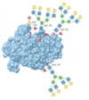
Whats the fluorescent protein used to visualise subcellular localisation?

What organism was it discovered in?

A

GFP

Discovered in Aequorea victoria (Nobel prize winning discovery)

5
Q

What structure does GFP have and when it’s mutated in different ways what can be produced?

A

GFP has a beta-barrel structure

Different colours can be obtained by mutating the Beta-barrel structure

e.g. mutating into YFP, BFP, CFP

This is because the wavelengths can be manipulated whilst moving through the structure and therefore produce different colours

6
Q

How else can different colours be obtained from GFP?

A

By using other organisms such as DsRed

7
Q

What can GFP fuse with?

A

Your proteins of interest (chimera- A chimera is essentially a single organism that’s made up of cells from two or more “individuals”)

8
Q

Whats a disadvantage of GFP?

A

Chimeric protein may not fold correctly/ act normally due to large size of FP

9
Q

The use of Immunofluorescence (IF) has the same kind of procedure as what?

A

Immunoblotting (detection of a protein on an antibody)

10
Q

What are the disadvantages of Immunofluorescence?

A
  • Cells have to be fixed as permeabilised to allow antibody into them i.e. dead
  • antibodies may give false signals via non-specific binding
11
Q

What are some other useful fluorescent dyes and what do they detect?

A
  • DAPI (4’,6-diamidino-2-phenylindole) and Hoechst stain bind to the minor groove of DNA and emit blue light when exposed to UV light
  • Phalloidin toxin from the death cap mushroom (Amanita phalloides)- binds to filamentous Actin (F-actin) and can be conjugated to different fluorophores
12
Q

What are some of the reasons that cells migrate?

What does it require a rapid change in?

What needs to be newly synthesised?

A
  • Wound healing, movement of WBCs to infection sites, metastasis
  • Requires rapid changes in the cytoskeleton
  • The leading edge of the cell has lots of actin microfilaments
  • These assemble/disassemble to push membrane forward
  • New synthesis of actin protein is also required to help in the migration process
13
Q

Whats a FISH?

A

A probe with a fluorophore to show where the RNAs are

14
Q

Whats are UTRs in eukaroytic mRNA?

A

They are Untranslated regions which are involved in regulation

15
Q

sequences in the 3’ UTR may be involved in what?

A

localisation

e.g. the beta-actin mRNA contains a “zip code” which has an essential ACACCC sequence

16
Q

What does the Zip-code in eukaryotic mRNA form?

A

A secondary structure via base-pairing within the 3’ UTR

17
Q

Tell me what the Zip code biding protein (ZBP1) contains?

And what do these bind to?

A
  • Contains 2x RRMs (RNA recognition motif) which has basic regions (positive charge) and interacts with negative charge on RNA
  • Also, 4x KH domains (K-homology, first found in hnRNPK) which bind to single-stranded RNA and DNA
18
Q

What does ZBP1 bind to?

What is this complex then transported through?

A

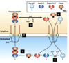
ZBP1 binds to nascent Beta-actin mRNA in nucleus as part of a large mRNP complex

These mRNPs are actively transported through the nuclear pore complex (NPC) with the aid of Ran GTPase

19
Q

How is beta-actin mRNA transported to the leading edge of a migrating cell?

A
20
Q

Where are many proteins made before being moved to where they need to be?

What do they therefore require?

A

Many proteins are made in the cytoplasm and then are moved to where they need to be

They require organelle-specific targeting sequences

21
Q

How can organellle-specific targeting sequences be identified?

A

Some can be identified from primary sequence (NLS or NES)

Some are more structural

  • amphiphilic helix (mitochondria)
  • Signal patch (lysosome)
22
Q

With organelle-specific targeting when entering the nucleus, what is required?

Tell me about them

A

A targeting sequence known as nuclear localisation signal (NLS) which is NOT removed following transport

There are two kinds of NLS which may be anywhere in the protein

  • Basic
  • Non-basic
23
Q

What are the two kinds of NLS?

A
  • Basic
  • Non-basic
24
Q

Whats a Basic NLS and give an example

A

Basic

e.g. SV40 large T antigen

  • rich in lysine (K) and arginine (R)
  • The exact sequence is not important instead it is the cluster of basic amino acids
  • can be bipartite (split into two parts)
25
Give an example of a Non-basic NLS?
**hnRNPA1** which is hydrophobic
26
With organelle specific targeting exiting the nucleus, what is required? Where are they in the sequence? Are they removed after transport?
**Nuclear export signals (NES)** These can be found anywhere in the sequence Not removed following transport
27
What are NESs rich in? How do they look in the sequence?
* Rich in hydrophobic residues (leucine, L and isoleucine, I) * In sequence: L X2-3 (F/I/L/V/M) X2-3 (L/I) X (L/I) The X represent any AA
28
What is the nuclear localisation signals **eIF4GI?**
A large protein that acts as a scaffold in translation initiation
29
Indirect immunofluorescence of HeLa cells in eIF4GI shows what?
Some of the endogenous protein is in the nucleus, but most is in the cytoplasm
30
What was nuclear localisation in eIF4GI dependent on and how was this discovered?
* We expressed fragments corresponding to known protease cleavage sites – N-terminal part goes to nucleus (detected by indirect IF) * N-terminal fragment sequence contains KRRRK * Made fusion proteins with GFP - whole fragment or just a small fragment of surrounding sequence (X5) * Mutated KRRRK to AAAAA (charge goes from positive to neutral) * Nuclear localisation was dependent on this sequence
31
How do ions, small metabolites and globular proteins move through the NPC (nuclear pore complex) ? What size are these ions?
Ions, small metabolites and globular proteins **20-40 kDa** move through the nuclear pore complex (NPC) by **passive diffusion**
32
How do moelcules greater than 40 kDa move through the NPC?
Bigger proteins and mRNAs (in mRNPs) need to be transported through pore- this is the central role of the **GTPase Ran**
33
What are the stages to the Nuclear **import** process?
1. Nuclear import receptor: heterodimer of **importin-alpha** (recognises NLS in “cargo”) and **importin-beta** (interacts with nucleoporins in NPC) 2. Nucleoporins contain FG-rich repeats (hydrophobic) 3. These help transport (shuttle) the complex into the nucleus 4. Nuclear **Ran-GTP** interacts with importin, releasing cargo 5. Ran-GTP: importin complex exits to cytoplasm 6. **Ran-GAP** stimulates Ran to hydrolyse GTP 7. Importin is now free for another round of import 8. Ran-GDP enters the nucleus to be “recycled” by **Ran-GEF** (guanine nucleotide exchange factor)
34
What are the stages to the Nuclear **export** pathway?
* Again, the **Ran GTPase is central** * In this case the cargo is included in a complex with **Ran-GTP** and a transport protein called an **exportin** (e.g. Crm1) * Exportins and importins are similar in both sequence and structure and are part of a family called **karyopherin’s**
35
What is **Mnk1?**
A kinase with an N-terminal NLS and a C-terminal NES
36
Leptomycine B can be used to prevent cancer What does it bind to and what does it do?
It binds to and alkylates **Exportin1** on a single cys residue, inhibiting the export activity
37
LMB (Leptomycin B) has been found to have anti-tumour activity in mouse models, where in the cell does it need to be any why? If not in the right location, what can this lead to? Provide an example.
Lots of proteins with tumour suppressor activity needs to be in the nucleus to carry out their function The mislocalisation of tumour suppressor proteins is a common feature of tumours e.g. RUNX3 (a transcription factor) is commonly mislocalised to the cytoplasm in breast and gastric tumours
38
Blocking Exportin 1-dependent nuclear export with LMB may help what?
may help to keep such proteins where they ought to be and prevent cancer progression
39
Lecture 4 conclusions
* Proteins in the cell need to be in the right place to perform their function * This can be achieved by movement of the mRNA via sequences located in the 3’ UTR (Pre-translation) * Movement of proteins Post-translation relies on targeting sequences * The simplest of these signals are those that mark out a protein for nuclear import or export * All these movements can be visualised in cells by various methods * Hybridisation of a fluorescent nucleotide probe to mRNA * Binding of fluorescently labelled antibodies to proteins * Expression of a protein of interest fused to a fluorescent protein
40
* Human cells expressing suspected GFP-tagged shuttling proteins can be fused to murine cells (no initial expression) * Human cell nuclei stain differently to those from mice (murine cells shown with arrow) * The photos were taken 6 hours after fusion of the cells Which of the two hnRNPs shuttles?
41
Overview of protein targeting and export
42
Movements in the cell can be visualised by what methods?
* Hybridisation of a fluorescent nucleotide probe to mRNA * Binding of fluorescently labelled antibodies to proteins * Expression of a protein of interest fused to a fluorescent protein
43
Whats the purpose of the nucleolus in the nucleus?
* not membrane bound * site of transcription of major rRNA genes by RNA Pol I * rRNA is processed and ribosomes are assembled in nucleolus
44
Whats Nucleolar localisation signals (NoLS)?
A longer version of NLS | (run of basic residues- K/R)
45
99% of mitochondrial protens are made from the nuclear genome. What are the functions of the mitochondrial proteins?
46
What is present in the mitochondrial matrix in multiple copies?
mtDNA is present within the matrix in multiple copies
47
What does the mitochondrial genome contain?
Mitochondrial genome (16,569 bp) contains 37 genes (13 proteins, 22 tRNAs and 2 rRNAs)
48
The 13 mitochondrial genes are subunits of what?
Subunits of the enzyme complexes of the oxidative phosphorylation system
49
When fractionating cellular components (differential centrifugation) tell me the different spins, pellets and supernatants formed
* can take these partially purified components and reconstitute them in the test tube in a cell-free system
50
Mitochondrial matrix targeting is a N-terminal matrix-targeting sequence. Tell me how mant aa it has? its polarity and why? What its process requires?
* **20-50 aa** * Rich in hydrophobic, positively charged (R/K) and hydroxylated (S/T) amino acids, forming an amphipathic alpha-helix – positively charged on one side, hydrophobic on other * Process requires **actively respiring mitochondria + ATP** * Once in matrix, targeting sequence is cleaved off by matrix protease
51
Mitochondrial targeting signals
52
What does the mitochondrial targeting peptide (mTP) have to be close to?
Has to be close to N-terminus of protein, rich in hydrophobic, positively charged (R/K) and hydroxylated (S/T) amino acids
53
mTP is a structural motif and forms what?
an amphipathic helix
54
what does mTP bind to?
binds to a receptor on mitochondrial outer surface and brought towards translocase of the outer membrane (TOM)
55
Mitochondrial import complexes
56
Tell me the steps to translocation
1. Unfolding helped by chaperones (e.g. Hsc70), which require ATP 2. mTP binds to receptor 3. Brings protein to TOM pore 4. Protein begins to pass through 5. Proximity of TIM complex enables passage through inner membrane helped by ATP turnover by matrix HSc70 6. mTP is removed by protease 7. Chaperones help refold protein to active state
57
During translocation, proteins are premade and folded in the cytoplasm. Why do most proteins have to be unfolded?
They have to be unfolded before they can pass into the mitochondria in order to thread through narrow translocase pores
58
Whats the size of TOM, TIM and nuclear pore in nm?
TOM = 2-2.5 nm TIM = 0.8–0.9 nm nuclear pore = 9-12 nm
59
The process of oxidative phosphorylation creates what?
a membrane potential
60
How do protons being pumped into the IMS drive ATP synthesis?
Protons pumped into IMS, reimport to matrix by ATP synthase drives ATP synthesis
61
Mitochondrial activity aids import, what is the chaerge of the matrix and IMS and why?
Matrix becomes negatively charged, with IMS positive due to high [H+] due to proton pumping from ATP synthesis
62
In an agarose gel, which electrode does DNA move towards and why?
DNA is negatively charged so it moves towards the positive electrode
63
Why is mitochondrial membrane potential (ΔΨ) thought to help drive import by 'electrophoresis'?
Due to the attraction of positively charged amino acids in mTP towards the matrix
64
Where do mitochondrial matrix targeting sequences/peptides have to be located?
at the N-terminus of the protein
65
The mitochrondrial matrix import is a structural motif, forming an amphiphatic helix. What is the 3˚ structure unwound by?
Unwound by chaperones to aid transit through pores
66
What is the mitochondrial matrix import recognised by?
A receptor and brought to the TOM pore on the OMM
67
Where is the mitochondrial matrix import fed through?
The protein is fed through the outer pore, then the inner pore Chaperones within the matrix help with the import and refolding The charge in the matrix also aids import by electrophoresis
68
Where do lots of mitochondrial diseases come from?
The maternal line due to inheritance of **mtDNA from mother**, but some are due to problems in nuclear genes some diseases are directly related to problems with mitochondrial import
69
Give an example of a mitochondrial disease and what it leads to? Whats this mutation caused by?
**Human deafness-dystonia syndrome** (recessive, X-linked) leads to hearing loss, mental retardation and blindness. Caused by mutations in **TIMM8A** (moving proteins to IMS and into IMM)
70
What does the mutation of **DNAJC19 (TIM14)** cause?
autosomal recessive **dilated cardiomyopathy with ataxia (DCMA) syndrome**
71
Aß peptide can plug into what? What link is it thought to be linked to but isnt clear?
Aβ peptide can plug up the **TOM and TIM pores**, but not yet clear of link with **Alzheimers** as Aβ peptide has many other possible mitochondrial targets e.g. inhibition of Cytochrome c oxidase
72
Like mitochondria, the chloroplast genome is very limited, cDNA has an average of how many genes?
120 genes
73
How many proteins are needed for function?
2-3,000 Therefore, most proteins are nuclear-encoded and imported
74
Chloroplast
75
Where is the key recognition signal of the chloroplast transit peptides (cTP)?
At the N-terminus of the protein
76
Unlike mTP, the cTP has no well-defined sequence/structure but common structural and physical features include what?
* serine rich * rarelt have acidic amino acids * lengths range from 20 to \>100 residues
77
What are prolines important in?
Keeping the cTP unfolded for certain proteins
78
What plays a key role in driving import in chloroplasts?
GTP and ATP
79
chloroplast import mechanism
* unfolding proteins in cytosol, GTP in TOC * Internal motors in the stroma as part of TIC
80
Lecture 5 conclusions
* Subcellular compartments can have further destinations, e.g. nucleolus within nucleus, and matrix/intermembrane space in mitochondria * The vast majority of proteins required by mitochondria and chloroplasts are made from the instructions present in the nuclear genome * Post-translational targeting of mitochondrial proteins relies on structural motifs, not just linear sequence * Mitochondrial diseases might be due to problems with import process * Import to mitochondria and chloroplasts facilitated by energy rich environment of the organelles * Next lecture, translocation to the endoplasmic reticulum DURING translation
81
Proteins that are destined for secretion are found where?
In the ER lumen
82
What is unable to pass through the lumen?
Ribosomes
83
As ribosomes are unable to pass through into the lumen, how are the secretory proteins transported there?
1. Start with intact cells and "pulse" with radiolabelled amino acids 2. Homogenise and separate out microsomal fraction 3. Allow protein synthesis to go to completion 4. Microsome membranes can be destroyed with detergents 5. Labelled proteins in intact microsomes are resistant to protease digestion
84
When fractionating cellular components, what gradient is used? What does it seperate the components by?
A sucrose gradient is used It fractionates by density rather than particle size
85
After fractionating cellular components, what are the different layers produced at the end?
Layer solutions of different concentrations on top of each otherm strongest at bottom (discontinuous gradient, but can also make continuous ones with special gradient mixers) Place sample on top of gradient and spin Denser particles sediment furthest
86
What type of proteins can free ribosomes encode?
* Soluble cytoplasmic proteins and enzymes * nuclear proteins * Mitochondrial proteins * Chloroplast proteins (except those encoded by organelle DNA) * Cytoskeletal proteins * glyoxysomal proteins
87
What do free ribosomes attach to?
They are proteins that attach to membranes through myristic acid and isoprene (ras, src)
88
What type of proteins can bound ribosomes encode?
* **Integral membrane proteins** Plasma membrane Nuclear membrane rough ER Golgi membrane Lysosomal membrane Endosomal membrane * **secreted proteins** (insulin, plasma proteins) * **Lysosomal** and **peroxisomal enzymes** * **Rough ER enzymes** * **Golgi enzymes** and complexes
89
How can ribosomes be extracted from microsomes?
By treating with detergent
90
How can you makes "stripped" microsomes?
By adding EDTA (**chelates** Mg2+: a type of bonding of ions and molecules to metal ions. it involves the formation or presence of two or more seperate coordinate bonds between a polydentate ligand and a single central atom. these ligands are called chelants) OR A mix of KCl and puromycin (causes premature chain termination by mimicking aminoacyl tRNA)
91
How can seperate components be reconsituted (formed again)?
In vitro
92
Whats In vitro vs In vivo?
**In vitro:** used to describe work thats performed outside of a living organism **In Vivo:** refers to when research or work is done with or within an entire living organism
93
Tell me about what needs to be added to bound ribosomes in order to be a translation of a protein?
Bound ribosomes that have been extracted from membranes + mRNA specifying soluble cytoplasmic, non-exported protein (e.g. GAPDH)= translation of protein
94
What needs to be added to free ribosomes in order for translation of the protein to be incorporated into microsome?
Free ribosomes +stripped microsomes + mRNA encoding secretory protein (e.g. preproinsulin= translation of protein that is incorporated into microsome
95
What must a protein determine when being synthesised?
Whether the ribosome binds to the ER or whether the ribosome remains free in the cytoplasm
96
What were the initial thoughts as to where ribosomes would attach? What has been discovered more recently?
"signal sequences" were proposed but initially, people were unsure if this was a signal in the mRNA or a signal in the protein Later, there was a discovery of the N-terminal sequence (leader sequence/signal peptide)- led to a Nobel prize
97
In 1972, what was discovered by Milstein and co-workers about immunoglobulin light chains?
Showed that immunoglobulin light-chains that were synthesised in vitro, contained an N-terminal segment that was no present in the mature protein that was synthesised in vivo
98
ER-localised proteins have what core amino acids?
6-12 hydrophobic amino acids (**bold**) preceded by one or more basic amino acids (R/K) (_underlined_)
99
What do mature proteins in the ER lose in regards to the N-terminal signal sequence? What is found in certain positions around the cleavage sites, and what positons are these?
The mature protein in the ER **loses the signal sequence** by cleaving between the residues marked with (downwards arrow) Residues with small side chains are found at positions **-3** and **-1** upstream of the cleavage site
100
Are we able to change the order in which components are added to a reconstitution experiment?
yes
101
mRNAs have been translated into proteins with the right signals, but when are they then unable to enter microsomes?
They cannot enter the microsomes AFTER they were made NB. The signal sequence which should help target the protein is NOT removed if they are not incorporated
102
The signal sequene protruding from the elongated ribosomes is recognised by what?
The signal recognition particle (SRP)
103
What is the SRP composed of?
**6 proteins** and a specific **300 nucleotide RNA** molecules that binds the ribosomal RNA
104
What does the **p54** protein contain?
A hydrophobic patch of methionine residues that interacts with the hydrophobic
105
Draw the signal-recognition particle (SRP) and label its components
106
What are the steps to translocation to the ER?
**https://www.ncbi.nlm.nih.gov/books/NBK21532/**: to understand SRP and SRP receptors and its involvment with translocation to the ER 1. The ribosome “pauses” in protein synthesis and the ribosome/protein/SRP complex “docks” with an **SRP receptor** in the ER membrane 2. The **translocon** (sometimes referred to as the Sec61 complex or translocator) is composed of 3 integral membrane proteins in a heterotrimeric complex 3. The SRP receptor facilitates the binding of the ribosome to the protein “translocon” pore 4. The translocon is composed of 3 integral membrane proteins in a heterotrimeric complex 5. An **α–helix** in one of the translocon proteins acts as a **“plug”** 6. The ribosome/protein/SRP/receptor complex binding to the translocon coupled with **hydrolysis of GTP** opens the pore 7. Translation elongation is restored, and the nascent protein is fed into the **ER lumen** 8. The signal sequence is cleaved by a signal **peptidase**
107
When is the signal sequence normally cleaved and by what?
The signal sequence is normally cleaved from the N-terminus by a membrane bound signal **peptidase** as it enters the ER lumen and before the protein C-terminus has been completed
108
Proteins with their signal sequences intact can be isolated from what?
They can be isolated from a cell free protein synthesis system at an early time in their synthesis
109
What do cleavage sites of the signal sequence conform to?
The -3, -1 rule where these residues are usually small and uncharged
110
What is the answer to the proposed question below?
111
Localisation mechanisms can be what ...
* **post-translational** (nucleocytoplasmic shuttling, targeting to the mitochondria) * **co-translational** (to get proteins into the ER) * **one-way only** (cleavage of signal after transport to mitochondria or ER)
112
What is the structure of the translocon? What can particular movements of the structure allow to occur?
* The translocon consists of **three Sec61 protein subunits (alpha, beta and gamma)** * A single **alpha helix** forms the **plug** at the base of the structure * The structure is able to **hinge** allowing proteins passing through to exit laterally into the membrane
113
What are transmembrane domains formed of? What do the regions outside of them tend to be?
Alpha helices containing hydrophobic amino acids The regions outside the membrane tend to be hydrophilic
114
What does the orientation and number of TM domains in the integral membranes result in these proteins being classified according to?
Their topology (study of place)
115
What are single pass and multi pass transmembrane proteins?
Single pass: cross the membrane only once Multi pass: cross the membrane multiple times
116
Is the Sec61 protein a single-pass or multi-pass protein?
The Sec61 proteins in the translocon are an example of a multiplass protein
117
Tell me about Type I membrane proteins?
* Stop-transfer sequences are approximately 22 amino acids long and hydrophobic * They will form part of the TM domain * They stop translocation through the channel * The translocon opens at the side and allows the transfer of the protein
118
Tell me about type II and III membrane proteins?
**Type II** * Has signal-anchor sequence: combination of signal sequence (that is NOT at the N-terminus) and a stop-transfer sequence * Signal-anchor is bound by the SRP during translation as before * +ve AA of N-terminus of signal-anchor (helps keep this part of protein in cytosol) * Anchor passes through side of translocon, rest of protein is passed into the ER lumen **Type III** * Type III have +ve AA at C-terminus of their singla anchor and so have opposite orientation
119
Tell me about Type IV membrane proteins?
* consists of several different polypeptides assembled together in a channel through the membrane
120
What are hydropathy/ hydrophobicity plot?
It is used to characterise or identify possible structures or domains of a protein Look at a "window" of amino acids and assign an average score based on amino acid content (hydrophobic residues give different positive score, hydrophilic give negative)
121
What do ER and membrane localisation mechanisms rely on?
Recognition of an N-terminal signal peptide by SRP
122
What does SRP bring the stalled translation complex to? What happens when its arrived at this location?
A receptor on the ER membrane Once at the membrane, translation resumes into the translocon pore It can only move one way due to cleavage of signal after transport to ER
123
Protein targeting from the ER
124
What is glycosylation?
The addition of a glycan (polysaccharide chain) to proteins An example of post-translational modification
125
What is almost all N-glycosylated at the same time and when does this occur?
Almost all soluble and membrane-bound proteins are N-glycosylated at the same time as they emerge through the ER membrane translocator pore
126
What are the two types of glycosylation?
**O linked** **N linked** **N-linked glycans** attached to a nitrogen of asparagine or arginine side-chains. N-linked glycosylation requires participation of a special lipid called dolichol phosphate. **O-linked glycans** attached to the hydroxyl oxygen of serine, threonine, tyrosine, hydroxylysine, or hydroxyproline side-chains, or to oxygens on lipids such as ceramide
127
What type of glycosylation can proteins be **after** they have been completely synthesised?
O-glycosylated
128
What does glycosylation target?
Proteins to specific locations or labels for secretion
129
What does glycosylation prevent?
Incorrect folding of proteins
130
What does glycosylation protect?
The protein against proteolytic degradation
131
What does glycosylation assist in?
The stabilisation of proteins
132
What does glycosylation help to stabilise?
it helps to stabilise or 'locks' protein topology within membranes
133
What does glycosylation confer?
Specific recognition properties inside and outside cells
134
What is N-linked glycosylation and what happens in this process?
* Preformed oligosaccharide transferred from a novel membrane lipid called **dolichol-phosphate** * The 14 residue oligosaccharide is made up of **2 N-acetylglucosamine, 9 mannose and 3 glucose residues** (for synthesis, see Lodish 8th Edn Fig 13.17) * The oligosaccharide is linked to asparagine **(Asn/N) residues** in the protein at sequences containing **Asn-X-Ser/Thr**
135
Proteins are rapidly glycosylated on Asn residues after passing through the pore. Explain what happens at each stage in the following diagram
(1)The 14-mer is transferred while the protein is still being translated (2-4) The 14-mer is further processed by glycosidase enzymes to remove the glucose residues and one of the mannose sugars. One of the glucose residues may be re-added later on to help with folding certain proteins (3a)
136
The core region in glycosylation is always the same, what is this core region?
137
Further processing can produce two classes of N-linked oligosaccharides, what are these two classes?
* Complex oligosaccharides * ​High-Mannose oligosaccharides
138
What are the components of the N-linked glycosylation assay?
* α-factor mRNA from S. cerevisiae, Rabbit Reticulocyte Lysate, * Canine Microsomal Membranes, amino acids (including [35S] methionine) * All reactions run for same time, only levels of CMM vary
139
In the following image of an N-linked glycosylation assay, which sample has the highest amount of microsomes?
The RHS (larger proteins on RHS due to glycosylation after entering microsomes)
140
Example data analysis question
141
Label the Golgi compartments
142
What are lysosomes?
Membrane bound organelles with an acidic lumen (pH5 cf pH7.2 in cytosol) containing acid hydrolases + other enzymes
143
What are lysosomes used for?
Used for digestions of extracellular and intracellular components e.g. wornout organelles or invading bacteria/ viruses
144
Proteins which are destined for transport to lysosomes are synthesised where?
In the ER and are targeted to the Golgi
145
Recognition of a lysosomal protein requires what?
A signal patch
146
How is lysosomal targeting achieved?
By initial N-glycosylation
147
The glucose residues and one mannose are next removed, and the terminal mannose is then phosphorylated. Explain what happens in stages 1 and 2 in the following image
1. N-Acetylglucosamine phosphotransferase recognises a “signal patch” and catalyses the reaction of the terminal mannose with UDP-N-acetylglucosamine forming an intermediate of protein-glycosyl-mannose 6P-N-acetylglucosamine 2. Phosphodiesterase catalyses the cleavage of N-acetylglucosamine from the intermediate leaving a protein-glycosyl-mannose 6P
148
Explain the stages of targeting of M6P-tagged proteins to lysosomes in the following image
149
Tell me about Autosomal recessive lysosomal storage disease
affecting about 1:326,000 UK births Severe developmental defects which lead to death by age 7
150
Tell me about I-cell diesease
* Deficiency of the **GlcNac phosphotransferase** enzyme so mannose 6-P “tags” are not assembled * Potential lysosomal hydrolytic enzymes therefore not recognised by the mannose 6-P receptor, instead exit the cell * The secreted **hydrolase** enzymes are inactive in the neutral pH of the blood * Glycolipids that would normally be processed by lysosomal enzymes form large inclusions (insoluble aggregates), hence the name inclusion-cell or I-cell disease
151
What does O-glycosylation involve?
Linking sugars to the OH serine or threonine and takes place exclusively in the golgi
152
Tell about the typical chain length produced from O-glycosylation
Typically, they are **short** chains, and unlike the 14-mer chain added in N-glycosylation, sugars are added specifically one at a time in various cellular compartments by **glycosyl transferases**
153
What does O-glycosylation define and how does it do this?
O-glycosylation defines your **blood group**, which is determined by whether you have the **transferase for N-Acetylgalactosamine (Group A), Galactose (Group B), both (AB) or neither (O)**
154
What process is O-glycosylation also important for?
secretion
155
Give an example of a protein which can be N- and O-glycosylated?
Erythropoetin
156
Whats **Erythropoetin (EPO)?**
This is a hormone secreted by the kidneys and encourages an increase in formation of erythrocytes (RBC)
157
What happens in mature EPO proteins?
Three Asn residues are N-glycosylated and one serine is O-glycosylated
158
What do the glycosylation modification's do to EPO?
increase the activity, stability and molecular weight of the protein
159
How is EPO tested for?
* uses 1-D isoelectric focusing to separate glycosylated forms from a blood sample by charge, not size * antibody recognises EPO with a certain migration pattern as being endogenous (so naturally occuring)
160
Another data analysis example question
161
Summary from lectures 6 and 7
**Protein amino acid sequence specifies:** * Nucleocytoplasmic shuttling, mitochondrial targeting * Passage to the ER by recognition of signal sequence by SRP * Membrane localisation via hydrophobic helices **Type of glycosylation (N- or O-) specifies:** * targeting to various organelles * folding and stability * retention in ER or Golgi (N-) or secretion (O-) **Further reading:** * Binding of chaperones (e.g. BiP) to aid protein folding in the ER during translation * Stabilisation of protein loops by cysteine-cysteine S-S bonds in the ER lumen catalysed by protein disulphide isomerase (PDI) * Other modifications e.g. addition of lipids – myristylation and isoprenylation