Pregnancy + Birth + Pregnancy Loss Flashcards

+ fetal growth restriction

1
Q

Give an overview of an average human pregnancy

A
  • last 40 weeks (9 months)
  • made up of three trimesters
    • 1st: <12 weeks
    • 2nd: 12 - 26 weeks
    • 3rd: 27 weeks- birth
2
Q

What are the things that change that the women body needs to change to accommodate for growing a child?

A
  • supplying the baby with enough nutrients
  • managing increased waste production: CO2, nitrogen compounds
  • change in hormones to support pregnancy and prepare for delivery
  • anatomical changes to accommodate the growing fetus ad preparing for labour
  • the stress of delivery and potential haemorrhage
  • postnatal recovery and breastfeeding require physiological changes
3
Q

What endocrine changes are seen in pregnancy?

  • what do these hormones do?
A
  • BHCG: dramatic rise in the first days-weeks - released from the corpus luteum then by the placenta
    • prevents corpus luteum involution,
    • may stimulate the maternal thyroid
  • Progesterone: maintains myometrial quiescence
    • inhibits other smooth muscles of the body GI and Urinary Tract
    • stimulates the appetite, fat storage and the resp. centre
  • Oestrogen: breast growth, areolar enlargement
    • promote uterine blood flow and myometrial growth
    • promote cervical softening and expression of myometrial receptors at term
4
Q

How does the placenta act as an endocrine organ?

  • hormones released
A
  • It releases the following hormones
  • hCG (human chorionic gonadotrophin) – produced by trophoblast, first detectable 8-9 days, peaks 8-9 weeks.
  • hPL (human placental lactogen) - Similar structure to prolactin and growth hormone.
    • a larger placenta produces more hPL.
    • Alters maternal carbohydrate and lipid metabolism to provide steady-state of glucose for fetal requirements
  • hPG (human placental gonadotrophin)- induces maternal insulin resistance to regulate fetal growth
    • this may become a pathological process seen as GDM
  • CRH (corticotropin-releasing hormone)- implicated in human labour
    • acts to increase prostaglandin synthesis
    • may also directly stimulate myometrial contractility
  • Progesterone
  • Oestrogen
5
Q

What is the role of hPL - human placental lactogen?

A
  • Similar structure to prolactin and growth hormone.
  • a larger placenta produces more hPL.
  • Alters maternal carbohydrate and lipid metabolism to provide steady-state of glucose for fetal requirements
    • antagonises the effect of insulin and promotes lipolysis
    • reduces glucose utilization and enhances amino acid transfer across the placenta
6
Q

What is the role of hPG - human placental gonadotropin?

A
  • released by interstitial trophoblastic cells
  • induces maternal insulin resistance to regulate fetal growth
  • this may become a pathological process seen as GDM
7
Q

What is the role of hCG-human chorionic gonadotropin

A
  • maintains corpus luteum secretion of prog & oest, decreases as the placental production of progesterone increases
    • beta unit forms the basis of pregnancy testing.
    • Alpha unit can mimic LH, FSH, and TSH
    • Large quantities are released in molar pregnancy and multiple pregnancies
    • High levels cause vomiting (hyperemesis)
8
Q

What is the role of Progesterone?

A
  • Relaxes smooth muscle – everywhere!
  • Maintains uterine quiescence by decreasing uterine electrical activity
  • causes Constipation, gastric reflux, supra-pubic dysfunction
  • acts as an Immune suppressor ( HLA )
  • causes Lobulo-alveolar development in breasts
  • Substrate for fetal adrenal corticoid synthesis eg cortisol
9
Q

What is the role of oestrogen?

A
  • Growth of the uterus, cervical changes
  • Development of ductal system of breasts
  • Stimulation of prolactin synthesis
10
Q

What changes are seen in the Haematological System?

A
  • 40% increase in plasma volume - 2.5L to 3.7L (8-10kg fluid weight gain)
  • 25% increase in RBC
    • these two factors leads to dilutional anaemia (peak at 32 weeks, exaggerated in multiple pregnancies)
  • Plasma colloid osmotic pressure falls – shift of fluid into the extracellular space
  • Increase clotting factors –> hypercoagulable state
    • Evolutionary balance between thrombosis and haemorrhage
    • Increase plasma fibrinogen (increased ESR), platelets, factor VIII & von willebrand factor
11
Q

What changes are seen in the Cardiovascular system?

A
  • Increased cardiac output
    • occurs early on in pregnancy and plateaus at 24-30 weeks of gestation
  • Decreased peripheral resistance
  • Blood pressure
    • proportional to change in CO and PR
    • as peripheral resistance is often greater than CO, blood pressure may decrease at various stages of pregnancy
12
Q

How is peripheral vascular resistance impacted bu pregnancy?

A
  • Peripheral vasodilatation (effect of progesterone)
  • Peripheral resistance decreases by 35%
  • Combined with increased cardiac output, results in slightly lower BP
  • Decreased vascular resistance leads to lower blood pressure
13
Q

What changes are seen in the Respiratory system in Pregnancy?

A
  • ventilation increased by 40% in first trimester
    • tidal volume is increased to meet oxygen demands (deeper breathing rather than faster breathing)
    • all done to meet increased O2 consumption
  • increased inspiratory and expiratory reserve
  • increased vital capacity
  • decreased residual volume and total lung volume
    • increased abdominal mass
  • increased hyperventilation –> decreased PCO2 –> decreased HCO in-order maintain pH
  • effects caused by progesterone and prostaglandin
    *
14
Q

What is the clinical impact of the physiological changes on women?

A
  • Splinting of diaphrgm, increased ventilation – sensation of increasing SOB
  • Raised HR leads to palpitations
  • Lots of cross over with symptoms for PE and known hypercoagulable state… leads to lots of investigations…!
  • Excess plasma volume shifts causing oedema – peripheral
  • Decreased exercise tolerance
  • Low BP causing fainting / dizziness
15
Q

What are the dermatological and Musculoskeletal impacts of pregnancy?

A
  • Increased lumbar lordosis
  • Ligamentous laxity – pelvic girdle pain / pubis dysfunction
  • Stretch marks
  • Changes in skin pigmentation - Linea Nigra, melasma,
  • darkened nipples
  • Carpal Tunnel
  • Sciatica
  • Cramps
16
Q

What urological changes are seen in pregnancy?

A
  • Kidney increases 1cm in size during normal pregnancy
  • increased renal flow by 50%
  • increased GFR (BUT tubular reabsorption capacity is unchanged) - decreased glucose reabsorption - glycosuria is common
  • Plasma levels of creatinine and urea decrease in pregnancy
  • Dilated ureters (progesterone)
  • Increased pressure (increased urine frequency)
17
Q

What changes to Thyroid function are seen in pregnancy?

A
  • Increased serum T3 and T4 levels
  • increase in thyroid-binding globulin (caused by oestrogen
    • levels of free T3 or T4 remain the same/ slightly fall as most are found in the bound form
18
Q

How is Labour initiated?

A
  • maternal anterior pituitary is stimulated by factors from the fetal hypothalamus
    • fetal hypothalamus-pituitary axis releases cortisol–> plays a role in cervical ripening
  • stimulates inhibition of pre-pregnancy hormones
    • progesterone
  • stimulates an increase in pro-labour hormones
    • oxytocin
  • the Decidua of the placenta release prostaglandins
    • this stimulates cervical ripening and stimulates uterine contractility
    • this is through a direct effect and by upregulation of oxytocin
  • there is also an inflammatory response that is thought to contribute to cervical ripening
19
Q

What is the effect of the initiation of labour?

A
  • uterine contractions are stimulated
  • there is mechanical stimulation of the uterus and cervix caused by stretching and stretching from the babies head causes the cervix to shorten and dilate?
20
Q

What are the stages of Labour?

A
  • Latent phase
  • 1st stage - 3rd stage of labour
21
Q

Explain the latent stage of labour

A
  • long exhausting stage of labour, usually a sleepless period for women
  • The cervix softens and shortens in response to prostaglandin
  • Contractions caused by oxytocin and prostaglandin
    • vary in intensity and refularity
22
Q

What are the stages of cervical effacement?

A
  • measured in percentages
  • when the cervix is completely effaced the cervical os starts to dilate
23
Q

How is Active labour Diagnosed?

A
  • Painful regular contractions
    • usually 3 every 10 minutes lasts 50-60seconds from beginning to end
    • necessary power in order to give birth
  • cervical effacement
  • dilatation of the cervix of 4cms or more (digital examination)
  • blood-stained mucous discharge, and spontaneous membrane rupture can also form part of the diagnosis
24
Q

Explain the First stage of Active Labour

A
  • first stage is considered from the time, regular uterine activity associated with progressive effacement and dilatation of the cervix (active labour) until the cervix becomes fully dilated at 10cm
  • progression of dilation is usually around 0.5cm/hour
    • vaginal examination every 4 hours to check the progress
    • can be faster if the mother is multiparous
25
Q

How is the descent of the fetal head measured?

A
  • progress is measured by the cervical dilation
  • and the descent of the fetal head in relation to the pelvic brim and the ischial spines
26
Q

Explain the Second stage of labour

A
  • begins when the cervix is fully dilated and ends at the delivery of the baby
  • the length can vary between nulliparous and parous women
    • the length in nulliparous can be within 3 hrs of the start of the second stage or within 2 for parous women
  • Progress is measured in by the descent and rotation of the fetal head on the examination and is in two distinct phases
    • Propulsive/passive phase
      • full dilatation until the head reaches the pelvic floor
      • the position of the head is usually occiputotransverse
      • there is no urge push
    • Expulsive/active phase
      • the head becomes visible and there is a strong involuntary desire to push
  • delivery is usually in OA position
27
Q

How does the fetus and the maternal pelvis work together to reach birth?

A
  • a baby is born via the mechanism of descent, flexion, rotation and extension
  • approaches pelvis facing either the mothers left or right pelvis as this is the widest point
  • as the baby descends further into the baby’s head flexes it’s chin into the chest and rotates it’s head into the anterior-posterior diameter
    • the coccyx is deflected backwards this is the space available during birth
28
Q

Review the fetal skull and how this relates to delivery

A
  • the biparietal diameter is the widest part of the skull
  • the separate bones of the skull mould together during the mechanics of birth
  • in normal birth, the occiput (back of the skull) presents anteriorly at the outlet of the pelvis
  • the head flexes to present the smallest diameter to accommodate the limited space this is the:
    • suboccipitobregmatic diameter
      • diameter from the back of the skull to the anterior fontanelle (bregma)
29
Q

How is the head position assessed during birth?

  • how would the structures be identified?
A
  • the anterior and posterior fontanelle can be digitally assessed in a vaginal examination
  • Anterior fontanelle (bregma)
    • larger, diamond-shaped at the intersection of 4 sutures
    • measures 2x3 cms
    • this closes at 18 months
  • Posterior fontanelle
    • ​y-shaped at the intersection of 3 sutures
    • closes at 6-8 weeks
30
Q

What are the different presentations of the fetal skull at the pelvis?

  • how would this affect birthing?
A
  • Suboccipitobregmatic (9.5cms) = OA position
  • Occitopitofrontal ( 11cms) = OP position
  • Supraoccipitomental ( 13.5 cms) = brow
  • Submentalbregmatic (9.5cms) = face
  • the pelvic outlet is approx. 12,5cm in diameter: therefore a brow position would not be possible to deliver vaginally
31
Q

Summaries the mechanism of birth

A
  • Head at pelvic brim Occipital transverse (OT) position
  • Flexion of neck (Suboccipitobregmatic)
  • Head descends and engages
  • Head reaches pelvic floor- rotates to Occipital Anterior
  • Head delivers by extension
  • Head “restitutes” (rotates to come in line with the shoulders)
  • Shoulders rotate into anterior/posterior diameter of the pelvis (the widest part)
  • Anterior shoulder delivered by lateral flexion from downward pressure on baby’s head
  • Posterior shoulder by upward lateral flexion
32
Q

Explain the Third stage of Labour

A
  • this is the delivery of the placenta and membranes
    • Chorion membrane
    • Amnion membrane
  • a blood loss of 300-500 mls is normal/ expected
  • This delivery can be carried out via
    • Active management
      • oxytocin is given i.m into the maternal thigh
      • control pull of the umbilical cord
      • aids delivery of placenta and contraction of the placental bed
      • decreases the risk of Postpartum haemorrhage (PPH)
    • Physiologically
      • a mother naturally expels the placenta and membranes with contractions
33
Q

What fetal monitoring is done during labour?

A
  • fetal monitoring is done to detect fetal hypoxia
    • FBS (fetal blood sampling)
      • small papillary stab on the scalp
  • fetal heart rate can be measured by
    • Intermittent auscultation by Pinard (ear trumpet) or Sonicaid
      • every 15 minutes before and after a contraction in the first stage
      • every 5 minutes in the second stage
    • CTG (cardiotocograph) (can be used if any abnormalities are heard during auscultation)
      • abdominal transducer can be used or a fetal scalp clip (FSE)
34
Q

What is FBS, and when would it be contraindicated?

A
  • A Fetal Blood Sample
    • used to monitor pH and bass excess to check for hypoxia
  • infection such as HIV and Hep B
  • Fetal Bleeding Disorder
  • Prematurity less than 32 weeks
35
Q

What influences the incidence of twin pregnancies?

A
  • increased maternal rate
  • ethnicity
  • increasing parity
  • family history
  • fertility treatment
36
Q

How are multiple pregnancies defined?

A
  • Zygosity
    • refers to whether the twins have developed from a single ovum or from different ova
    • dizygotic pregnancies have separate placentae
  • Chorionicity
    • this refers to the number of placentae
  • Amnionicity
    • refers to the number of amnionic sacs
37
Q

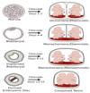
Explain the different types of chorionicity that would form from various stages of embryo development

  • 0-4 days
  • 4-8 days
  • 8- 14 days

> 14days

A
  • 0-4 days
    • Dichorionic diamniotic (joined placentae)
  • 4-8 days
    • Monochorionic diamniotic
  • 8- 14 days
    • monochorionic monoamniotic
  • > 14days
    • conjoined twins
38
Q

Briefly explain Dichorionic twin pregnancies

A
  • All dizygous twins are dichorionic
  • Dichorionic twins must be diamniotic
  • Dichorionic = separate circulations
  • 1:3 monozygous twins are dichorionic
39
Q

Briefly explain Monochorionic twin pregnancies

A
  • Monozygous twins may be monochorionic (2/3) or dichorionic
  • MC have vascularly joined placentae
    • twin to twin transfusion (net blood flow from one twin to another)
  • MC twins 3x increased loss rate
  • MC twins usually diamniotic
  • there are more fetal malformations and more fetal growth restriction
40
Q

How are twin pregnancies diagnosed?

A
  • uterine size
  • ultrasound
    • also used to determine chorionicity of the fetuses
41
Q

What are some complications of Mulitple pregnancies?

A
  • “everything except post-dates”
  • Symptoms of pregnancy
    • hyperemesis (severe and prolonged vomiting)
    • increased risk of pre-eclampsia
  • Anaemia
  • Hypertension
  • Intrauterine growth restriction
  • Pre-term labour
  • Delivery problems
  • Perinatal mortality
42
Q

What is twin-to-twin transfusion syndrome?

  • management?
A
  • there is a net blood flow from one twin to another through arterial to venous anastomoses in the shared placenta
    • circulation of recipient becomes hyperdynamic - risk of hight output cardiac failure and polyhydramnios
    • donor develops oliguria (low urine output) and polyhydramnios
  • occurs in 10-15% of monochorionic multiple pregnancies and accounts for 15% of perinatal mortality in twins
  • manage by early delivery by caesarean
43
Q

What is the management of twin pregnancies in the first trimester?

A
  • Discuss screening for chromosomal anomalies
  • Determine chorionicity
  • Discuss fetal reduction if triplets or more
44
Q

What is the management of twin pregnancies in the second trimester?

A
  • Detection of fetal abnormality
  • Serial scans for growth for all
    • DC monthly from 24 weeks
  • Serial scans for TTTS if MC twins
    • 2 weekly 16-28 weeks then monthly
  • Maternal complications
45
Q

What is the management for twin pregnancies in the third trimester?

A
  • Scanning as in 2nd trimester
  • Monitor blood pressure
    • serial scans for growth of all fetuses
    • serial scans for TTs if MC twins
  • Pre-term labour
  • Delivery planning
46
Q

Define the below terminology used in pregnancy loss

  • Threatened miscarriage
  • Inevitable miscarriage
  • Incomplete miscarriage
  • Complete miscarriage
  • Missed miscarriage
  • Molar pregnancy
A
  • Threatened miscarriage – symptoms of bleeding /pain but intra-uterine pregnancy still present
  • Inevitable miscarriage – symptoms of bleeding /pain, cervical os is open
  • Incomplete miscarriage – symptoms of bleeding, os open, some tissue remaining
  • Complete miscarriage – no tissue remaining
  • Missed miscarriage (early fetal demise/ anembryonic pregnancy) – diagnosed at scan with no symptoms
  • Molar pregnancy - fetus doesn’t form properly in the womb and abnormal cells develop instead of a normal fetus. Known as hydatidiform mole
    • *​*complete mole, where there’s a mass of abnormal cells in the womb and no foetus develops
    • partial mole, where an abnormal fetus starts to form, but it can’t survive or develop into a baby
47
Q

What factors increase the risk of miscarriage?

A
  • older Age mothers
  • Obesity
    • 25% increase risk with BMI > 30 compared with normal BMI
  • Antiphospholipid syndrome / SLE
  • Parental chromosomal translocation
  • Poorly controlled diabetes
  • Alcohol, smoking, recreational drug use
  • Uterine anomaly
    • i.e. bicornuate nucleus
  • (High levels of natural killer cells)
48
Q

What are some typical presentations of miscarriage?

A
  • Bleeding
  • Pain
  • Found at time of routine scan
    • (i.e. missed miscarriage)
  • (Loss of pregnancy symptoms)
  • Acute collapse
    • Cervical shock, hypotension, tachycardia, bleeding
  • Sepsis
    • Pyrexia, hypotension, tachycardia, raised resp rate, confusion
49
Q

How is miscarriage managed?

A
  • Conservative / expectant (40-80% success)
  • Medical management – can be managed at home or on the ward dependent on size of pregnancy (CRL length) / patient preference
    • Patients must be counselled re: expectations for bleeding and discomfort, potential risk of retained tissue
  • Surgical management (SMM)
    • Surgical risks include infection, bleeding, uterine perforation, cervical damage, retained tissue and need for a repeat procedure.
  • Follow up - support/counselling,
50
Q

What is recurrent miscarriage and how is this managed?

A
  • 3 consecutive pregnancy losses <24 weeks
    • affects 1% of couples
  • on third or later miscarriage offer karyotyping of the tissue
    • chromosomal abnormalities affect 2-5% of couples
    • attend miscarriage clinic
    • Blood tests - thrombophilia screen, anti-cardiolipin antibodies, lupus anticoagulant
      • Antiphospholipid syndrome - 15% of patients with recurrent miscarriage
      • treatment (aspirin and LMWH) improves outcome
    • check for Uterine anomalies: Ultrasound scan (3D scan) or hysteroscopy/laparoscopy
51
Q

What are the risk factors for ectopic pregnancies?

A
  • Previous ectopic pregnancy (15% risk after one)
  • Tubal damage
    • Previous tubal surgery
    • History of pelvic infection / PID
    • History of endometriosis
  • History of subfertility/ IVF
  • Presence of IUCD
  • Use of progesterone-only contraception
  • Cystic fibrosis
52
Q

What are the ‘Classic’ Clinical presentations of an ectopic pregnancy?

  • management
A
  • Pain and bleeding 6-8 weeks gestation
  • Positive pregnancy test
  • Empty uterus on TV ultrasound scan
  • Given options for conservative/expectant, medical (methotrexate), or surgical management
53
Q

What is the ‘modern’ presentation of an ectopic pregnancy?

  • management?
A
  • Seen with EPAU minor symptoms in early pregnancy
  • May be monitored over 48 hours to confirm diagnosis (using scans and hcg levels)
  • Expectant management / medical / elective surgical
54
Q

What would the hCG monitoring of a suspected ectopic or pregnancy of unknown location show?

A
  • a normal intrauterine pregnancy would b expected to double in concentration every 48 hours
  • in pregnancy of unknown location (not visible on the USS) a sub-optimal rise in hCG should provoke strong suspicion of an ectopic
55
Q

What are the types of second trimester miscarriage?

  • when is this considered a second trimester
A
  • loss of a baby between 12-24 weeks
  • Intra-uterine death or
  • Premature labour/delivery
56
Q

What are causes of intrauterine death?

A
  • Fetal abnormality - structural/ chromosomal
  • Infection
  • Placental dysfunction, growth restriction
    • SLE/ anti-phospholipid syndrome
57
Q

What are causes of very Pre-term Labour?

A
  • Cervical weakness /incompetence
    • (e.g. due to multiple LLETZ- used to remove abnormal cell changes in the cervix)
  • Uterine abnormality
    • anomaly vs. acquired
  • Infection
    • inflammatory response causing uterine irritability and contractions
  • Rupture of membranes
    • can be a result of infection
  • Bleeding
    • e.g. from placenta = abruption (emergency)
58
Q

When would cervical sutures be indicated?

A

Indicated when

  • Past history of mid-trimester pregnancy loss
  • Past history of cervical surgery
  • Known uterine anomaly
  • Cervical dilatation noted - on speculum examination or TV scan
  • Usually inserted after 12 weeks gestation
    • can be later in an emergency
  • Can be inserted pre-pregnancy
  • Transvaginal (most) or transabdominal
  • risk of infection
59
Q

What is considered a stillbirth?

A
  • Baby delivered with no signs of life after 24 completed weeks of gestation
60
Q

What are the causes of Stillbirth?

A
  • Antepartum/ intrapartum bleeding
  • Intrauterine growth restriction (eg. caused by maternal medical problems)
  • Fetal abnormality- structural/ chromosomal
  • Placental factors
  • Infection
  • Poorly controlled diabetes
61
Q

What is Neonatal death?

A
  • Early neonatal death: baby dies within 7 days of delivery
  • Late neonatal death: baby dies within 7-28 completed days
62
Q

What are causes of Neonatal death?

A
  • Prematurity
  • Congenital abnormalities
  • Infection
  • Intrapartum asphyxia
63
Q

Why does diagnosing maternal hyperglycaemia matter?

A
  • Morbidity in the offspring “from the uterus to the grave”
  • prevents the exacerbation of the obesity & Type 2 diabetes epidemic
  • Future Type 2 diabetes in the mother
64
Q

What are the two types of Hyperglycaemia in pregnancy?

A
  • Women with Normal Glucose tolerance
  • Women with Abnormal Glucose tolerance
    • Known Diabetes
    • Unknown Diabetes or IGT
  • there i
65
Q

What are the possible scenarios that present hyperglycaemia during pregnancy?

A
  • Pre-gestational Hyperglycaemia
    • Type 1 Diabetes
    • Type 2 Diabetes (known and unknown)
    • Monogenic Diabetes
    • Impaired Glucose Tolerance (IGT)
  • “Gestational Diabetes” (GDM)
    • Any newly found Abnormal GTT after the
      • 1st trimester of pregnancy ( i.e. Diabetes or IGT )
66
Q

What is Gestational Diabetes?

A
  • Diabetes or impaired glucose tolerance
    • fasting glucose =/> 5.6 mmol/l
    • 2 hour GTT glucos =/> 7.8mmo/l
  • HAPO study shows that a 75g GTT with the following results would diagnose gestational diabetes based on the risk it presents to the fetus
    • fasting 5.1 mmol/l
    • 1 hour 10.0 mmol/l
    • 2 hours 8.5 mmol/l
67
Q

What changes are seen in maternal metabolism changes as the pregnancy progresses?

A
  • Early pregnancy = Facilitated Anabolism
    • Increased Insulin sensitivity
    • Glucose concentration slightly lower
    • Increased maternal energy stores
  • Later Pregnancy = Facilitated Catabolism
    • Increased Insulin resistance
    • Increased transplacental passage of nutrients leading to
    • Rapid fetal growth
68
Q

What is the effect of maternal hyperglycaemia in the 1st trimester?

A
  • Increased Fetal abnormalities
    • Fuel Mediated Teratogenesis
  • Abnormal placental programming & development
    • Increased risk of Pre-eclampsia
    • Excessive glucose transport
69
Q

What is the effect of MAternal Hyperglycaemia in the late 2nd and 3rd trimester?

A
  • Excessive fat deposition
    • and rapid growth in excess of what is desired
  • adverse fetal programming
    • epigenetic changes (increased methylation of certain genes)
70
Q

What congenital malformations are due to maternal hyperglycemia?

A
  • Neural tube defects
  • Congenital cardiac abnormalities
  • Sacral agenesis
    • failure of the sacrum to develop - effects walking later on
  • Renal problems
  • high dose folic acid (5mg daily) to reduce this
71
Q

What interventions can be done in the 1st trimester to get good diabetic control?

A
  • Prepregnancy counselling
    • Lifestyle Modification
    • Intensive glucose monitoring
    • If not on Insulin commence Insulin
    • Optimize Insulin Regimen
      • Basal Bolus or Pump
      • Freestyle Libre or continuous glucose monitoring
72
Q

What are risk factors for Hyperglycaemia of pregnancy?

A
  • Previous gestastional diabetes
  • Obesity
  • PCOS
  • Family hx of T2DM
  • High-risk racial groups
    • Hispanic people
    • ppl from an Asian background
73
Q

What problems are seen in late 2nd and 3rd-trimester pregnancies in maternal hyperglycaemia?

A
  • Macrosomia causing
    • difficult birth
    • shoulder dystocia (difficult to deliver shoulder)
    • breathing problems
    • jaundice
    • hypoglycemia
    • hypocalcemia
    • polycythemia
74
Q

What are the lifelong/ long term effects of Hyperglycaemia in pregnancy on the fetus?

A
  • Obesity
  • Insulin resistance
  • Type 2 diabetes
  • Dyslipidaemia
  • Hypertension
  • Vascular disease
  • Adverse neurodevelopmental outcomes?
75
Q

What are the blood glucose targets for women with maternal hyperglycemia?

A
  • Fasting glucose < 5.1 mmo/l
  • 1 hour postprandial glucose < 7 (7.8) mmol/l
  • Fetal Abdominal girth < 70th centile (based on caucasian individuals)
    • Less in Asians
76
Q

What drug treatment is given to achieve glucose control in pregnancy

A
  • Prepregnancy /1st-trimester hyperglycaemia
    • switch to a Basal bolus Insulin regimen, (far better control)
  • “Gestational” diabetes - diagnosed later in pregnancy
    • lifestyle changes can usually be effective if not the following medication is appropriate
      • Metformin
      • Basal Insulin
      • Basal bolus Insulin
      • Glibenclamide (ONLY IF NO ALTERNATIVE)
77
Q

How is gestational diabetes managed postpartum?

A
  • Encourage Breast Feeding
    • reduce inulin administration by 25% as they are more sensitive
  • Maintain good Glycaemic control
    • To prevent excess glucose in milk
    • Reduce maternal weight gain
  • Advice on next pregnancy
  • Contraception advice - present random pregnancy
  • Encourage long term glycemic control
78
Q

How are those with Specific gestational diabetes mellitus managed postpartum in the long term?

A
  • Screen for diabetes at 12 weeks postpartum
    • HbA1c +/- Fasting glucose, ( or GTT )
  • Review GAD ect. antibody status if done (if they are at risk with other risk factors i.e weight or family presentations of endocrine diseases)
  • Lifestyle advice
  • Advice re next pregnancy
    • Optimize exercise & Nutrition
    • Pre-pregnancy GTT
  • Annual glucose screening
    • 50% develop type 2 diabetes at 10 years
79
Q

What is the role of the Synctiotrophoblast layer in the early embryo?

A
  • formed from cytotrophoblast cells (CTB)
  • Syncytiotrophoblast burrows into the myometrium of the uterus
  • it invades the maternal spiral arteries and starting the formation of the primary/secondary and tertiary villi
    • which begins the process of forming the placenta
  • they are in direct contract with the maternal blood and act as a barrier between the fetal and maternal blood (CTB cells also do this but they decrease as the pregnancy progresses)
  • produce placental hormones
80
Q

How is the placenta formed?

  • what abnormality can arise from this?
A
  • Syncytiotrophoblast invades decidua (endometrium)
  • Cytotrophoblast cells erodes maternal spiral arteries and veins
  • Spaces (lacunae) between the fill up with maternal blood
    • very big spaces to allow lots of blood flow
  • Followed by mesoderm that develops into fetal vessels
  • Aiding the transfer of nutrients, O2, across a simple cellular barrier
  • if the syncytiotrophoblast cells don’t invade deeply enough into the maternal myometrium there is a narrow lacunae
    • leads to reduced blood flow causing pre-eclampsia
81
Q

How does the placenta act as an endocrine organ?

A
  • Human chorionic gonadotrophin (HCG)
    • maintenance of corpus luteum of pregnancy
    • progesterone and oestrogen
  • Human placental lactogen HPL
    • growth, lactation
    • carbohydrate and lipid

various others

82
Q

What is transferred across the placenta?

A
  • Gases – oxygen and carbon dioxide by simple diffusion
  • Water and electrolytes
  • Steroid hormones
  • Proteins poor – only by pinocytosis
  • Transfer of maternal antibodies IgG -starts at 12 weeks – mainly after 34 weeks
    • therefore lack of protection for premature infants
83
Q

What is this an image of?

  • explain its significance
A
  • Vasa praevia
    • when the fetal blood vessels run across the cervical os
  • As the internal os dilates in labour the vessels are stretched and exposed and can rupture leading to massive fetal blood loss and death.
  • Diagnosed on Ultrasound using colour dopplers
  • Management: delivery by Caesarean Section when the fetus is above 34 weeks.
84
Q

Where is the position of the placenta in the uterus?

  • what clinical presentation can there be?
A
  • mainly fundal in the uterus (at the top)
  • on the anterior or posterior walls
  • can be “low-lying” or placenta praevia
    • this is when it is near the cervical os
    • this can sometimes manage itself in the third trimester of pregnancy, stretching of the lower part of the uterus causes it to thin and “move away”
  • this is checked in the 20 week scan
85
Q

What is this an image of?

  • what is the clinical significance?
A
  • Placenta Praevia

can cause

  • massive bleeding in pregnancy
    • usually painless bleeding
  • fetal death
  • maternal death
86
Q

What are the clinical complications during pregnancy assocaited with the placenta?

- what is the main cause of this?

A
  • poor maternal-fetal mixing of blood
  • lack of oxygen and nutrients to the fetus
    • causes fetal growth restriction
    • and pre-eclampsia (raised blood pressure)
  • failure of trophoblastic invasion into maternal circulation at 12 & 18 weeks
87
Q

What is Placenta Accreta?

  • treatment and risk factors
A
  • When the placenta is unable to separate at birth
    • the uterus cannot contract down and causes massive bleeding
  • this occurs when the placenta has invaded too deep into the myometrium
  • Treatment: hysterectomy
  • Risk factors: scar in the uterus
    • maybe due to a previous c-section
    • fibroid
    • surgically managed abortion
88
Q

What is a placental abruption?

  • management and risk factors
A
  • seperation of the placenta during pregnancy
    • massive bleeding in pregnancy (often concealed)
      • extremely painful
      • concealed as the uterus is filled with blood, not the vagina as in placenta praevia
  • can cause fetal and maternal death
89
Q

How does the placenta present in multiple pregnancies?

A
90
Q

What is the difference between growth and development in pregnancy?

A
  • Development mostly happens in the first 12 weeks of pregnancy - when organs are being formed
  • Growth - is when the baby gets bigger
91
Q

What does it mean if a fetus is SGA?

A
  • means they are Small-for Gestational-Age
    • this is using their estimated fetal weight (EFW) or abdominal circumference (AC)
    • they are below the 10th centile for fetal growth scans
  • not all small babies are synonymous with fetal growth restriction
    • may be constitutionally small baby (look at parents)
    • normal-sized baby that is showing reduced growth
92
Q

What ultrasound measurements are down to monitor fetal growth?

A
  • Abdominal circumference (AC)
  • Head circumference (HC)
  • Femur length (FL)
  • Liquor volume (LV)
  • additional Doppler tests
93
Q

What are the three main causes of SGA fetuses?

A
  • (1) “Normal” baby ie. constitutionally small
    • Based on maternal size & ethnicity
  • (2) Non-placenta mediated growth restriction
    • eg. structural or chromosomal problem, fetal, infection, inborn errors of metabolism
  • (3) Placenta mediated growth restriction
    • eg. PET, hypertension, autoimmune disease (lupus etc.), thrombophilias, renal disease, diabetes
  • groups 1 and 2 would show symmetrical UGR, whilst group 3 would show asymmetrical IUGR (reduced abdominal circumference)
94
Q

What are the risk factors of IUGR?

A
  • Maternal age > 40yrs
  • Nulliparity
  • Low or high maternal BMI
  • Diabetes / Renal disease / APLS
  • Smoking (nicotine is a vasoconstrictor)
  • Maternal SGA
  • IVF
  • Previous SGA infant
  • Previous stillbirth
  • PAPP-A < 0.4 MoM at combined screening
  • Hypertension/PET/recurrent APH this pregnancy
95
Q

What is the effect of fetal growth restriction

A
  • Deficient placental invasion –> Reduced placental reserve –> Fetal need exceeds supply –> IUGR
  • Hypoxia
  • Fetal vascular redistribution to try and compensate
  • Oliguria
  • Abnormal CTG (Cardiotocography fetal heart monitor)
  • ultimately Fetal death after compensation fails
96
Q

How is fetal growth restriction diagnosed?

A
  • Clinical measurement of uterine size: Symphysis - fundal height (SFH)
    • should roughly match the number of weeks pregnant they are
  • Ultrasound scan gives a more accurate measurement of growth
97
Q

What does this fetal growth chart show?

  • what does it indicate?
A
  • asymmetrical growth restriction as only the abdominal circumference is reduced
  • indicates a type 3 SGA fetus
98
Q

What does this fetal growth chart show?

  • what does it indicate?
A
  • symmetrical growth restriction as
    • the head circumference
    • the biparietal diameter of the head and
    • the abdominal circumference are all reduced
  • indicates either type 1 or type 2 SGA fetus
99
Q

Why is fetal abdominal circumference a good indicator of fetal growth?

A
  • it’s where the fetal liver is and the liver stores glycogen
  • if the fetus is struggling it won’t be able to store glycogen therefore eh abdominal circumference will be smaller
100
Q

What are the consequences of hypoxia in the fetus?

A
  • blood flow redirected to more important areas –> brain
  • blood flow redirected away from other less important areas
    • Gut
    • Kidneys
      • low fluid levels may indicate IUGR
    • Lungs
101
Q

What are ultrasound findings in a fetus with IUGR?

A
  • small abdominal circumference (liver)
  • decreased amniotic fluid (produced by the kidneys, however their function has been reduced)
  • increased blood flow to the brain
    • looking at eh middle cerebral arteries in the brain using the doppler test
102
Q

What are the clinical features of IUGR?

A
  • SFH (symphysis-fundal height) smaller than expected
  • Baby’s movements lessen to conserve energy
  • Fetal heart rate changes as hypoxia develop (as seen on CTG)
  • Fetal death
103
Q

What is the management of a FGR baby?

A
  • Serial ultrasound evaluation of fetal growth, liquor volume and fetal doppler
  • Timing delivery depends on
    • Gestational age
    • Doppler studies
    • Other risk factors
  • aim to maximize fetal maturity and growth but minimize the risks of perinatal mortality and morbidity
104
Q

Explain fetal dopplers

  • why are they used
  • what do they look at
A
  • used to differentiate the different type of SGA
  • helps guides the timing of interventions
    • intensive monitoring, antenatal glucocorticoid, early delivery
  • Looks at blood flow and resistance in
    • Umbilical artery
      • should have low resistance
    • Middle cerebral artery
      • are usually constricted - can dilate if the baby is struggling as they increase blood flow to the brain
    • Ductus venousus
  • if blood flow is normal monitoring can continue, otherwise abnormal blood flow cay indicate expedited delivery
105
Q

What does this doppler show?

  • what does it indicate?
A
  • normal blood flow in the middle cerebral artery in the brain
  • the peak = systole of the heart and during diastole flow is negative
    • this scan shows an appropriate pressure difference in systole and diastole
106
Q

What does this doppler show?

  • what does it indicate?
A
  • shows increased blood flow as there is maintained blood flow during both systole and diastole rather than a pressure difference
  • indicates there is fetal growth restriction and that the baby is compensating
107
Q

What is Betamethasone/ dexamethasone?

  • when are they used
  • what are their actions in pregnancy
A
  • Steroids are given to mother x2 24hrs apart, if the baby needs to be delivered early
    • this will cross the placenta and stimulate the aveoli cells to produce surfactant gene
  • Surfactant stops the collapse of the aveoli cells by coating the cells and reducing the surface tension
  • Helps prevent Respiratory Distress Syndrome which leads to neonatal death in premature babies
  • Produced from 24- 34 weeks and usually the baby will have enough by 34 weeks in preparation for a term delivery
    • hence this will be lacking in premature babies