Anatomy Flashcards

1
Q

What are modalities are used to image the pelvis?

A
  • MRI
  • U/S
2
Q

What are the advantages and disadvantages of MRI?

A
  • No Ionising radiation
  • Better soft-tissue contrast
    • useful for staging cancers/tumours
  • Longer examination time (45 mins)
  • More expensive
3
Q

When would a CT be used to image the pelvis?

A
  • In an emergency or acute settings
    • produces a lot of artefact due to the pelvic girdle
  • also has poor soft-tissue contrast
4
Q

What key structures are in the male pelvis at this level?

A
  • Prostate
  • Rectum
  • Ischio-rectal fossae
    • cancers of the rectum erode into this area - looking for a clean line of fat (not present in this image)
  • Obturator internus
    • provides lateral rotation and the abduction of the hip
5
Q

What key structures are in the male pelvis at this level?

A
  • rectus sheath at the front
  • the bladder
  • the seminal vesicles (bowtie)
6
Q

What key structures are in the female pelvis at this level?

A
  • Uterus
    • Endometrium (looks bright white in T2)
    • Myometrium (same density as skeletal muscle)
    • Junctional zone
      • if this is narrowed or thickened –> suggest endometrial cancer
  • Ovary- fluid-filled and bright on the T2 weighted image
  • Bladder
  • Cervix
7
Q

What can be seen in this Sagittal MRI of the female pelvis?

A
  • Bladder
  • Uterus (OM)
    • (anteverted uterus in this image- no functional difference)
  • Cervix
  • Rectum
    • the sacral space should have a clear line fo fat - sigmoid colon cancer can infiltrate into this space
8
Q

How are the ovaries imaged?

A
    • HSG: Hysterosalpingogram
      • very similar to a smear test
      • you want to see a blurring from the contrast - shows the infundibulum is clear
9
Q

The Peritoneum in the female pelvis - sagittal MRI -

A
  • The peritoneal reflections in the pelvis consist of the peritoneum wrapping around
    • the bladder
    • the uterus
    • the rectum
  • this creates two pouches/spaces
    • vesicouterine pouch
    • rectouterine pouch (pouch of Douglas)
  • the pouches are a common site of infection/ abscess
10
Q

The peritoneum in the male pelvis - sagittal MRI -

A
  • there is only the vesicorectal pouch - as the perineum wraps around the bladder and down the front of the rectum
    • common site of abcist formation
11
Q

What types of Ultrasound imaging can be down to image the pelvis?

A
  • Transabdominal
  • Transvaginal
  • Transrectal
12
Q

What is measured in an Ante-natal ultrasound?

A
  • 12 week scan (dating scan)
  • 20-week scan (looking for congenital abnormalities)
    • growth rate: crown-rump length, head measurements etc.
13
Q

How is the vascular anatomy of the pelvis?

A
  • Magnetic Resonance Angiography (using contrast (e.g gadolinium), similar to MRI)
  • Duplex U/S (doppler effect)
    • plots the wave form blood flow of the artery
  • CT angiogram
    • iodinated intra-venous contrast through the peripheral cannula with imaging
  • Angiography (direct catheter angio)
14
Q

What type of imaging is this?

A

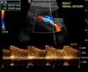
Duplex ultrasound of the right renal artery

15
Q

What type of imaging is this?

A

CT Angiogram

16
Q

What type of imaging is this?

A

Catheter angiogram

17
Q

At what level does the aorta and vena cava bifurcate?

A
  • aorta is L4
  • vena cava is L5:
    • the right CIA crosses the origin of the Left CIV –> left iliac DVT
    • May-Thurner syndrome
18
Q

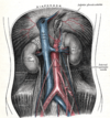
Anatomy of the Internal Iliac artery

A
  • Divides into the Anterior and posterior division
    • the posterior division is iliolumbar
    • lumbosacral and superior gluteal
19
Q

What pathology is this?

A

Unifying Pseudoaneurysm of the Right Common Femoral Artery

  • the blood vessel isn’t dilating there is just a hole in it
20
Q

Labelled anatomy of the sagittal female pelvis

21
Q

What is the blood supply and nerve supply of the ovaries?

A
  • supplied by the ovarian artery and the vein within the suspensory ligament
  • Nerve supply of the ovarian plexus
22
Q

Describe the structure of the uterine tube

  • VAN
A
  • Infandibulum
  • Ampulla
    • often the site of fertilization
  • Isthmus
  • Uterine
  • VAN
    • V: ovarian arteries pass laterally and drain into the left renal vein (on the left side), drain into the inferior vena cava (on right side)
    • A: ovarian artery
    • N: sympathetic supply from the ovarian nerve and parasympathetic from the pelvic splanchnic nerve
23
Q

Where is pain from the ovaries felt in which dermatome region?

A
  • ischaemia and pain which is referred to the dermatome region of T10
24
Q

Describe the structure of the uterus

A
  • made up of the fundus, body of the uterus and cervix
  • Formed of 3 layered walls
    • Perimetrium – outer serous wall covering the uterus
    • Myometrium – thick muscular layer, responsible for the process of parturition
    • Endometrium – inner mucous layer; site of implantation; thickness changes through menstrual cycle
25
What is this innervation and supply of the uterus?
* Innervation: symp from the hypogastric plexus (along uterine artery); parasymp via the pelvic splanchnic n. * Arterial supply: uterine artery * Venous drainage: via the uterine plexuses to the internal iliac veins * Touch and Pain (birth): via somatic afferents to S2-S4
26
What are the key uterine ligaments?
* Ligament of ovary * **Round ligament of uterus** * remnants of the gubernaculum * from the uterus to the labia majora- the route the process the ovaries would have taken if it was male and would have formed the scrotum * women who are pregnant may report a pulling * **Broad ligament** * covers over the uterine body and ovaries * forms the **mesosalpinx** when it pinches around the uterine tube * forms the **mesovarium** when it pinches around the ovaries * Transverse cervical ligament * Uterosacral ligament * **Peritoneal pouches**- trapped fluid where infection can take place * Vesicouterine pouch * Rectouterine pouch
27
Describe the structure of the vagina - VAN
* musculomembranous tube 7-9cm long * has to recesses superiorly * Posterior fornix * Anterior fornix * VAN * V: drainage though the vaginal plexus to the **internal iliac veins** * A: by branches from the **internal iliac arteries** * N: autonomic nerves, sensory nerves convey pain messages to an area of the abdomen supplied by **T12 and L1 spinal nerves**
28
Review the vasculature of the female reproductive tract organs
29
What structures make up the external female genitalia?
Consists of: * **Mons pubis,** * **Labia majoria,** * **Bulb of vestibule** * **Clitoris**- erectile organ consists of: root, body and glans * **Bulb of vestibule**- erectile tissue over bulbospongiosus muscle * **Vestibular glands**- secrete mucus * **Labia minora** encloses the vestibule (openings for vagina and urethra) and the clitoris * Blood supply/drainage: _pudendal artery (erectile) & vein_ * Innervation: branches of _genitofemoral and pudendal_ n (e.g. dorsal n of clitoris)
30
What changes happen to the female reproductive tract during pregnancy?
* Uterus expands from 50g to 950g. * Uterus extends to the xiphisternum. * Centre of gravity is altered-increased lumbar lordosis. * Sacroiliac joint and pubic symphysis relaxes * With uterine expansion the ovaries and uterine tubes are displaced laterally. The cervix becomes softer and swollen.
31
What is the VAN of the testis?
* V: form the pampiniform plexus * forms around the testis that keep it cool * A: supplied by the testicular artery * N: spermatic plexus
32
Describe the structure of the testis
* The dorsolateral surface of each testis is overlain by the **epididymis** which has an expanded ‘head’ connected to the superior part of the testis. * The **seminiferous tubules** in each testis drain, via a series of tubules, into the single duct of the epididymis at the head of the epididymis. * In the epididymis, the duct of the epididymis is tightly coiled and it continues as the uncoiled **ductus (vas) deferens** at the inferior pole of the epididymis.
33
What are the three fascial layers of the testis?
these are continuous with the layers of the surrounding spermatic cord * The outer layer, the **external spermatic fascia,** is derived from the _external oblique aponeurosis._ * The middle layer, the **cremaster muscle and fascia**, is derived from the _internal oblique and transversus abdominis_. * the cremaster muscle lifts the testis back into the abdomen when it isunder threat or if it's cold * The inner layer, the **internal spermatic fascia**, is derived from _transversalis fascia_.
34
What is the microscopic structure of the seminiferous tubules?
* Each testis is subdivided into approx. 250 pyramid-shaped lobules contain seminiferous tubules * Function- production of spermatozoa (spermatogenesis) * Structure- highly tortuous, lined by seminiferous epithelium * Tunica propria-loose C.T, fibroblasts and myoid cells * Stroma-surrounds seminiferous tubules, loose C.T, vascular and houses small clusters of large interstitial cells of Leydig * _Sertoli Cells_ * Stretch from basal lamina to the lumen, linked by tight junctions * perform many roles in the testis
35
What is the role of Sertoli Cells?
* Stretch from basal lamina to the lumen, linked by tight junctions * Enfold developing spermatozoa-acting to * Protect them from antibodies in the blood * Provide nutrients * Phagocytose excess cytoplasm * Secrete androgen binding protein * Secrete inhibin * Add fluid to the lumen
36
What is the VAN of the Ductus Deferens?
* Arteries: those that primarily supply the lower bladder – inferior vesical– and rectum – middle rectal. * Venous: blood follows the arteries and so have the same name.
37
What is the function of eh vas deferens and where is it found?
* the tube continuous with epididymis that is joined by duct seminal gland =\> ejaculatory duct * The ductus deferens (vas deferens) ascends on the medial side of the epididymis, * enters the spermatic cord and then passes through the inguinal canal on its way to the urethra. * Spermatozoa, with their accompanying fluid (from the seminal vesicles and prostate), are then discharged by a closed duct system
38
What are the accessory glands of the Ductus deferens? - what are their roles?
* **Seminal vesicles** – just inferior to rectovesical pouch, _secretes alkaline fluid + fructose + coagulate_ * **Prostate** – secretes prostatic fluid (20% total volume) * **Bulbourethral glands** – pea sized glands; a.k.a. Cowpers glands; located inferior to prostate; _secretion assists lubrication of ducts and removes any urine that would have been in the tract_
39
What is the VAN of the Prostate?
* Supplied by prostatic arteries and veins, * Innervated by sympathetic from _inferior hypogastric_ and parasympathetic from _pelvic splanchnic nerves_
40
What is the VAN of the Seminal Vesicles?
* Wall of sac contains smooth muscle, which when stimulated by sympathetic nerves expels fluid out-ejaculation. * Blood: Inf vesicle + prostatic vessels (int iliac a)
41
What is the VAN of the bulbourethral gland?
* inferior vesicles + prostatic vessels and the internal iliac arteries
42
What is the VAN of the penis?
* A: Internal pudendal arteries * V: External pudendal veins. * N: by S2/4 Pudendal nerve, dorsal nerve to penis, ilioinguinal nerve
43
Describe the gross anatomy of the penis
* Consists of * root, * body and glands, * the body contains 3 cylindrical bodies * Paired corpora cavernosa and 1 corpus spongiosum
44
What are the various parts of the male Urethra?
* Prostatic * Membranous * Penile
45
Explain the stages of Erection Emission and Ejaculation in males during sexual stimulation
* **Erection-** Deep arteries-supply the corpora-in flaccid state A/V shunts * Increased parasympathetic nerve supply, increases blood flow- diverted to vascular spaces * this causes increased pressure in erectile tissue thus decreasing venous return * **Emission-** semen is delivered to the prostatic urethra, prostatic fluid added. * Emission results from sympathetic activity. * **Ejaculation**-semen expelled through the urethra * Inhibition of sympathetic supply-return to flaccid state
46
What clinical pathologies arise from the male reproductive tract?
* Testicular Torsion * Cryptorchidism * Patent processus vaginalis * Hydrocele * Hematocele