Physiology 1 22 Flashcards

1
Q

There are two components to respiration what are they ?

A

The two compenents of respiration = Internal and External

External respiration = external environment to tissue

  • Ventilation = bulk transport of external media across a gas exchange surface
  • respiratory exchange - diffusion
  • circulation
  • cellular exchange

Internal respiration = cellular metabolism

2
Q

What is Dalton’s law ?

A

Daltons law

  • the partial pressue of gas
  • the total pressure of a gas is equal to the sum of the partial pressures of the individual gases in a mixture

With altitude as the total pressure decreases, the partial pressure of a particular gas will decrease.

Partial pressure of oxygen = PO2

The partial pressure of any gas will change with environment.

3
Q

What is Henry’s law ?

A

Henrys law

The amount of gas dissolved in a liquid is afftected by the gases partial pressure and solubility coefficient of that gas.

gas dissolved = pressure of gas in the media * solubility coefficient

  • gas and liquid are in equilibrium
  • only free gas molecules (physically dissolved contribute to the partial pressure of gas in liquid)
  • gases will always diffuse down their partial pressure gradients.
4
Q

What is Fick’s law, and what four factors affect the rate of diffusion ?

A

Describes the net diffusion rate of a gas across a fluid membrane.

  • gases move down their partial pressure across a semi permeable membrane

Rate of diffusion

  • surface area
  • thickness or distance (T)
  • pressure gradient (P)
  • Diffusion coefficient (D)
5
Q

What is Boyle’s law ?

A

Boyle’s law

For a fixed amount of an ideal gas kept at a fixed temperature, pressure and volume are inversly proportional.

6
Q

What is tidal breathing and flow through breathing ?

A

Flow through breathing

  • unidirectional flow of media, yeilds greater O2 uptake
  • birds, fish and some crocodiles

Tidal breathing

  • relatively inefficient as external medium moves in and out through the same opening
  • mammals (bidirectional).
  • fresh media is mixed with depleted media
7
Q

Describe concurrent, cross current and countercurrent gas exchange ?

A

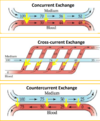
Concurrent exchange

  • external medium and blood flows in the same direction
  • partial pressure gradient declines
  • does not exist in a biological system

Cross current exchange

  • external medium and blood travels at right angles to each other
  • diffusion occurs through-out declines gradually
  • PO2 is higher in blood then external medium at the end
  • birds

Coutercurrent exchange

  • external medium and blood flows in the opposite direction and parallel to each other
  • partial pressure gradient is maintained and diffusiin is constant
  • fish highly efficient
8
Q

Describe gas exchange in fish through the use of gills ?2

A

Gills

  • countercurrent exchange system
  • flow through
  • at every point O2 is diffused into blood - great efficiency in gas exchange
  • four gill arches - filaments - lamella
  • large surface area which is proportional to how active the fish is

Buccal pressure pump - fish opens and closes mouth

Opercular suction pump - fish open and close opercular flaps

9
Q

Describe the upper and lower respiratory tract, what are these systems purpose?

A

Upper respiratory tract

  • located outside the chest cavity
  • no gas exchange anatomical deadspace
  • covered with mucus and ciliated epithelium
  • trachea and bronchi supported by cartilage
  • smooth muscle lines trachea, bronchi and bronchioles.

Function = conditioning of air (warm, humidification), and filtration muco-ciliary escalator

Lower respiratory tract

  • alveolar surface thin layer of squamous epithelial cells (type one)

Contains three cell types

  • Type one alveolar cell - assist in gas exchange
  • Type two - large cuboidal produce surfactant
  • Alveolar macrophage - phagocytic patrol the alveolar surface.
10
Q

Hoe does air move into the lungs ?

A

By creating negative pressure. Increasing volume by contraction of the diaphragm and external intercostal muscles.

11
Q

Describe the histological sections of the upper respiratory tract ?

A

Trachea = C shaped cartilage

Bronchi = cartiliginous plates

Bronchioles do not have cartilage

The amount of smooth muscle increase from the trachea to the bronchioles.

12
Q

Describe the histology of the alveolar ?

A

Alveolar

  • Alveolar surface covered by a dense capillary surface, lung interstitium has mast cells, connective tissue and elastic tissues to hold the alveolar open.
  • mainly composed of type one alveolar cells - single layer of a single layer of sqaumous epithelial cells.
  • Type two alveolar cells are cuboidal and secrete surfactant and the fluid layer which lines the lungs
  • Alveolar macrophage
13
Q

What effect does the parasympathetic and sympathetic nervous system have on the lungs ?

A

Sympathetic

  • air way relaxation
  • blood vessel constriction
  • inhibition of glandular secretion through beta 2 receptors

Parasympathetic

  • air way constriction
  • blood vessel dilation
  • increased glandular secretion
  • vagus nerve
14
Q

Describe the intrapleural space and the intrapleural pressure, and what they do?

A

Intrapleural space

  • pleura anatomically seperates the lungs from the thoracic wall
  • enables friction free movement between the lungs and the thorax
  • lung is mechanically connected to the thoracic cage and pleura by the pleural fluid
  • pressure of fluid is negative (intrathoracic pressure) holds the lungs open
  • -3mmhg
  • intrapleural space of the two lungs is seperated in most species
  • cause for negativity is the mechanical interaction between the lungs and chest wall.

During quiet inspiration the intrapleural pressure becomes more negative, while during expiration it becomes less negative.

15
Q

What is the intrapulmonary pressure (Palv) ?

A

Alveolar or intrapulmonary pressure is the pressure of the air inside the lung alveoli

Normal respiration alveolar pressure decreases to about -1 mmhg (inspiration) and rises to about 1mmhg during expiration.

16
Q

What is the transpulmonary pressure ?

A

Transpulmonary pressure

Is the difference between the alveolar pressure and the pleural pressure (distending pressure)

  • the more positive the distending pressure ptp becomes the more the lungs are distended or inflated
  • ptp= Palv -Ppl
17
Q

Describe Pulmonary ventilation in mammals ?

A

Inspiration

  • diaphragm and intercostal muscles contract
  • lung volume increases
  • lung pressure decreases belwo atmospheric pressure
  • air flows down its concentration gradient
  • inspiration is always active as it requires muscle contraction
  • 1-2% of the total energy metabolism

Expiration

  • diaphragm and intercostal muscles recoil
  • lung volume decreases
  • lung pressure increases
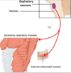
  • internal intercostal muscles may contract to assist with expiration
  • passive process in rest
18
Q

What is eupnea ?

A

normal quiet breathing

19
Q

What is Hyperpnea ?

A

increased depth and frequency of breathing during exercise

20
Q

What is Tachypnea ?

A

excessive rapidity of breathing

21
Q

What is bradypnea ?

A

abnormal slowness of breathing

22
Q

What is polypnea ?

A

rapid shallow breathing, as occurs during panting

23
Q

What is dyspnea ?

A

Difficult, painful or laboured breathing - pathological

24
Q

What is apnea ?

A

cessation of breathing, in clinic refers to transient state of cessation of breathing

25
Q

What are adventitious sounds ?

A

crackles and wheezes abnormal sound superimposed on normal breath sounds.

26
Q

Why do birds need a more efficient gas exchange system when compared to mammals ?

A

Birds are exothermic with a higher body temperature - 40-41 degrees

Highly active - flying high altitudes where the partial pressure of oxygen is lower.

27
Q

How do birds achieve a greater efficiency in respiration when compared to humans ?

A

Greater efficiency of respiration in birds

  • narrow diffusion distance air capillaries
  • flow through breathing unidirectional
  • cross current breathing
28
Q

How does the bird’s respiration system work ?

A

Bird

  • During inspriration both the anterior and posterior air sacs expand
  • During expiration both the anterior and exterior air sacs deflate
  • allows for flow through breathing
  • lung relatively rigid and the air sacs act as bellows
  • both inspiration and expiration are active even at rest
  • respiratory muscles act upon the sternum to reduce or increase pressure within the air sacs
  • air exchange occurs in the parabronchi ( air capillaries)

Requires two cycles to move a single bolus of air through the entire system.

29
Q

What is lung compliance ?

A

Lung compliance is a measure of how much force is required to distend the lung.

  • the greater the change in volume, with a smaller force = greater compliance
  • lung and thoracic structures
  • alveolar surface tension
30
Q

Why is the surfactant produced by type two alveolar cells so crucial ?

A

Surfactant

Surface tension acts to reduce the surface area collapsing the alveolar.

Surfactant reduces surface tension .

  • prevents alveolar from collapsing
  • reduces effort to expand lungs
  • prevenst adhesion of adjacent respiratory surfaces

Barker syndrome = in piglets and horses is caused by too low a production of surfactant during gestation. The animal requires excessive force to extend lungs and rapid expiration.

31
Q

What is a restrictive disease of the lungs, provide an example and type of breathing pattern this will generally cause ?

A

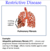
Restrictive disease

  • results in restriction of lung expansion
  • pulmonary fibrosis - progressive scarring of the lungs seen in older terriers
  • causes rapid shallow breathing
32
Q

What is an obstructive lung disease, provide an example and what type of breathing pattern this will generally cause ?

A

Obstructive lung disease

  • Causes obstruction within the airways - decreasing airway diameter
  • decrease in airway diameter occurs during expiration (due to positive intrapleural pressure)
  • expiratory dyspnoea
  • animals abnormally slow and deep breathing pattern.

Examples

  • Heaves in horses = hypersensitive to allergens
  • Feline asthma inflammation and constriction of the bronchioles
33
Q

In a spirogram we can obtain four lung volumes of TV, IRV, ERV and RV difine these volumes.

A

The four volumes obtained in a spirogram.

Tidal volume (TV or VT) = volume of air inhaled or exhaled per breath

Inspiratory reserve volume (IRV) = amount of additional air that can be taken into the lungs after a normal inspiration.

ERV = amount of additional air than can be taken from the lungs after a normal expiration.

Residual volume = amount of air remaining in the lungs after maximal expiration (can not be measured by spirometry).

34
Q

Describe what will happen if lung compliance becomes abnormal ?

A

Higher than normal compliance

  • Lung inflates easily but due to reduced elastic recoil it becomes difficult to expire
  • emphysema

Lower than normal compliance

  • Requires increased force to expand alveoli
  • pulmonary fibrosis
35
Q

Define the four lung capacites inspiratory capacity, functional residual volume, vital capacity and total lung capacity obtained in a spirogram ?

A

Four lung capacity

Inspiratory capacity = IRV + TV

Functional residual capacity = ERV + TV

Vital capacity = ERV + IRV + TV

Total lung capacity (TLC) = VC + RV

36
Q

Define PEFR and PIFR ?

A

Peak flow rate can be taken as a quick measurement and is useful to monitor obstructive diseases.

PIFR = Peak inspiratory flow rate

The fastest flow rate achieved during inspiration

PEFR = Peak flow rate achieved with maximal force during expiration after a complete inspiration.

37
Q

Define minute ventilation ?

A

Minute ventilation

Minute ventilation = The volume of air breathed in or out within one minute.

mV = RR * TV

  • increases during physical activity
  • gives the volume flow throughout the entire respiratory tract including lungs
38
Q

Define respiratory dead space, anatomical dead space, and physiological dead space ?

A

Respiratory dead space = air that enters the lungs but dose not participate in gas exchange.

Anatomical dead space = air which fills conducting airways, conditioning, thermoregulation

Alveolar dead space = gases which enter the alveolar but do not participate in gas exchange eg no perfusion.

Physiological dead space = alveloar dead space + anatomical dead space

In a normal healthy lung the physiological dead space = anatomical dead space as a healthy lung has no alveoli dead space.

39
Q

Define alveolar ventilation ?

A

Alveolar ventilation accounts for the fact that not all minute ventilation contributes to gas exchange

Alveoli ventilation = (TV - dead space volume) * RR

It is the rate at which the alveolar air is replaced by atmospheric air.

40
Q

What is the Va / Q ration and why is it important in respiration ?a

A

For gas exchange to occur efficiently the ventilation of th lungs to perfusion ratio must be matched

Va / Q = 1 Maximum gas exchange

  • If perfusion of the lungs increases the bronchioles, alveoli will dilate in response to increase ventilation: and vice versa
  • local control mechanisms
  • bronchiole dilates - arterioles dilate
  • bronchiole constricts - arterioles constrict

Mismatching of the Va and Q are the main causes of hypoxemia

41
Q

What happens during mismatching of ventalation and perfusion, and provide an example ?

A

Ventalation Va and perfusion Q mismatching occurs in abnormal conditions

  • healthy perfusion ventilation ratio = 1
  • as the degree of mismatching increases, O2 exchange becomes less efficient
  • most common cause of hypoxemia decreased 02 in the blood

Eaxamples

  • Shunt = blockage of the airway (mucous plug) so perfusion without ventilation
  • embolism = blockage of an artiole ventilation without perfusion
42
Q

Why would impairment of partial pressure gradient affect 02 but not CO2?

A

Diffusion coefficient is higher in CO2, than O2

43
Q

Describe how O2, and CO2 are transported around the body and it what proportions ?

A

Blood needs to carry more gas than can dissolve in plasma.

Oxygen is carried in two forms

  • bound to haemoglobin 98.5%
  • unbound dissolved 1.5%

Carbon dioxide is carried in two forms

  • carbaminohemoglobin (3) as bicarbonate form

Oxygen bound to hemoglobin dose not contribute to PO2

  • this maintains oxygen gradient in the lung
  • the amount of Hb in blood determines the oxygen carrying capacity of blood
  • each HB molecule binds four O2 molecules when fully saturated
44
Q

What shape is the hemoglobin oxygen disociation curve, why dose the flat part occur ?

A

The oxygen haemoglobin dissociation curve is sigmoid in shape.

The saturation of HB is related to the PO2 of the blood

The flat part

  • Favors loading of oxygen and is due to a high PO2
  • occurs in the lungs
  • no significant change in % saturation between 60 - 100 mmHg
  • at normal PO2 complete saturation of hemaglobin occurs
  • large plateau of curve provides a large safety margin.
45
Q

The sigmoid oxygen haemoglobin dissociation curve has a steep part why dose this occur.

A

Steep part

  • curve shows steep decline in % saturation between 40 to 10 mmHg
  • helps dissociate oxygen from the haemoglobin molecule
46
Q

What four factors affect the affinity of Hb for O2, how would these facotors be changed to cause a decrease in affinity ?

A

The four factors

  • temperature
  • PH
  • 2,3 DPG
  • PO2

To cause a decrease in affinity

  • temperature increases
  • PH decreases
  • 2,3 DPG increases
  • PCO2 increases
47
Q

What is the BOHr affect ?

A

An increase in carbon dioxide in the blood and a decrease in PH results in a reduction of the affinity of haemoglobin for O2.

48
Q

What affect will exercise have on the oxygen - haemoglobin dissociation curve why dose this occur ?

A

Exercise

right shift of sigmoid curve decreased affinity - favoring unloading of oxygen to the tissues

note = at rest only 25% of oxygen bound to HB is released to the tissues in one circuit - so there is a large reserve of oxygen for increased metabolic rate.

Factors

  • PO2 reduced metabolically active tissues / reduced saturation of HB 10-40mmHG
  • CO2 increases in tissues
  • acidity increases
  • when tissues need more they get more
49
Q

Dose the form of haemoglobin vary, and why would this occur ?

A

Haemoglobin form varies between species and life stages

Animals have varying form of Hb suited to their metabolic rate and environment

  • small animals with fast metabolism have HB with low affinity - favoring oxygen unloading in tissues
  • animals at high altitudes - HB higher affinity of for oxygen as the partial pressure of oxygen is lower
  • The effect of CO2, H+ causes different affects on HB depending upon its form
  • effect of 2,3 DPG varys depending on form
  • Fetal HB often has a lower affinity for oxygen than in its adult form.
50
Q

Describe the diffusion of oxygen at the tissue level ?

A

O2 diffusion tissue level

  • Tissue PO2 at rest = 40 mmhg
  • partial pressure of O2 systemic 100 mmhg
  • partial pressure gradient = 60 mmhg

Oxygen diffuses from blood into the tissue down its partial pressure gradient.

Tissues with a higher demand for oxygen will be more highly vascularised.

51
Q

Describe the diffusion of CO2 at tissue level ?

A

Tissue diffusion of PCO2

  • tissue partial pressure = 45mmhg
  • systemic blood partial pressure = 40mmhg

CO2 moves into blood from the tissues down its partial pressure gradient

52
Q

Describe the three forms carbon dioxide is transported in, within the blood ?

A
  • Bicarbonate 60%
  • Bound to HB 30%
  • Dissolved 10%
53
Q

What does carbonic anhydrase do?

A

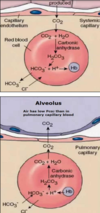
Carbonic anhydrase located within red blood cells

  • CO2 + H2O - H2CO3 - H+ and HC)3-
  • Carbonic anhydrase converts CO2 into HCO3- within the red blood cell close to the tissue
  • This liberates H+ ions (increasing acidity) which bind to HB causing the further release of O2 (Bohr effect)

Chloride shift = the HCO3- ions are transported out of the cell in exchange for CL-

Note the reverse happens at alveoli.
54
Q

What is the Haldane effect ?

A

Haldane effect

  • CO2 binds to a different site then oxygen = CO2 binds to the globin site where as O2 binds to the heme component.
  • removal of oxygen from HB in tissues increases affinity for CO2 - favouring the loading of CO2 in tissues
55
Q

What is hypoxemia ?

A

A condition where arterial oxygen tension or partial pressure (PAO2) is below normal.

56
Q

What is hypoxia ?

A

Inefficient O2 at the cellular level

57
Q

What is cyanosis ?

A

refers to a blue-purple discolouration of the skin and mucous membranes, caused by elevated concentration of deoxygenated hemoglobin.

58
Q

What is Hypercapnia ?

A
  • excess CO2 in the artial blood
  • affects acid base balance
  • occurs during hyperventilation
59
Q

Name the three components that regulate the respiratory system ?

A

Respiratory system

Regulated tightly to maintain concentration of O2, CO2 and H+ at constant levels independant of varying external conditions

  • Control centre = rhythmic neural discharge for automatic control
  • Chemical receptors = regulate the magnitude of ventilation to match physiological needs
  • modification of respiration to meet other physiological needs
60
Q

Describe the central respiratory centre

A

Pons and medulla central respiratory centre

  • rhythmic neural discharge for automatic control
  • respiratory rhythmicity originates in the brain stem : respiratory central pattern generator located pons and medulla
  • neurons in medulla signals inspiratory muscles
  • Stimulate diaphragm via phrenic nerve
  • stimulate intercostal muscles by the intercostal nerve.
61
Q

How does the central respiratory centre regulate expiration ?

A

Expiration

  • at rest expiration is due to inhibition of inspiratory neurons
  • when physiological demand increases expiratory neurons become activated to enhance ventaltion
  • Signal nerves innervate the internal intercostal muscles and abdominal muscles
62
Q

Describe how the higher brain regions can act on the respiratory centre to alter respiration ?

A

Voluntary control is innervated by the cerebral cortex

  • phonation, swallowing, partuition and defacation have altered respiratory signals voluntary control
  • cerebral cortex
63
Q

Where are respiratory central and peripheral chemoreceptors located, and what affect do theses receptors have ?

A

Central chemoreceptors = medulla close to respiratory centre

Peripheral chemoreceptors = carotid bodies and aortic arch

Chemical chemoreceptors regulate the magnitude of ventilation to match physiological needs

  • goverened by PO2, PCO2 and H+ in arterial blood which is detected by chemoreceptors.
64
Q

Describe central chemoreceptors and how they are activated ?

A

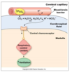
Central chemoreceptors

  • situated in the medulla close to the respiratory centre
  • Hypercapnia (high CO2) is the primary driver for activation of central chemoreceptors its action is carried out through H+
  • CO2 can not cross the blood brain barrier, where as H+ can
  • Increased H+ in interstitual fluid of the brain stimulates central chemoreceptors
  • stimulates hyperventilation
65
Q

Describe peripheral chemoreceptors and how they are activated ?

A

Peripheral chemoreceptors

  • Located the aortic arch and carotid bodies
  • stimulated by a decrease in arterial PO2, increase in H+ (especially non CO2 generated as in metabolic acidosis) and increase in PCO2
  • hyperventilation by stimulating the respiratory center in the medulla
  • increases ventilation

Note - adjustment for acid base balance stimulated by an increase in H+. will stimulate peripheral chemoreceptors to a major extent and to a lesser extent central chemoreceptors. Diabetes mellitus

66
Q

Describe the evolution of the cardiovascular system ?

A

Evolution of the cardiovascular system

  • simple small organsisms simple diffusion
  • multicellular flow through system eg sponges
  • Fish simple two chambered heart
  • Amphibians three chambered heart - lead to a seperate circuit for the first time - one ventricle two atrium - some mixing of deoxygenated blood and oxygenated blood in the one atrium
  • mammals, birds and crocodiles eveolved a four chambered heart - two atria and two ventricles - oxygenated and deoxygenated blood completely seperated
67
Q

Name the four layers of the heart ?

A

Pericardium - contains fluid

Epicardium

Myocardium

endocardium

68
Q

Describe the heart valves of the mammalian heart and their function - location ?

A

Heart valves

Functions to ensure one way flow of blood and prevent mixing of oxygenated and deoxygenated blood

Pulmonary valves =

Atriventricular valves = prevented from closing by chordae tendineae supported by papillary muscles. (tricupsid valve)

  • the valves open and close passively
  • forward pressure gradient opens valves
  • backward pressure gradient closes valves
  • valvular disease will reduce cardiac output
69
Q

Describe the structure of the myocardium in the mammalian heart ?

A

Myocardium

  • middle layer the thickest
  • consits of myocardial cells which comprise 99% of the heart

Myocardial cells

  • cardiac muscle cells
  • excitable, striated, mononucleated and has abundant mitochondria
  • intercalated disc = gap junctions which allow molecules and small ions to move between cells
  • The intercalated disk allows quick spread of action potentials

The myocardium acts as functional syncytium contracts as a single unit.

Atrial and ventricular syncytia are seperated by a layer of connective tissue

70
Q

Describe auto-rythmic cells location and their pathway through the heart ?

A

The heart depends on auto-rythmic (pacemaker cells) for rythmic beating, and does not require activation by the nervous system.

  • situated in the sinoidal node, aventricular node and conduction pathways of the heart
  • do not have a constant resting membrane potential
  • pacemaker activity
  • slowly depolarises between action potentials - declines to the firing level - pace maker potential or pre potential

The SA node is the usual pace maker as HR is determined by the fastest pacemaker.

71
Q

Describe phase four action potential in the SA node ?

A

Phase four = pre potential

Funny Na+ channels open allowing Na+ into the cell - this is unusual as it occurs while the cell is relatively polarised (if)

This causes transient Ca2+ channels to open which causes further depolarisation (ica), which occurs during the second half of phase four.

72
Q

Describe what happens during phase 0 and 3 during the action potential of the SA node ?

A

Phase 0 rapid depolarisation

  • occurs due to opening of long lasting voltage gated CA2+ channels once the threshold potential is meet -40mV

Phase 3 repolarisation

  • Due to opening of K+ channels and closing of Ca2+ channels
  • as the membrane reaches -65mV, the if Na+ channel start to reopen.
73
Q

Describe the parasympathetic activity on the SA node ?

A

Parasympathetic

  • Acetylcholine
  • Parasympathetic nerves cause a slower rate of rise in the pacemaker cells
  • phase four SA node prolonged
  • deacrease in HR
74
Q

Describe sympathetic activity on the heart ?

A

Sympathetic

  • catecholomines
  • sympathetic nerves release catacholomines which act on the beta receptors of heart muscle
  • increase the rate of depolarisation
  • increase heart rate
75
Q

Describe the conduction pathways within the heart ?

A

Conduction pathway within the heart

  • SA node - internodal path - AV node - AV bundle (HIS bundle) - bundle branches and purkinje fibres
  • atrioventricular node delays the impulse (AV node delay)
  • AV node and AV bundle provide the only route for the propagation of action potentials from the atria to ventricles
  • AV bundle splits into left and right bundle branches
  • purkinje fibres arising from the bundle spread impulse over the ventricular wall
  • rapid to allow synchronous depolarisation.
76
Q

Describe the five phases of the action potential in the cardiac contractile cell ?

A

Contractile cell

  • Phase 0 rapid depolarisation
  • opening of voltage gated Na+ channels
  • Phase 1 initial rapid repolarisation
  • opening of transient K+ channels
  • Phase 2 plateau phase
  • opening of long lasting Ca 2+ channels
  • Phase 3 rapid repolarisation
  • K+ influx by the opening of voltage gated K+ channels
  • Phase 4 resting phase
77
Q

In cardiac muscle what is the refractory period, compare this to the refractory period in skeletal muscle ?

A

Refractory period

Occurs immediately after the initiation of an action potential, and last until the action potential is complete

  • no response to further excitation until the refractory period has ended.

Contrast skeletal and cardiac refractory period

  • skelatal short 5 msec
  • prolonged in cardiac muscle 300msec
  • contractile time in cardiac muscle is longer than in skeletal muscle (as action potential duration is longer) this ensures adequate time for the ejection of blood
78
Q

What are the benefits to having a prolonged refractory period in cardiac muscle ?

A

Refractory period

  • cardiac muscle can not be restimulated until contraction is over - unable to have sustained contraction or summation of contraction
  • can not fatigue cardiac muscle
  • provides sufficient time to empty and refill cardiac chambers
79
Q

What physiological differences are apparent in cardiac muscle excitation coupling compared to skeletal muscle ?

A
  • T tubules are wider
  • sarcoplasmic reticulum less extensive
  • Ca2+ from the ECF induces the release of Ca2+ from SR
80
Q

What is an electrocardiogram ECG, and how do we attch it ?

A

ECG is the record of electrical activity of the heart

  • word ‘lead’ in ECG recording refers to the tracing of the voltage difference between two electrodes
  • ECG waveforms are produced by momentary changes in voltage differences during the spread of cardiac excitation.

Connection

  • Lead 1 right foreleg to left foreleg
  • Lead 2 right foreleg to left hindleg
  • Lead three left foreleg to left foreleg
81
Q

Describe normal ECG waves ?

A

Waves

P wave

  • atrial depolaristation - small positive deflection
  • atrial repolarization is a slow process, hence dose not usually produce a significant visible wave (may be obsecured by QRS)

QRS complex

  • ventricular depolarisation
  • Q first negative deflection, R large positive deflection and S negative deflection following R.

T wave

  • ventricular repolarisation
  • pattern of ventricular repolarisation varies between animals and may be positive or negative.
82
Q

Describe the mechanical events which follow the electrical events on an ECG ?

A

Mechanical events which follow

  • p wave followed by atrial contraction
  • QRS followed by ventricular contraction
  • T wave followed by ventricular relaxation

The delay of cardiac impulse conduction at the AV node prevents overlap of atrial and ventricular contraction

83
Q

In an ECG describe the PR interval, if this interval was prolonged what disease would you expect ?

A

PR interval

  • Longer than normal signifies a delay in impulse from SA node to ventricles
  • Duration of conduction from the SA node to ventricles
84
Q

QT interval in an ECG what does this signifiy ?

A

QT interval

  • electrical activity in ventricles - repolarisation and depolarisation
  • interval of QRS to T wave
  • influenced by electrolyte balance, drugs, ischemia
85
Q

Describe the RR interval what does it signify ?

A

RR interval

  • interval between two successive RR
  • duration is equal to one cardiac cycle
  • HR/min = 60/RR interval
86
Q

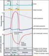
What is a Wiggers diagram, and describe the four phases of a Wiggers diagram ?

A

The four phases of a Wiggers diagram

1 = ventricular filling + gradual rise in left ventricular pressure

2a = isovolumetric contraction phase = steep rise in pressure as both valves remain closed

2b = ejection phase + left ventricular pressure is higher than in the aorta

3 = isovolumetric relaxation phase + no change in volume + both valves remain closed + steep drop in ventricular pressure

87
Q

Describe a first degree AV block - conduction block

A

First degree block

  • AV node block is a common dysfunction of cardiac impulse conduction
  • may be caused by cardiac trauma, toxins, bacterial infections etc
  • Three dgrees of severety of AV block are recognised

First degree AV block

slowed conduction through AV node

PR interval prolonged

88
Q

Describe a second degree, and third degree AV block ?

A

Second degree AV block

  • some P waves are not followed by a QRS complex

As the degree of AV block increases until the AV node fails completely and no QRS wave is seen

Third degree AV block

  • complete conduction block
  • no impulse goes through the AV node
  • the atrium and ventricles beat at their own intrinsic rate
  • purkinje fibres initiate ventricular contraction
  • cardiac out put and blood pressure is compromised - animals will be weak and may collapse.
  • treat by using a pace maker
89
Q

Describe a cardiac arrhythmia and how they may be identified on an ECG ?

A

Cardiac arrhythmias

Any variation in the normal rythm of the heart , requires treatment if animal develops clinical signs such as syncope

  • impaired impulse formation
  • impaired impulse conduction

As seen on an ECG

  • no P wave
  • P wave not followed by a QRS
  • abnormal P wave
  • prolonged PR interval
  • irregular waves as in ventricular fibrillation
90
Q

Describe a cardiac arrhythmia - ectopic pacemakers and atrial fibrillation ?

A

Atrial fibrillation

  • frequency of the generation of action potentials is too high
  • common in older animals and horses
  • continuous random passage of action potentials within the atrium
  • well tolerated, few AP manage to get through the AV node

Complications = blood may pool in parts of the atria forming clots

91
Q

Describe ectopic pacemakers - ventricular fibrillation ?

A

Ventricular fibrillation

  • electric chaos, random voltage fluctuations generated by fibrillating ventricles
  • no coordinated ventricular contraction
  • reduced cardiac out put and arterial pressure

Defibrillation = CPR

92
Q

Describe parasympathetic innervation of the heart invlude function, receptors and the location of where the nerves act ?

A

Parasympathetic innervation

  • right vagus supplys SA node
  • left vagus supplys AV node and Bundle of His
  • Inervation of atrial muscles, but very few connections to ventricular muscle

Function

  • reduce HR
  • reduce conduction of impulses
  • Increase delay in AV node

Vagus nerve acts by releasing acetylcholine which acts upon muscarinic M2 receptors on the heart muscle

93
Q

Describe the sympathetic innervation of the heart, including function, what receptors it acts upon and where the nerves are located ?

A

Sympathetic innervation

  • SA and AV node
  • Bundle of HIS branches and ventricular muscle

Function

  • quicker, stronger and faster
  • increase HR
  • increase force of contraction
  • speeds up relaxation
  • increases cardiac conduction velocity of impulses

Sympathetic nerevs act upon beta one and two in ventricular muscle, which respond to circulating adrenaline and noradrenaline

94
Q

What creates the arterial pulse, and where can it be measured ?

A

Arterial pulse

A pressure wave travels along the artery when blood is forced into the aorta during systole

  • pressure wave extends arteriole walls as it travels and this expansion is palpable
  • felt at femoral artery, facial artery, radial artery and carotid artery
95
Q

Describe S1 and S2 of the four heart sounds ?

A

Heart sounds

S1

  • Lub closure of atriventricular valves
  • denotes the onset of ventricular systole
  • low pitch, soft, relatively large Lub

S2

  • closure of aortic valves and pulmonary valves
  • signals onset of ventricular diastole
  • shorter, sharper and of a slightly higher pitch DUB
96
Q

What is a heart murmer ?

A

Heart murmer

Abnormal heart sounds resulting from turbulant flow of blood through heart defects

  • narrowed stenotic valves
  • injured, leaky insufficient valves
97
Q

Define cardiac output and how is it determined ?

A

Cardiac out put

It is the volume of blood pumped out by each ventricle per minute

  • both ventricles simulaneously pump the same amount of blood
  • continually adjusted to meet biological needs

cardiac out put (vol/min) = stroke vol (vol pumped) * HR (beats/min)

98
Q

Define cardiac reserve ?

A

Cardiac reserve

The maximum percentage that the cardiac output can increase above normal is known as the cardiac reserve

  • cardiac out put can be increased up to a limit
  • low cardiac reserve may indicate heart disease such as cardiac failure
99
Q

Define ejection fraction ?

A

Ejection fraction

This can be defined as the end fraction of duystole that is ejected juring systole

SV/EDV = EF

  • normal EF values between 50% and 80%
  • index of ventricular function
100
Q

What is stroke volume and what three factors dose it depend on ?

A

Stroke volume = is the volume of blood pumped by each ventricle per beat.

SV = EDV (end dystolic vol) * ESV (end systolic volume

Stroke volume depends upon three factors

  • pre load
  • after load
  • cardiac muscle contractility
101
Q

Describe preload and what factor affects it?

A

Pre load = extent of diastolic filling or end diastolic volume is preload.

  • the strength of contraction is increased by increased filling of the heart
  • increase in preload increases force of contraction
  • within limits stretching of the ventricular fibres optimizes overlap between myosin and actin filaments increasing the strength of contraction.
  • also increases the amount of CA2+ released from the sacroplasmic reticulum.
102
Q

What is afterload, and how can it be adjusted ?

A

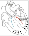
Afterload = is the tension developed in the ventricular wall during ejection

  • determined by arterial resistance (arterial pressure)
  • increase in afterload - the more difficult it becomes for ventricles to eject blood
  • sustained high arterial pressure increases left ventricular workload - eventually leading to heart failure

Higher afterload reduses stroke volume in a heart that is failing.

103
Q

Name the four organ systems involved in excretion ?

A

Four organ systems

  • respiratory system
  • digestive system
  • integument glands and skin
  • renal organs
104
Q

State the four functions of the kidney ?

A

The four functions of the kidney ?

  • removal of waste materials
  • maintenance of the composition of body fluids
  • production of hormones (vitamin D3, renin and erythropoietin)
  • production of pheramones
105
Q

What is a nephron, and what is its functional components ?

A

The nephron is the function component of the kidney.

It is composed of

  • glomeralus
  • proximal collecting tubule
  • distal collecting tubule
  • collecting duct
  • medulla and cortex of the kidney
  • macula denser
  • Bowmans capsule
106
Q

Name and describe the two different types of nephron ?

A

Two types of nephron

Juxtamedullary

  • specialised for the formation of concentrated urine
  • renal corpscles lie just above the junction of the cortex and medulla
  • long hoop of Henle extends into the inner medulla
  • supplied by vasa recta which runs parallel to the loop of Henle
  • varies between species eg cat 100%, pig 3%

Cortical nephron

  • renal corpuscles lie in the outer layer of the cortex
  • short loop of Henle
  • % of cortical nephrons varies between species
107
Q

How is the renal system stimulated ?

A

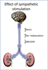
Stimulation of the renal system

  • supplied by sympathetic nerves
  • primarily supply the afferent and efferent arterioles
  • also proximal and distal tubules, loop of Henle and juxtaglomerula cells
  • powerful stimulation decreses bllod flow

The effect is a increase in renin, increase in Na+ reabsorption and decrease in EBF/GRF

108
Q

How is the vascularisation of the kidney unique, and what is the function of this unique system?

A

Vascularisation of the kidney and its function

  • kidney highest blood flow per unit mass among major organs 20-25%
  • mainly for the rate of glomerula filtration
  • double capillary network in series

Glomerulus = high pressure capillary bed favors filtration (60mmhg)

Pertubular capillary network = low pressure capillary network (20mmhg) permits rapid reabsorption.

Note afferent arterioles are shorter and wider than efferent arterioles, which is required for the high pressure within the glomerulus.

109
Q

Describe the three basic steps of urine formation ?

A

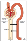
Glomerular filtration

tubular reabsorption

tubular secretion

110
Q

How does the glomerular barrier prevent the filtration of proteins and cells?

A

All molecules and ions are filtered at the same concentration as in plasma except protein and cells.

What prevents protein and cells being filtered the glomerular barrier

  • endothelium capillary (fenestrated negative charge)
  • Basement membrane or basal laminar capillary (negatively charged, collagen and proteoglycans)
  • epithelial cells of the Bowman’s capsule (negatively charged) which have podocytes
111
Q

What is a podocyte ?

A

Podocyte

  • Within the epithelial cells of the Bowman’s capsule
  • has foot processes
  • negatively charged
  • prevents the filtration of proteins and cells
112
Q

What determines the net filtration pressure ?

A

Net filtration pressure

  • glomerula hydrostatic pressure
  • Glomerula protein osmotic pressure (colloid osmotic pressure opposes filtration).
  • hydrostatic pressure of the Bowman’s capsule (opposes filtration)

The colloid osmotic pressure of the Bowman’s capsule is negligible as proteins are not filterred in a healthy kidney.

The changes in glomerular hydrostatic pressure is the main means for physiological control regulation of GFR.

113
Q

Describe the glomerular filtration ?

A

Glomerular filtration

  • fenestrated capillaries
  • high capillarie hydrostatic pressure
  • capillary tuft inbetween two arterioles

The afferent arterioles are shorter and wider than efferent arterioles - which is required for high pressure within the glomerulas

  • glomerular filtration is a passive process
  • filtrate is identical to plasma (PH, osmolality) but protein free
    *
114
Q

Describe the juxta glomerular apparatus ?

A

Juxtar glomerular apparatus

  • autoregulation
  • GFR and RBF are primarily determined by the glomerular hydroastaic pressure
  • constriction of afferent arteriole increases GFR, but severe constriction decreases GFR

Composed of -ve feedback mechanism

  • juxta glomerular cells (granular cells). Located mostly around the afferent arteriole, and act to secrete renin
  • Lacis cells which are located between the afferent and efferent arteriole. These cells are involved in the renin-angiotensin-aldosterone system.
  • Macula densa - afferent arteriole or modified distal tubule. The macular densa detects changes in tubular fluid Na+ and Cl-
115
Q

Desribe how GFR may be measured ?

A

Glomerular filtration rate

GFR is the sum of filtration rates of all nephrons = total glomerular filtration rate .

Measurement of GFR is the best single means of assessing kidney function.

Criteria for a substance to be used to measure GFR are

  • freely filtered (not reabsorbed or secreted)
  • non toxic, not metabolised or stored within the kidney
  • substance which meet these criteria inulin, iohexol and endogenous creatine.
  • Plasma creatine concentration is a good indicator of GFR as it is freely filtered, produced continuously and excreted in urine.
116
Q

What is renal clearance ?

A

Renal clearance

It is a measure of the kidneys ability to remove substances from the plasma

  • of a substance is the volume of plasma from which that substance is removed by the kidneys per minute.
117
Q

Describe how you could obtain a estimate of GFR ?

A

Estimate of GFR

Creatine

Criteria for a substance to be used in estimation of GFR

  • freely filtered (not secreted or absorbed)
  • non toxic, not metabolised in the body or stored in the kidney
  • inulin, iohexol and endogenous creatine

Why is creatine suitable

  • continuous production, being a byproduct of muscular metabolism
  • freely filtered
  • excreted in urine
  • frequently used to measure GFR
118
Q

Describe tubular function ?

A

Tubular function

Glomerular filtration is non selective (to a large extent) process but tubular reabsorption and secretion are highly selective processes.

  • modulating the volume and composition of urine
  • structure of each tubule section is adapted to its specific function
  • transport proteins present on the cell membrane of tubular lining in each part of the tubule varies - which is the reason for the different function by different parts of the tubule.
119
Q

Describe the process behind tubular reabsorption ?

A

Process of tubular reabsorption

Process

  • driven by sodium - potassium ATPase (generates Na+ gradient inside of the cell)
  • reasorption and secretion of most substances inside the tubule is driven by ATPase system.
  • Cotransport - Na+ and solute same direction
  • Counter transport - Na+ and solute moves in opposing directions

Process of solute transport

  • paracellular reabsorption
  • transcellular reabsorption
  • carrier mediated transport
120
Q

Describe carrier mediated transport across the apical membrane ?

A

Carrier mediated transport in the kidney

  • 5-10% of mammalian genes code for transport proteins
  • lack of causes many kidney diseases
  • numerous transport proteins are crucial drug targets
121
Q

Describe the glucose reabsorption and where it occurs ?

A

Glucose reabsorption in the proximal tubule

Normally glucose is completely reabsorbed by the proximal tubule

  • secondary active transport, which prevents nutritive loss
  • sodium ion and glucosetransported into PT cell by sodium-glucose transporter (SGLT) on luminal side
  • glucose is then transported out of the cell by Glut2 transporter
  • eventually glucose enters the blood
122
Q

What is glycosuria, and what dose it cause ?

A

Glycosuria

It is the pressence of glucose in urine

  • a common cause is diabettes mellitus due to rise in blood glucose conc
  • may also occur in tubular two dysfunction (SGLT mutations)
  • also causes an excessive water loss into the urine, a process called osmotic diuresis (osmotic effect of glucose)
  • carrier mediated transport is saturable - renal threshold

filtered load > reasorbing capacity of the proximal tubule

123
Q

Describe the function of the proximal tubule ?

A

Function proximal tubule

  • 65% of filtered Na+, CL- and water reabsorbed
  • osmotically reabsorbed
  • reabsorbs glucose, amino acids, Na+, K+, CL-, Ca++ and HCO3-
  • 40% of urea reabsorbed via pinocytosis
  • secretes organic anions and cations.
124
Q

Describe the function of the loop of Henle

A

Describe the function of the loop of Henle

Descending

  • highly permeable to water and moderately to urea

Ascending part

  • thin impermeable to water, but permeable to sodium
  • thick reabsorbs 25% of Na+, Cl- and K+

Both thin and thick ascending parts are impermeable to water

125
Q

Describe the function of the distal part of the tubule ?

A

Function of distal tubule

The first part of the distal tubule resembles that odf the ascending loop of henle - permeable to water and reabsorbing Na+ and Cl-

The late distal tubule and collecting duct reabsorb sodium and is impermeable to water.

Avid absorption of salts without water absorption produces a hypotonic tubule fluid, thus, these segments are sometimes called diluting segments.

126
Q

Describe the function of the medullary collecting duct ?

A

Medullary collecting duct

Crucial in determining the final output of urine and solutes

  • permeability to water depends on ADH
  • permeable to urea contains urea transporters
  • secretes hydrogen ions against a lrage concentration gradient
127
Q

Describe the function of principle cells, intercalated disks and where are they located ?

A

Both principle cells and intercalated disks are located in the late distal tubule and collecting tubule

Principal cells

  • reabsorb sodium from the lumen
  • secretes potassium
  • may become permeable to water in the pressence of ADH

Intercalated disks

  • key role acid base balance
  • alpha cells secrete H+, reabsorb bicarbonate
  • Beta cells secree bicarbonate and reabsorb H+
128
Q

The kidney acts to regulate four physiological parameters simultaneously, what are these parameters ?

A

Four physiological parameters

  • water balance
  • osmolality
  • ECF volume
  • blood pressure
129
Q

Discuss Na+ reabsorption within the proximal tubule ?

A

Proximal tubule

  • 65% of the mass of filtered sodium is reabsorbed
  • isosmotic tubular fluid

Angiotensin 2

  • enhances Na+ reabsorption from the proximal tubule
  • early Na+ reabsoprtion is linked to nutrient transport and H+ secretion
130
Q

Describe Na+ reabsorption by The thick ascending limb of loop of Henle and early distal tubule ?

A

Ascending limb and distal tubule.

Thick ascending limb reabsorbs Na+ and Cl- but is virtually impermeable to water.

  • hypo-osmotic (more dilute fluid) tubular fluid when compared to the plasma

ADH vasopressin = enhances Na+ reabsorption from thick ascending limb

131
Q

Describe the reabsorption of Na+ by the late distal tubule and collecting tube ?

A

Na+ reabsorption in the late distile tubule and collecting duct

Principal cells = Na+ entry from the lumen is via an ion channel epithelial sodium channel (ENaC)

Aldosterone = increases the absorption of Na+ and increases K+ secretion in principal cells

132
Q

What affect does Natriuretic peptide have on sodium reabsorption ?

A

Natriuretic peptide reduses sodium reabsorption in the principal cells of the later distal tubule and collecting duct.

In response to a high ECF volume the atrial wall becomes distended. This results in the release of atrial natriuretic peptide (ANP) which decreases sodium absorption from principal cells.

133
Q

Describe the RAAS Renin-angiotensin, aldosterone system and what it achieves ?

A

Renin-angiotensin aldosterone system

  • important salt conserving system
  • important role in regulating Na+ balance
  • ECF palsma volume
  • regulation of arterial pressure.

Angiotensin two stimulates the production of

  • ADH vasopressin, acts upon the ascending loop of Henle to incrase Na+ absorption
  • aldosterone acts upon principal cells to increase Na+ absorption.
134
Q

Identify the six effects of Angiotensin 2.

A

Angiotensin two acts to increase ECF volume in several ways. (defends against a decrease in ECF volume).

  • increase water retension
  • increase thirst
  • vasocontriction of the systemic circulation
  • stimulate secretion of ADH posterior pituitary
  • stimulate secretion of aldosterone secretion
  • increase proximal tubule Na+ absorption + increase action of principle cells to increase Na+ absorption

Aldosterone and ADH is released from the zona glomerulosa

135
Q

Describe the actions of ADH (antidiuretic hormone) on the renal tubule ?

A

ADH

Acts to decrease volume and increase concentration

  • increase in plasma osmolality (concentrated) results in increased secretion of ADH
  • inserts urea transporters increasing urea uptake
  • stimulates reabsorption of Na+ thick ascending loop of Henle
  • Inserts aqau porins in the luminal membrane of principal cells increasing water reuptake
  • negative feed-back mechanism
136
Q

What is the affect of aldosterone on the kidney?

A

Aldosterone