Anatomy semester two Flashcards

1
Q

Describe the muscle innervation of the toungue ?

A

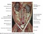
Muscles of the toungue

Extrinsic muscles

yellow = styloglossus

green = hyoglossus, attachment of the toungue

pink = genioglossus

Intrinsic muscles of the toungue

Proper lingual muscle

2
Q

Describe the sensory and motor innervation of the toungue ?

A

Inervation

Sensory = lingual nerve and glassopharngeal nerve

Motor = hypoglossal nerve

3
Q

Describe the structure and topography of the toungue ?

A

Toungue

  • apex cranial part free and highly mobile
  • body fixed thick
  • caudal part fixed and attached to the hyoid bones, soft palate and the pharynx
4
Q

Describe the topography of the heart ?

A

Heart

  • lies between the 3rd and the 6th rib
  • it is located between the two lobes of the mediastinum
  • The base lies more dorsally, and the apex more ventrally
  • The left face of the heart lies caudally
  • the right side of the heart lies cranially
  • the heart lies above the sternum just infront of the diaphragm
  • in domesticated species its lies more to the left than the right
5
Q

Describe the structure of the right face of the heart externally ?

A

Right face of the heart

The right side of the heart provides the small circulation of deoxygenated blood into the lungs.

Right atrium

  • receives the cranial and caudal vena cava on the right side (large orifices without any valve)
  • floor almost entirely occupied by the right atrioventricular orifice and tricupsid valve

Right ventricle

  • two compartments pulmonary infundibulum and atrioventicular chamber
  • right atrioventricular orrifice tricupsid valve
  • pulmonary orifice semilunar valves
  • blood exits through the pulmonary artery
6
Q

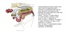
Identify the structures numbered on the right side of the heart ?

A
  • 1 right ventricle
  • 9 cranial vena cava
  • 14 caudal vena cava
  • 7 aorta
  • 10 and 11 left and right pulmonary arteries
  • 13 left azygous vein
7
Q

Describe the structure of the left external heart ?

A

Left side of the heart

Left atrium

  • receives the orifices of five-six large pulmonary veins
  • floor almost entirely occupied by the left atrioventricular orifice

Left ventricle

  • two compartments the atriventricular chamber and the aortic infundibulum
  • aortic orifice bicupsid valves
8
Q

Identify the structures on the left side of the heart ?

A
  • 10 left and right pulmonary arteries
  • 11 left and right pulmonary veins
  • 12 left azygous vein
  • 7 aorta
  • 8 ligamentum arteriosum
9
Q

Describe the division of the ventricles ?

A

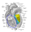
The medial and lateral division of the ventricles

Atrioventricular chamber

  • veinous chamber yellow

Aortic infundibulum

  • aortic chamber
  • leads to the aortic orifice
  • blue
10
Q

Describe the structures within the heart ?

A

Heart inside

  • yellow chordae tendinae
  • green papillary muscle
  • blue cusp of atrioventricular valve
  • note right auricle above atrioventricular valve
  • orange trabeculae carnae
  • pink semilunar valve

craniolateral view of the internal right face of the heart.

11
Q

Describe the location of the fossa ovalis ?

A

The fossa ovalis

It is a depression (Fossa ovalis) a remnant of the foramen ovale from. It is a communication between the right and left sides of the heart. In fetal life it allows the blood to bypass the pulmonary circulation as at this time the lungs are unfunctional.

12
Q

Describe the three tissues which constitute the heart ?

A
  • Fibrous structures (connective tissue
  • myocardium
  • nodal tissue (special nervous tissue)
13
Q

Describe the layers of the pericardium ?

A

The layers of the pericardium

  • 1 heart
  • 2 great vessels
  • 3 visceral pericardium
  • pericardial cavity
  • 5 parietal pericardium
  • 6 connective tissue layer of the pericardium
  • 7 mediastinal pleura
  • 8 sternopericardial ligament
14
Q

Describe the topography and structure of the nose ?

A

The nose

  • The nostrils nares are the openings above the upper lip on each side of the surface called the nose
  • nasal vestibule = entrance to each nasal cavity
  • Horse = nasal diverticulum (cul de sac) false nostril
  • cartiliginous skeleton strengthens openings, muscle and skin
  • animal may increase and deacrease size of apetures
  • nasal septum = median wall.
15
Q

Describe the function, structure of the nasal cavity ?

A

Structure of the nasal cavity

  • ensures the passage of air and is the site of olfaction
  • the structure is bone and cartilage
  • lined by the nasal mucosa (pituitary membrane)

Structure

  • olfaction
  • thermoregulation, filter, humidity (condition the air)
  • respiration
  • pheromone, behaviour
  • Vomero nasal organ - detects pheromones
16
Q

Identify the nasal cartilages in the nose ?

A

dorsal lateral nasal cartilage

alar plate of cartilage

ventral lateral nasal cartilage

17
Q

Describe the topography of the nasal cavity ?

A

Topography of the nasal cavity

  • There are two nasal cavities seperated by a median partition the nasal septum
  • seperated from the oral cavity by the hard palate ( maxillary, incisive and palatine bone.
  • corresponds with the nasopharynx through the corresponding choanae.
  • contains anfractious annexes (which give attachment for the paranasal sinuses.)
  • Nasal choncae define longitudinal gutters called meatus.
18
Q

Identify the following structures within the nasal cavity ?

A

Identify the following structures of the nasal cavity ?

  • dorsal meatus
  • middle meatus
  • ventral meatus
  • ethmoid
  • pharynx
  • dorsal nasal chonae
  • ventral nasal choncae
19
Q

Describe chonchae ?

A

Nasal choncae

  • thin coiled bone blades, supplemented by cartilage and lined by the nasal mucosa
  • simple choncae delimit the narrow recess that communicates with the nasal cavity where air can circulate freely
  • increases surface area

Bulla - if the coiled blade is joined to itself forming a cavity it is named bulla

There is a dorsal, ventral and middle choncae

20
Q

Describe the nasal meatus ?

A

Nasal meatus

  • Longitudinal gutters
  • 1 dorsal, 1 ventral and one middle
  • communicate along the medial side of the nasal septum by a vertical gap the common nasal meatus
  • The middle nasal meatus is located between the dorsal and ventral nasal choncae, and ends caudally at the entrance of the ethmoid bone.
21
Q

Name the paranasal sinuses of the dog, horse and Ox ?

A

Paranasal sinuses

Horse = 5 frontal, sphenoid, ethmoid, rostral, maxillary and caudal maxillary

Ox = 4 frontal , sphenoid, ethmoid and maxillary sinus

Dog = 3 frontal, sphenoid and maxillary

22
Q

What are the paranasal sinuses ?

A

Paranasal sinuses ?

The paranasal sinuses are anfractious cavities that are attached to the nasal cavities and communicate with them.

They are carved into the bones of the head and arranged around the ethmoid bones.

  • lighten the bones of the head
  • structural bony elements protect the brain and sensory organs
  • allow for the conditioning of inspired air
  • increase olfactory mucosa surface area in carnivores.
23
Q

Describe the uniqueness of the equine choncae ?

A

Equine choncae

Dorsal nasal choncae not weel developed, but very long. Its caudal part forms the dorsal choncal sinus that communicates with the frontal sinus.

Middle nasal choncae not very well developed. Has a bulla conformation and dose not open into the middle meatus.

Ventral nasal choncae (maxillary choncae) developed but short. Its caudal part forms a ventral conchal sinus, that communicates with the rostral maxillary sinus forming its medial compartment.

24
Q

Describe the uniqueness of the equine meatus ?

A

Equine meatus

Dorsal meatus = is very narrow

Middle meatus = caudally there is a narrow sinuso-nasal slot which caudal and rostral parts communicate with the caudal and rostral maxillary sinus.

Ventral equine meatus = height and width make it the preferred site for the naso gastric tube in the horse.

25
Describe the equine paranasal sinuses ?
There are five paranasal sinuses in the horse **Caudal maxillary sinus** * largest * carved into the maxillary, lacrimal and zygomatic bone * seperated from the cranial maxillary sinus via **intersinusal blade** * reach surgically above facial crest * lateral and medial compartments seperated by the **infraorbital duct** **Rostral maxillary sinus** * carved into maxillary bone * extends into ventral choncal sinus * lateral and medial compartments ( infraorbital duct seperates them) * may contain the dome of the first molar * inaccessable surgically due to masseter muscle * medial compartment is formed by the ventral nasal choncae * communicates with coressponding nasal cavity by a slot **Chonco frontal sinus** * two portions true dug into frontal bone (true), rostral formed via the dorsal choncae sinus **Sphenoidal sinus** * sphenoid and palatine bones * opens into the rostral part of the caudal maxillarysinus **Ethmoidal sinus** * It is represented by a large cavity formed in the volute of the ethmoid.
26
Describe the uniqueness of the ox meatus ?
OX meatus In the Ox the median septum dose not seperate completely **Dorsal nasal choncae** * elongated, narrow, and somewhat triangular * weakly wrapped posterior end **Ventral nasal choncae** * more developed * basal laminae attached to the maxillary detaches two divergent spiral blades **Middle choncae** * is shaped by the great volute of the ethmoid
27
Describe the paranasal sinuses of the OX ?
**Frontal sinus** - detailed card made **Ethmoidal sinus -**is represented by the cavity of the large volute of the ethmoid **sphenoidal sinus -** it is carved in the orbital sphenoid wing **Maxillary sinus -** detailed card made
28
Description of the Ox frontal sinus ?
Ox frontal sinus * highly developed with numerus diverticulum * A complete median septum seperates the two frontal sinuses - together they completely surround the skull frontal sinus is completely seperated from the other sinuses Three diverticulum * orbital diverticulum * cornual diverticulum * nuchal diverticulum (accessory does not communicate with the main compartment) **Aditus naso-frontalis =** An orifice located in the middle meatus used to communicate between the nasal cavities and the frontal sinus
29
Describe the maxillary sinus of the Ox ?
Ox maxillary sinus * single maxillary sinus which is well developed into the palatine vault * carved into the maxillary, zygomatic and lacrimal bones A bone blad supporting on its free border divides the infraorbital canal into two compartments * supra dental portion (lateral) * maxillo-palate portion (medial) The naso-maxillaris aditus is located 5-6 cm infront of the medial cantus of the eye.
30
Describe the choncae of the dog ?
Nasal choncae of the dog **Dorsal nasal choncae** * reduced * caudal part rolled and delegates into two extensions (one caudal, one ventral) **Middle nasal choncae** * very wide + complicated * corresponds to the large volute of the ethmoid **Ventral nasal choncae** * very wide but short * multiple longitudinal rolled strips
31
Describe the paranasal sinuses in the dog ?
Three paranasal sinuses in the dog **Frontal sinus** * very developed with free compartments rostral, medial and larger lateral compartment * the different compartments of the frontal sinuses communicate with the ethmoid by their own openings. **Sphenoid sinus** * carved into the sphenoid bone and filled by the last endoturbinal of the ethmoid **Maxillary sinus** * shallow * carved into the maxillary, palatine and lacrimal bones * infraorbital canal dose not protude inside * communicates directly with the middle and ventral meatus * no medial and lateral compartment * near the nasopharynx and ventral choncha
32
Describe the communication with the paranasal sinuses of the horse ?
Paranasal sinuses with in the horse * maxillary sinuses * divided into rostral and caudal complete division * incomplete seperation into lateral and medial compartments * communicates with the middle meatus, through the caudal medial compartment * Frontal sinus complete seperation left to right side * communicates with the dorsal choncae Communication * dorsal choncae - frontal sinus * ventral choncae - caudal maxillary sinus * rostral/ medial compartment of maxillary sinus communicates with the ethmoid sinus, sphenoid sinus, frontal sinus * caudal maxillary sinus directly to the nasal septum
33
Describe the topography of the pharynx ?
Pharynx * musculo membraneous passage * 7 openings - 2 choanae, 2 auditory tubes, 1 oral cavity, 1 oesophagus, 1 larynx ( trachea) * cranial side constituted by the soft palate * caudal side at the midline of the pharynx where several muscles fit the organ * lateral sides inclosed by several cranial nerves and in contact with the carotid branches * upperfloor nasopharynx * lower floor oropharynx
34
Identify and name the muscles of the pharynx
6 muscles of the pharynx * palatopharngeus * hyopharyngeus * thyropharyngeus * pterygopharyngeus * cricopharygeus * stylopharyngeus
35
Name and identify the five cartilages of the larynx ?
Five cartilages of the larynx * 1 epiglottis * 1 thyroid * 1 cricoid * 2 arytenoid
36
Describe the structure of the larynx ?
Larynx It is a complex valvular mechanism of interconnected cartilages, muscles, fibrous tissue and a mucous membrane. * attached to hyoid bones * lies in the midline down between the rami of the two mandibles
37
Describe the topography of the trachea ?
Topography of the trachea * permenantly open tube attached to the cricoid cartilages of the larynx * seperated from the skin ventrall by the sternothyroideus and hyoideus muscles. * oesophagus runs on its dorsal left lateral side * thoracic part passes through the mediastinum and terminates at the left and right bronchi (4-6 rib) * note in pig and ox extra branch (**right apical bronchus)**
38
Describe the structure of the trachea and any species differences ?
Structure of the trachea * incomplete tracheal cartilages open dorsally * connected via fibrous tissue and smooth muscle (trachealis muscle) * respiratory mucous membrane is continuous with the larynx and bronchi Species differences * carnivores trachealis muscle is attached on the dorsal side * Ox trachealis muscle is attached to the deep surface of the tracheal rings + dorsal median crest * horse flattened dorsoventrally + muscle inside
39
What is stroma and parachyma tissue with respect to the lungs ?
Parenchyma = responsible for the specific function of the tissue Stroma = tissue which supports the air passages and blood vessels.
40
Describe the topography of the lungs ?
Lungs The left and right lung fill the majority of the thoracic cavity * right lung is larger * bronchi and associated blood vessels form the root of the long * Hilus where the lung attaches to the root * in some species the lung tissue may be completely divided by fissures * Apex - is directed craniallyand lies within the thoracic inlet * Base - caudo-ventrally and lies upon the diaphragm Three surface of the lung * costal surface lies against the ribs * medial surface lies against the thoracic vertebrae * diaphragmatic lies against the diaphragm The medial surface meets the costal surface dorsally to form the rounded dorsal border.
41
Describe the species differences with respect to the lung ?
The species share a general lung pattern as seen in the pig and the carnivore * 4 lobes right 1 cranial, 1 caudal, 1 middle and 1 accessory * 2 lobes left 1 cranial, 1 caudal, Ox = the same except the cranial lobe in the right lung has a cranial and caudal part Horse = same except no observable external lobation
42
Describe the topography and structure of the pleura ?
Pleura * thin and transparent serous of the lungs * 1 pleura per lung * the two pleura sacs are backed in the midline to form the mediastinum * each pleura is comprised of a parietal pleura and visceral pleura **Mesopulmonum =** located between visceral and parietal pleura a pulmonary ligament **Pleural cavity =** contains pleural fluid which allows for the sliding of pleural layers and prevents their seperation via cohesion.
43
Describe the topography and structure of the mediastinum ?
Mediastinum It is composed via the backing of the two pleural sacs Regionalised Cranial * Cranial to the heart rib 1-3 * contains trachea, oesophagus, blood vessels etc Middle * contains the heart rib 3-6 * oesophagus, thoracic duct, phrenic and vagus nerves Caudal * caudal to the heart ribs 6-9 * oesophagus, azygous vein, thoracic duct, phrenic and vagus nerves
44
Characterise the differeneces between veins compared to arteries ?
Veins * branch into tributaries and roots * more numerous (often two veins to one artery) * often satelite to an artery but also located subcutaneously network (important) * pressence of valves **Veinous anastomoses** * more common and larger than that of the arterial systemallow connection between deep veins and subcutaneous networks
45
Describe the characteristics of a portel system ?
Portal system Consist of a vein which is divided by a capillary network, which is then followed by one or more veins eg. the portal vein between liver and intestine.
46
Descroibe the topography of the cranial aorta ?
Cranial vena cava The venous blood of the high systemic circulation goes to the heart through the cranial and caudal veina cava. Extends from the entrance to the thorax to the right atrium Roots = 4 large veins two axillary (horse, cows), two subclavian (carnivores) two jugular
47
Locate and describe the tributaries of the cranial vena cava (8)?
Tributaries of the cranial vena cava * right azygous vein * costocervical vein * vertebral vein * right and left jugular vein * right and left axillary vein * internal thoracic vein
48
Describe the species differences in the tributaries comming from the cranial vena cava ?
Species Ruminant and horse * 5 internal thoracic, vertebral, deep cervical, costo-cervical, cervical vein and right azygous * except cows left azygous Dog * thyroid vein, internal thoracic, vertebral vein, costo cervical vein and right azygous - no deep cervical vein Horses and carnivores have a right azygous vein and no corresponding artery OX have a left azygous vein and no corresponding artery
49
Describe the basic topography of the veins of the lower forelimb ?
Veins of the lower forelimb * lateral metacarpel vein * medial metacarpel vein * digital vein * coronary vein * coronary veinous plexus
50
Describe how species vary with structure and topography of the jugular veins.
Jugular veins * site of blood punction * head and neck drainage * normally there are two jugular veins each side (superficial and deep jugular veins). Species * Horses only have an external jugular vein * Carnivores external jugular vein is superficial due to the absence of a jugular groove - vein crosses the sternocephalicus muscleto get to the base of the neck * Ox External jugular vein extends into the jugular groove - seperated from the deep jugular vein by the sternocephalicus and amohyoidean muscle.
51
Describe the topography of the facial veins including the facial vein, caudal auricular vein and occipital vein ? + triangle of Viborg
Tributaries of the jugular vein
52
Identify the veins of the upper forelimb ?
Veins of the upper forelimb * axillary vein * brachial vein * median vein * antibrachial vein * median metacarpal vein
53
Describe the topography of the caudal vena cava ?
Caudal vena cava * The caudal vena cava is a large vessel that extends from the entrance to the pelvis to the right atrium of the heart * it collect the veins of the abdomen, hindlimbs and pelvis * its roots are the confluence of the 2 large common iliac veins * it runs forward under the body of the lumbar vertebrae at the right of the caudal aorta
54
Describe the topography of the caudal vena cava tributaries ?
Tributaries of the caudal vena cava * caudal vena cava * right common iliac vein * femoral vein * right azygous vein * renal vein * phrenic vein * portal vein * splenic vein * caudal mesenteric vein * cranial mesenteric vein
55
Describe the topography of the veins to the mammary gland ?
Veins of the mammary * subcutaneous abdominal vein (fountain of milk) * caudal mammary vein * Pudendal vein
56
Describe the topography of the hindlimb veins ?
Hind limb veins * medial saphenous vein * femoral vein * caudal tibial vein * tibial retanaculum The lateral saphenous vein is a good site for vein punction
57
Describe the topography of the portal vein ?
Portal vein * collects blood from the abdominal portion of the digestive system and spleen * liver blood is distributed by the celiac artery and both mesenteric arteries * TWO LARGE ROOTS = cranial and caudal mesenteric vein * moves forward slightly to the right * reaches the top side of the liver * it descends behind the organ on the right edge of the lesser omentum and ends at a portal fissure or hilum of the liver. * there is a capillary network at both its start and end
58
Describe the topography of the horses digestive system ?
59
Describe the topography of the equine oesophagus ?
Horse oesophagus * it starts above the cricoid cartilage * travels down the neck on the dorsally to the trachea, and then on its left side. * It travels within the mediastinum when inside the thorax passing through the oesophageal hiatus * it terminates in the abdomen at the cardia of the stomach * caudal third is particularly narrow and rigid
60
Describe the equine stomach ?
Equine stomach Topography * 9-15 intercostal space + 10-15 L * the stomach is slightly left of the median plane * (parietal) cranial convex face lies in opposition with the liver and the phrenic centre of the diaphragm * (visceral) caudal face oposition to the ascending colon, pancreas and small intestine * margo plicatus is about half way oesophageal non glandular area of the stomach above. Function = short transition time limited digestion in the horse
61
Describe the omentum of the horse ?
62
Describe the four components which make up the urinary system ?
The urinary system is comprised of * paired kidneys which produce urine * ureters which transport the urine to the bladder * urinary bladder that stores urine * urethra the expels urine to the exterior
63
Describe the topography of the kidneys (7)?
Describe the topography of the kidneys * situated against the dorsal wall of the abdomen in the thoracolumbar region * one kidney is located each side of the vertebral column * situated in the **retroperitoneal space** covered by a protective perirenial fat * the kidneys are asymetrical with the right kidney being cranial to the left (except in the pig) * the cranial pole of the right kidney sits in the renal fossa of the caudate process of the kidney Left kidney * left is pendulous in the feline * the left lies behind the right in the ruminant
64
Describe species differences in the external shape of the kidney ?
Kidney Firm, reddish brown colour whose appearance varies considerable among species 1. Large ruminant (B) = are oval shaped with multiple lobes visible on the surface - retains embryonic multipyramidal structure +surface divided by **fissures** 2. Pigs (C) are smooth elongated and flattened 3. Horses (E) the right is heart shaped, while the left is bean shaped 4. Dogs, cats, sheep (D) have a smooth external surface and are bean shaped.
65
Provide the external conformation of the kidney ?
Kidney conformation * medial concave border, with an indentation called the renal hilus * lateral convex border * dorsal surface * ventral surface * cranial pole * caudal pole
66
Describe the topography of the adrenal glands ?
Adrenal glands * paired endocrine glands * situated within the retroperitoneal space at the thoracolumbar junction * lie cranialmedial to the corresponding kidneys * (except in the horse where they lay medially) * phrenicoabdominal vein passes over the ventral surface of the adrenal gland.
67
Describe the four regions of the kidney ?
Four regions of the kidney **capsule** * strong fibrous capsule, which may be easily removed **cortex** * reddish brown, with a fine granular appearance * contains renal corpsicles and distal convuluted tubles * medullary rays arebundles of renal tubules which cross over into the medulla **medulla** * outer dark zone, and pale inner zone * striated in appearance * triangular shaped pyramidal lobes may be seen * apex of each lobe faces the renal sinus * the renal papilla drains the urine into the calyx or renal pelvis **renal sinus** * contains the calyces, renal pelvis and fatty tissue
68
Provides details of the shape of a pigs kidney ?
Pig kidney * multipyramidal structure * but there is some fusion of the pyramids * the outer surface of the kidney is smooth
69
Provide details of the structure of the bovine kidney ?
Bovine kidney shape The bovine kidney retains a multipyramidal structure internally and the renal surface is divided by fissures. * during embryonic development all mammalian species pass through a multipyrimidal phase - in most species the number of lobes drastically reduce * a renal lobe consist of a medullary pyramid and its associated cortex
70
Provide details of the internal structure of a sheep, dog, cat kidney?
Dog/cat/sheep/horse kidney * unipyramidal or unilobular species * the pyramids fuse to form a single medullary mass confining the cortex to the periphery * cortex forms a smooth shell * **renal crest - fusion of the medullary pyramids**
71
Describe the vascularisation and innervation of the kidney ?
Kidney Vascularisation * the renal artery and vein supply vascularistion to the kidney * interlobular artery and arcuate artery * segmental artery Innervation * sympathetic innervation
72
Disscuss what is the renal pelvis ?
Renal pelvis The proximal ureter begins with the renal pelvis (excepts ruminants) * located within the renal sinus * basin shaped and made of fibrous connective tissue, which gives it its white appearance * terminates at the ureter * pictured dog renal pelvis
73
Describe the spcecies differences in the shape of the renal pelvis ?
Species differences in the renal pelvis Dog and cat * renal crest (converging renal medullary pyramids) projects into the renal pelvis * the pelvis extends dorsally and ventrally to form the renal reccesses Horse (pictured below) * central expansion * two large recesses directed into the cranial and caudal lobe * most of the papilla drain into the recessses * mucosa of the renal pelvis produces a mucous - which is responsible for the protein found in the urine and its turbid appearnce Bovine (pictured on front of card) * **no renal pelvis** * the apex of each medullary pyramid fits into a small calyx * these branches unite to form large calyces * large calyxes then converge to the ureter
74
Describe the topography of the ureter ?
Ureter Muscular tube which transports the urine from the kidney to the bladder * passes caudally from the kidney along the dorsal abdominal wall in the retroperitoneal space * as it reaches the pelvis it bends medially to enter the broad ligamnet of the female or the genital fold of the male * the ureter then travelswithin the lateral ligament of the bladder to insert into the dorso lateral surface of the urinary bladder near its neck To prevent reflux of urine into the ureter - the ureter runs obliquely through the bladder wall. * this forms a valve * peristalic contractions of the ureteric wall overcome the resistance of this angle so urine may fill the bladder.
75
Describe the topography and structure of the urinary bladder ?
Urinary bladder * hollow musculutaneous organ * the **detrusor** muscle is the muscle found within the kidney wall * function to store urine * size shape and position varies greatly depending upon the fullness of the bladder * empty - pelvic cavity * full - extends cranially into abdominal cavity Can be divided into three areas * apex cranial blind end * body intermediate area * neck the funnel shaped caudal end which connects with the urethra
76
Describe the topography and structure of the urethral crest and trigone?
Urinary crest two urethral folds originating at the orifices of the ureters converge at the excit of the bladder = **urethral crest** * here they join to form the median urethral crest * this continues into the urethra The triangular muscular structure located between the uretic orifices and the urethral opening is called the **Trigone.**
77
Describe the ligaments which support the bladder ?
Bladder ligaments * lateral vesicular ligament - left and right attached to the abdominal wall * median vesicular ligament - attached ventrally to the abdominal wall
78
Identify the pouches formed by the supporting ligament between the tail and pubis of a dog ?
* rectogenital pouch * pubovesical pouch * vesicogenital pouch
79
Provide the innervation and vascularisation of the urinary bladder ?
Urinary bladder Vascularisation * cranial vesical artery - branch of the umbilical artery * caudal vesical artery - branch of the internal iliac artery Innervation * hypogastric nerve = sympathetic innervation * pelvic nerve = parasympathetic innervation * pudendal nerve = somatic innervation
80
Describe the structure and topography of the urethra ?
Urethra The tube which transports urine from the urinary bladder to the vestibule in females and externally in males. Female * female the urethra runs caudally on the pelvic floor ventral to the reproductive organs to open at the external urethral orifice on the floor of the vestibule * the length of the female urethra varies depending upon species * mares urethra is short with a wide opening * cow and sow suburethral diverticulum lies under the external urethral orifice * bitch the orifice is raised on the urethral turbicle Male * in the male the urethra is divided into two parts * pelvic urethra * penile urethra The urethra is surrounded by striated muscle which is innervated by the pudendal nerve Male *
81
Describe the structure, function of the ovaries ?
Ovaries * paired structures * usually ellipsoidal in shape (except mare kidney shape). Primary functions * produce gametes * hormones - estrogen and progesterone Structure * Outer layer is the tunica albuginea * The ovarian cortex is the parachymatous zone, and contains follicles of various sizes and the corpus lutea * ovarian medulla is the central part it contains all the vasculature, nerves and lymphatics.
82
Describe why the mare's ovary is unique ?
The uniqueness of the mares ovary * ovary is kidney shaped * the cortex forms the centre and it is enclosed by the medulla The **ovulation fossa** a deep indentation at the free margin of the organ is where the follicles rupture
83
Describe the position of the ovary in horses, dogs, ruminants and pigs ?
Ovary topography in a number of species. **Horse/dog/cat** * Dorsal abdomen caudal to the kidneys **Ruminant** * lie close to the ventral abdominal wall near the pelvic inlet **Pig** * middle of the abdomen
84
Describe the structure and function of the uterine tubes ?
Uterine tubes There are paired uterine tubes, also known as the salpinx, oviducts and fallopian tubes **Function** * Receive and transport the ova through to the uterus * Convey sperm * site of fertilisation (ampulla) **Structure -** **Infundibulum** * located ovary extremeity funnel shaped * contains many finger like projections fimbriae * fimbriae increase surface area to slip over the ovary during ovulation * ova transported through the **abdominal ostium** to the ampulla **Ampulla** * it is the site of fertilisation * half the length of the oviduct * wider in diameter than the isthmus * lined with ciliated epithelium **Isthmus** * transfers the fertilised egg to the uterine horn through the **uterine ostium**
85
What is the function of the smooth muscle layer within the uterine horn ?
Function of smooth muscle in the oviduct ? The primary function of the smooth muscle in the oviduct is too transport the newly ovulated oocytes and sperm to the ampulla site of fertilization. This requires sperm and ova to move in opposite directions. The muscosa secretes substances to support the ova and sperm. After fertilisation the newly formed zygote must remain within the oviduct for a few days before entering the uterus.
86
Describe how the connective tissues act to support the uterus ?
Uterine support **Mesovarium and mesosalpinx** * the ovaries and oviducts are suspended within the mesovarium and mesosalpinx respectively * the cranial portion of the broad ligament, the common suspension of the female genitalia * the ligament conveys the blood vessels, lymph and nerves to the organ **ovarian bursa** * The mesovarium, mesosalpinx and proper ligament enclose a small peritoneal cavity called the ovarian bursa.
87
Describe how the ligaments supporting the female reproductive system varies between species ?
Suspensory of the female genitalia **Dog** * contains an additional ligament the suspensory ligament, which forms the cranial portion of the free border of the broad ligament. * suspensory ligament continues caudally as the proper ligamnet of the ovary. * ovary is completely hidden commonication with the peritoneal cavity is through a slit like opening **Horse** * the ovary is too large to be contained within the ovarian bursa **Ruminant and Pig** * the ovary is partially hidden
88
Describe the structure and function of the uterus ?
Uterus The uterus is a hughly expandible tubular organ * sperm transport * gestation / implantation * expulsion of the foetus The uterus consist of the cervix * constricted caudal part joining the uterus to the vagina * located in the pelvic cavity * variable length * Cranial canal - uterine ostium opens cranially into the body of the uterus, and caudally into the vagina external uterine opening. * the caudal part of the cervix projects into the vagina in the cow and horse - surrounded by the vaginal fornix.
89
Describe the structure and function of the cervix ?
Cervix - thick walled non compliant **Function** * the cervical mucosa produces a mucous secretion which forms a plug, which assists in closing the cervix and isolating the conceptus during pregnancy. the plug is easily expelled during oestrus and partuition. * flush out of foreign material during copulation **Structure Species variations** Cows - rings form finger like interlocking projections - multiple rings Sows - rings interdigitate in a very intimate fashion, due to this the boar has a cockskrew penis. Semen is deposited directly into the cervix quickly entering the uterus - multiple rings Horses - loose folds of mucosa protrude into the vagina and the cervix is soft during oestrus - multiple rings Dog - the cervix protrudes caudally into the vagina is smooth with no elaborate folds Cat - small cervix which is thick walled smooth with no elaborate folds.
90
Describe the structure and topography of the carnivores uterus ?
91
Discuss the unique features of a sows uterus
92
Describe the unique features of a ruminant uterus ?
93
Describe the unique faetures of an equine uterus ?
94
Name and provide the topography of the ligaments of the female reproductive tract ?
95
Describe the structure and function of the vagina ?
Vagina This is a musculumembraneous organ connecting the cervix to the vulva The female copulatory organ and birth canal. Vagina proper * extends tfrom the cervix to the entramnce of the urethra * purely a reproductive passage * vaginal fornix - a cranioventral recess formed by the cervix bulging into the vagina in the mare and bitch * cow and ewe have a cranial dorsal recess Vestibular bulbs * erect venous plexus in the mare and bitch Vestibule * belonging to both the urinary and genital systems * caudal part of vagina is shorter * connects vagina with vulva * the hymen is a poorly developed mucosal fold at the junction of the vagina and the vestibule Vulva * the external orifice which ends the female genital system * the vertical vulvar is bounded by labia which meet at ventral fissures
96
Describe the topography of the female reproductive system ?
97
Describe the blood supply to the female reproductive system ?
Blood suuply to the female reproductive system * cranial uterine artery - branch from ovarian artery * middle uterine artery - branch from external iliac artery in the mare, and umbilical artery in the cow * caudal uterine artery - branched from the pudendal artery * ovarian artery is a collateral of the caudal aorta.
98
Describe the structure, function and topography of the rumen ?
Rumen * devoid of any gland * 130-180 L huge reservoir * almost occupies the entire left side of the abdominal cavity * craniocaudally elongated External conformation * 2 sides, 2edges and 2 extremeties * craniolateral lobgitudinal groove - on the right incontact with the otherstomaches, the intestine and pregnant uterus * upper edge supports the spleen * dorsal (4 last ribs) and ventral lobe * caudodorsal and caudoventral blind sac The openings of the dorsal sac = the oesophagus the cardia is on the left of the midline, it communicates with the reticulum through a large opening 20cm.
99
Describe the inner conformation of the rumen ?
Inner conformation of the rumen * fleshy pillars reinforce the muscles protruding at the bottom of the grooves which delimit the sacs and blind sacs * the mucosa is covered with numerous papillae
100
Describe the topography and structure of the reticulum ?
Reticulum the smallest of the reservoirs * most part is left of the midline * lower third of the 6-7-8 intercostal spaces * greater curvature is based upon the xiphoid process in contact with the diaphragm * Right part = reticulum cal de sac dead end where the foreign bodies are stopped * lesser curvature (concave) in contact with the omasum on the right side Inner conformation * honeycomb appearance - polyhedral alveoli * two openings the ruminoreticular orifice and the reticular omasal orifice
101
Describe the structure and topography of the omasum ?
Omasum - nutrients recycle * greater curvature above the lower part of the right 7th intercostal space * cranial in contact with liver and diaphrapm * caudal side in contact with the ventral lobe of the rumen * ventral side based on the abomasum Inner conformation * openings at the end of its lesser curvature * reticuloomasum opening is narrow, omasoabomasum opening is wider * omasal folds
102
103
Describe the structure and topography of the abomasum ?
Abomasum * paer shaped reservoir * true stomach contains glands * craniocauadally elongated * located within the long right costal arch from the lower end of the 7-11th rib * lower curvature receives the insertion of the greater omentum * uppercurvature recives detached the hepato gastric ligament (lesser omentum) * the end is reflected up and extends to the duodenum *
104
Describe the topography and structure of the small intestine.
Intestine The entire intestinal mass occupies some spec on the right side of the badominal cavity. * right of ruman back to liver abomasum and omasum * convulutions of the jujenum rest on the floor of the abdominal cavity