PGY4 - EKGs Flashcards
1
Q
A
A Fib with WPW
- Rate > 200 bpm
- Irregularly irregular
- Changing QRS morphology
- Pathway:
- Down normal- narrow QRS
- Down accessory- wide QRS
- Treatment
- No AV Nodal Blockers → VF
- CCB, BB, dig, adenosine or amio
- If unstable- cardioversion
- If stable- procainamide or cardioversion
- No AV Nodal Blockers → VF
2
Q
VT vs AFib with WPW
A

3
Q

A
Antidromic atrioventricular re-entry tachycardia (AVRT)
= WPW + SVT in antidromic pattern
Age clues you in
4
Q
Brugada ALgorithm - I dunno, just review this shit

A

5
Q

A
AAI
- Atrial paced and sensing
- If native activity sensed then pacing is inhibited
- If no native activity sensed for predetermined time then atrial pacing initiated
- Used in sinus node dysfunction with intact AV conduction
6
Q

A
VVI
Ventricular pacing and sensing
Similar to AAI but ventricular
Used in chronic atrial impairment
Note LBBB Morphology
7
Q

A
DDD- pacing and sensing the atria and ventricles
- Most common
- Atrial pacing if no native atrial activity for set time
- Ventricular pacing if no native ventricle activity
- Atrial channel function suspended during a fixed periods following atrial and ventricular activity to prevent sensing activity or retrograde p waves as native atrial activity
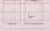
A-V sequential pacing:
- Atrial and ventricular pacing spikes are visible before each QRS complex.
- There is 100% atrial capture — small P waves are seen following each atrial pacing spike.
- There is 100% ventricular capture — a QRS complex follows each ventricular pacing spike.
- QRS complexes are broad with a LBBB morphology, indicating the presence of a ventricular pacing electrode in the right ventricle.
8
Q
Magnet Mode
For Pacemaker
For AICD
A
Pacemaker - Goes to asynchronous mode
ICD - Deactivates ICD
9
Q

A
Undersensing
- Undersensing occurs when the pacemaker fails to sense native cardiac activity.
- Results in asynchronous pacing.
- Causes include increased stimulation threshold at electrode site (exit block), poor lead contact, new bundle branch block or programming problems.
- ECG findings may be minimal, although presence of pacing spikes within QRS complexes is suggestive of undersensing.

10
Q

A
Oversensing
- Oversensing means that the pacemaker thinks it is sensing myocardial electrical discharges but it’s a LIE.
- The pacemaker appropriately inhibits itself, which leads to inappropriate underpacing
- Oversensing = Underpacing
- Causes:
- Large P or T waves, skeletal muscle activity, lead contact
*
- Large P or T waves, skeletal muscle activity, lead contact

11
Q

A
Failure to Capture
- This means the pacemaker discharges a stimulus, but the myocardium does not depolarize.
- EKG = pacer spikes without an associated QRS complex
- Can be caused by…
- lead dislodgement or malplacement
- fibrosis or inflammation at the interface of the lead and the myocardium
- pacemaker setting problems
- capture threshold set too high
- lead failure
- battery depletion
- recording system failure
12
Q

A
RVH
- RAD of +100 or more
- Dominant R wave in V1 (>7mm tall or R/S >1)
- Dominant S wave in V5 or 6 (>7mm deep or R/S <1)
- QRS duration < 120 ms

13
Q

A
Normal Peds EKG
- Heart rate > 100 beats/min
- Apparent right ventricular strain pattern:
- T wave inversions in V1-3 (“juvenile T-wave pattern”)
- Right axis deviation
- Dominant R wave in V1
- RSR’ pattern in V1
- Marked sinus arrhythmia
- Short PR interval (< 120ms) and QRS duration (<80ms)
- Slightly peaked P waves (< 3mm in height is normal if ≤ 6 months)
- Slightly prolonged QTc (≤ 490ms in infants ≤ 6 months)
- Q waves in the inferior and left precordial leads
14
Q

A
TCA
- Widened QRS
- QT prolongation (>100-sz, >160- ventricular arrhythmia),
- Right axis deviation of terminal QRS
- Terminal R > 3 mm in aVR
- R:S >0.7 in aVR

15
Q
LBBB STEMI Equivalent
A
LBBB is STEMI equivalent if:
- Hemodynamically unstable, acute HF
- Sgarbossa (a or b- c is questionable)
- A. Concordant STE > 1 mm any lead
- B. Concordant STD > 1 mm in V1-3
- C. Discordant STE > 5 mm
- Smith/Revised Sgarbossa
- Lead with > 1mm STE and proportionally excessive discordant STE (>25% of the depth ofpreceding S wave)

16
Q

A
Left Ventricular Aneurysm
- ECG Features
- Easy = Ant STE + Path Q Waves > 2 weeks after STEMI
- T-waves have a relatively small amplitude in comparison to the QRS complex (unlike the hyperacute T-waves of acute STEMI)
- Usually associated with well-formed Q- or QS waves
- May exhibit concave or convex morphology
- Most commonly seen in the precordial leads
- ST elevation seen > 2 weeks following an acute myocardial infarction
- Clinical Significance
- Ventricular aneurysms predispose patients to an increased risk of:
- Ventricular arrhythmias and sudden cardiac death (myocardial scar tissue is arrhythmogenic)
- Congestive cardiac failure
- Mural thrombus and subsequent embolisation

17
Q

A
Cor Pulmonale
- RBBB
- Extreme right axis deviation (+180 degrees)
- S1 Q3 T3
- T-wave inversions in V1-4 and lead III
- Clockwise rotation with persistent S wave in V6

18
Q

A
Arrhythmogenic Right Ventricular Dysplasia (ARVD)
EKG Findings
- Epsilon wave (30%)
- T wave inversions in V1-2 (85%) Prolonged S-wave of 55 ms in V1-3
General
- Inherited myocardial disease associated with paroxysmal ventricular arrhythmias and sudden cardiac death
- Fibro-fatty replacement of RV myocardium, autosomal dominant
Clinical
- Symptoms due to ventricular ectopic beats or sustained V tach
- palpitations, syncope, cardiac arrest, over time RV failure

