Peripheral Vascular System & Ischemia Part 2 Flashcards
(33 cards)
What occurs when one of the coronary arteries becomes totally occluded?
Myocardial Infarction
What may cause an MI? (6)
Occlusion – thrombosis
Coronary artery spasm – smooth muscle constriction
Decreased coronary artery blood flow
Increased myocardial workload
Decreased O2 levels
Toxic exposure to cocaine and ethanol
What are some signs when diagnosing an MI?
Prolonged substernal chest pain with radiation of pain to jaw or left arm (>30min), nausea, diaphoresis, and shortness of breath
Or, pt can have a “Silent MI”
What cardiac enzymes are affected in an MI? (3)
Troponin I: rise early and stay elevated for days*
CK-MB: 6 hours to rise and then normalize in 48 hours
CK
KNOW
What ECG changes when diagnosing an MI? (3)
T wave changes: first peak then inversion
ST segment elevation/depression: usually elevation with MI
Q waves: indicate irreversible myocardial cell death
What is the first enzyme indicator of a MI? How long will it remain elevated?
troponin
5-7 days
What cardiac enzyme will be elevated within 6 hours?
CK-MB
What are the stages of an AMI? (Acute MI)
(4)
T wave peaking
ST segment elevation
Appearance of new Q waves
T wave inversion

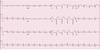
What is occuring with this waveform?

ISCHEMIA
Note: T wave inversion is not diagnostic of MI. If true infarct occurs – inversion can persist for months to years.
What part of the ECG reflects myocardial injury?
ST segment elevation

Is ST segment elevation reversible?
Yes, may reflect some damage, but still reversible and can return to normal.
What is a type of ST segment elevation seen in normal hearts?
J Point – where the ST segment takes off from the QRS complex

How do you distinguish a J point from MI? (2)
T wave maintains its independent waveform
In MI, elevation is bowed upward and tends to merge with T wave
Over ___ mm of ST segment elevation is clinically significant.
1
What ECG wave indicates irreversible myocardial death, is diagnostic of MI, usually appears within hours of infarct, and is persistent for a lifetime?
Q waves

During an infarction, ST segment usually goes back to baseline by the time Q waves appear. True or false?
True
Q wave > ____ sec in duration and depth at least ___ height of R wave signifies MI.
0.04
1/3
In leads distant from the site of infarction, will see these ECG changes: (2)
Tall R waves
ST segment depression
What artery supplies the anterior portion of the heart and most of the interventricular septum?
LAD
What part of the heart does the LAD supply?
supplies the lateral wall of the left ventricle
What is caused by occlusion of right coronary artery or descending branch of left coronary artery?
What leads will show changes?
inferior infarcts
II, III, AVF

Reciprocal changes will be seen in these leads in inferior infarcts: (2)
anterior
lateral leads
Note: also will observed peaked R waves.

For lateral wall infarcts, will observe changes in what leads? (4)
Note: you will also see reciprocal changes in inferior leads.
I
AVL
V5
V6

What type of MI is caused by the occlusion of the left circumflex?
lateral wall infarct