Pericarditis & Non-Atherosclerotic Vascular Disease Flashcards
The provided image is an example of what pathology?
Prominent features of this pathology?

Acute Fibrinous Viral Pericarditis (type of pericarditis isn’t as important)
- Shaggy appearance (white arrows) of thickened pericardium due to fibrin deposition
- Extensive deposits of fibrin (black curved arrow) on the surface adn scant vascular proliferation (open arrow, center)
What is the “bread and butter” of fibrinous pericarditis of acute rheumatic fever?
Fibrin deposition & shaggy appearance

The provided image is an example of what pathology?

Acute Supperative Pericarditis
- Fibrinous & presence of pus
- Possible causes
- adjacent pneumonia
- can be infectious
- Photo on left
- extensive shaggy, purulent pericardial exudate arising from direct extensive from adjacent pneumonia
- Photo on right
- extensive deposition of fibrin (black curved arrows) on the pericardial surface, focal hemorrhage, and many neutrophils (black open arrow, top)
What is constrictive pericarditis?
fibrous adhesions that obliterate the cardiac sac
adhesions are dense & widespread & will encase the heart in the scar with calsium deposits in it (up to 1 cm in thickness)
will cause constrictive cardiomyopathy that limits heart function
What are the most common causes of pericardial effusion?
congestive heart failure
malignancy
hypothyroidism
nephotic conditions, cirrhosis, etc. (decreased oncotic pressure)

What is the role of speed in size of cardiac effusion?
If it is developed quickly, even a small effusion can cause cardiac compromise
whereas, larger ones that develop slowly, they body can compensate so you can develop a larger effusion if it is developed slowly
What are the conditions that cause transudative periardial effustion?
Color?
gravity?
protein?
Fluid: serum protein? LDH? glucose?
- Transudative (non-inflammatory)
- CHF, radiation, reemia, hypothyroidism, trauma
- Color: clear
- Spec gravity: < 1.015
- Protein: < 3.0 g/dL
- Fluid
- serum protein: < 0.5
- serum LDH: < 0.6
- serum glucose: > 1.0
What are the conditions that cause transudative periardial effustion?
Color?
gravity?
protein?
Fluid: serum protein? LDH? glucose?
- Exudative (inflammatory)
- malignancy, infection, autoimmune
- Color: cloudy
- gravity: > 1.015
- protein: > 3.0 g/dL
- Fluid:
- serum protein: > 0.5
- serum LDH: > 0.6
- serum glucose: < 1.0
- low b/c inflammatory cells using up glucose
What is the stereotypical ascular response to injury?
Describe this 3-step process
intimal thickening
- precursors of smooth muscles that are circulating or medial smooth muscles get triggerd to migrate to the intima
- once they are in the intima, they will proliferate & elicit a variety of proteins they do not usually make & recruits other cells
- collagen, elastin, proteoglycan, etc.
- Develop an elaborate extracellular matrix – the end result, a thickening of the intima, that if thick enough can encroach up on the lumen

What are possible causes of vasculitis?
- immune-mediated
- cell-mediated
- antibody-mediated (immune complexes, ANCA, anti-endothelial)
- Direct infection
- bacterial, rickettsial, viral, fungal, mycobacterial
- Paraneoplastic
- trauma, radiation, etc.
- unknown
What are the two ways vasculitis can be categorized?
by mechanism or by vascular bed effected

What are the different types of immune-mediated vasculitis for large vessels, medium vessels, & small vessels?
- Large vessels
- Giant Cell arteritis
- Takayasu arteritis
- Medium vessels
- Polyarteritis nodosa
- kawasaki disease
- Small vessels
- Granulomatosis with polyangiitis
- Churg-Strauss syndrome
- Microscopic polyangiitis

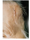
The provided image is an example of what pathology?

Giant Cell (Temporal) Arteritis
thickened, nodular segment of a vessel on the surface of the head
What vessels are most affected by Giant Cell Arteritis?
Clinical presentation?
Most commonly affected demographics?
Treatment?
has a propensity to affect the arteries of the head and neck, including the brain, but especially ocular arteries
aorta & its major branches but a predalection for carotid arteries, & superficial temoral arteries
- Presentation
- temporal artery will be prominent & thickened
- will typically be tender to palpation
- temporary loss of vision (Amaurosis fugax )
- Flu-like symptoms: fever, malaise, jaw pain
- Demographics
- Most common form of vasculitis seen in patients over 50 in the United States
- 2x as common in women
- Most common in white caucasians
- Treatment: immunosuppression
What is depicted in the provided image?

a normal large muscular artery (aorta)
What pathology is shown in the provided image?

- Left image (H&E)
- granulomatous inflammation – lymphocytes & giant cells
- disruption internal elastic lamina
- Right Image (elastic stain)
- notice destruciton of internal elastic lamina at the black arrow

What vessels are most commonly affected by Takayasu Arteritis
What is the clinical presentation?
Whta demographic is most commonly affected?
Treatment?
- Large artery arteritis
- aortic arch & large vessels coming off of it
- Demographics
- almost exclusively affects women
- younger women, usually 25 (under 50)
- Southeast Asia, Northern African, Mexican
- Presentation
- can cause severe occlusion of large arteries
- cause pulseless disease
- flu-like symptoms: fever, malaise, weight loss etc.
- upper body blood pressure will be lower than that of the lower body (large different)
- can have high disability rates b/c strokes, renal failure, etc.
- Treatment
- steroids
- immunosuppression
The provided angiogram shows an example of what pathology?

Tokayasu Arteritis
marked narrowing of brachiocephalic, carotid and subclavian arteries
The provided cross sections of the right carotid artery indicated what pathology?

Tokayasu Arteritis
- Cross section
- lumen drastically is narrowed
- marked intimal thickening
- Microscopic
- giant cells
- granulomatous inflammation
- destruction & firbrosis of arterial media
What pathology is characterized by the following photos?

Kawasaki Disease
most often presents after a viral illness
Rash, conjunctival and oral erythema, and blistering
What type of arteries are most commonly affected by Kawasaki Disease?
What is the major concern with this disease?
Clinical presentation?
What is the most commonly affected demographic?
- Medium-sized vessels (coronary artery)
- Can cause MI, sudden death, and coronary aneurysms
- Clinical presentation
- fever for at least 5 days
- non-purulent conjunctivitis
- oral & palmar erythema and blistering
- enlarged cervical lymph nodes
- Demographic: children (under 4)
- most common cause of acquired heart disease in children in the US


