Hypertensive & Atherosclerotic CV Disease Flashcards

1
Q

What are the functions of the normal vascular endothelium in the resting/basal state?

A
  1. maintain a permeability barrier
  2. provide a non-thrombogenic surface
  3. elaboration of mediators for hemostasis
  4. produce extracellular matriz
  5. modulate medial smooth muscle tone
  6. regulate inflammtion, immunity
  7. regulate cell growth
  8. metabolize some hormones (angiotensin)
  9. oxidize LDL
2
Q

What happens when you have some kind or injury/stimulus to the endothelium?

A
  • Activated State
    • increased expression of procoagulants, adhesion molecules & proinflammatory factors
    • altered expression of chemokines, cytokines, and growth factors
3
Q

What situations can lead to injury/stimulate the endothelial cells to the activated state?

A
  • turbulent flow
  • hypertension
  • cytokines
  • complememt
  • bacterial products
  • lipid products
  • advanced glycation end-products
  • hypoxia, acidosis
  • viruses
  • cigarette smoke
4
Q

What is the general response of the vascular wall to injury?

A

intimal thickening

recruitment of smooth muscles cells into the intima where they will proliferate (SM in the media will not be proliferating)

also end up recruiting macrophages & inflammatory cells into the intima as well

leads to deposition of lipids & elaboration of extracellular matrix

5
Q

What blood pressure levels are considered:

normal

elevated

stage I

stage II

hypertensive crisis

A
  • normal
    • less than 120/80 mmHg
  • elevated
    • systolic between 120-129 mmHg
    • diastolic less than 80 mmHg
  • stage I
    • systolic between 130-139 mmHg
    • diastolic between 80-19 mmHg
  • stage II
    • systolic at least 140 mmHg OR
    • diastolic at least 90 mmHg
  • hypertensive crisis
    • systolic over 180 mmHg and/or diastolic over 120 mmHg
6
Q

What is the equation for blood pressure?

A

BP = CO x PVR

7
Q

What factors directly influence cardiac output?

A
  • Blood volume
    • sodium
    • mineralcorticoids
    • atrial natruietic peptide
  • Cardiac Factors
    • heart rate
    • contractility
8
Q

What facrots directly impact peripheral resistance?

A
  • Humoral factors
    • constrictors
      • angiotensin II
      • catecholamines
      • thromboxanes
      • leukotrienes
      • endothelin
    • dilators
      • prostaglandins
      • kinins
      • NO
  • Neural factors
    • constriction
      • alpha-adrenergic
    • dilators
      • beta-adrenergic
  • Local factors
    • autoregulation pH
    • hypoxia
9
Q

Check this out! Will be helpful to understand the pathologies

A
10
Q

What are the two major types of hypertension? Which is most common?

A
  • Essential hypertension (90-95%)
  • Secondary hypertension
    • renal
    • endocrine
    • cardiovascular
    • neurologic
11
Q

What factors are known to play a role in essential hypertension?

Have any specific factors been identified?

A
  • Factors known to play a role
    • genetic factors
    • reduced renal sodium in the setting of normal arterial pressure
    • vasoconstrictive influences
      • increased peripheral resistance
    • environental factors
      • stress
      • obesity
      • smoking
      • high salt intake
      • inactivity
  • No specific triggers are known
12
Q

What hypertensive vascular diseases affect large to medium arteries?

Arteries larger than arterioles?

Small arteries / arterioles?

A
  • Large to medium arteries
    • degernative vascular wall changes
    • accelerate atherogenesis
  • Arteries larger than arterioles
    • fibromuscular intimal hyperplasia
  • Small arteries / arterioles (arteriolosclerosis)
    • hyaline arteriolosclerosis
13
Q

What pathology is shown in the provided image?

Describe the features of the vessel & in what conditions this pathology occurs.

A

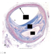
Hyaline arteriolosclerosis

Thickened arteriolar wall with increased plasma protein deposition and narrowed luman

very homogenously pink

Congo red stain will be negative

occurs in chronic hypertension & diabetes mellitus

14
Q

What pathology is shown in the provided image?

Describe the features of the vessel & in what conditions this pathology occurs.

A

Hyperplastic arteriolosclerosis

“onion-skinning” causes luminal obliteration. Laminations are composed of smooth muscle cells with reduplicated, thickened baement membranes

occurs in severe (malignant hypertension) – usually have a history of chronic hypertension

15
Q

What pathology is shown in the provided image?

A

Monckeberg Medial Calcific Sclerosis

notice calcium deposits centered on internal elastic lamina in the muscularis (dark purple at top of image)

calcium deposits: agranular, amorphous, plate-like deposition

very rarely encroacing on the lumen

16
Q

What is Monckeberg Medial Calcific Sclerosis?

It is most common in what demographics?

A
  • calcium deposits centered on internal elastic lamina in the muscularis of medium-sized muscular arteries
    • not usually severe enough to significantly narrow the lumen
  • Demographics
    • post-menopausal women
    • can sometimes show up on mammograms but ahve to be fairly numerous
17
Q

Describe the the process of atherosclerosis

A
  • Smooth muscle cells become activated & migrate into the intima from the muscularis
    • precursors to the smooth muscles can also be recruited to enter the intima when they will differentiate into smooth muscle cells & proliferate
    • deposition of extracellular matrix in the intima
    • deposition of extracellular lipids in the intima
    • may have some necrotic & inflammatory cells – get recruitment of inflammatory cells
      • macrophages (envibing lipids & will have foamy appearance) & lymphocytes
18
Q

What conditions can lead to atherosclerosis?

How long can this take to develop?

A
  • Conditions
    • hyperlipidemia
    • hypertension
    • smoking
    • toxins
    • hemodynamic factors
    • immune reactions
    • viruses
  • years to decades to develop
19
Q

What pathology is shownin the provided image?

A

Fatty streak

one of the first signs of atherosclerosis

20
Q

What pathology is show in the provided image?

A

Intimal Foam Cells

early intimal thickening, don’t really see muscle cells – very subtle

Atherosclerosis

21
Q

What pathology is shown in the provided image?

A

Mild atherosclerosis

Aorta with fibrous plaque (arrow)

Fatty yellow apearance, instead of streaks, you get plaques – can see some early streaks as well

22
Q

What pathology is shown in the provided image?

A

Severe atherosclerosis:

Aorta with plaque rupture and thrombosis

b/c the aorta is such a large vessel, thrombus will not frequently occlude the vessel

“grungy” appearance rather than fatty yellow appearance

however, that thrombus product can break off & go to other organs

23
Q

Describe the structure & components of an atherosclerotic plaque

A
  • Plaque
    • yellow area is thickened intima above the media
      • lipid
      • cholesterol crystals (pointy shapes)
      • foam cells (macrophages)
      • necrotic debris
      • deposition of calcium
    • typically will have a fibrous cap
      • collagen (blue strands)
      • smooth muscles cells
      • macrophages
24
Q

What pathology is seen in the provided image?

identify the specific parts.

A

Atherosclerotic plaque

L indicates lumen

arrow = normal vascular wall

F= fibrous cap

C = central lipid core

this fibrous cap is very well developed (lots of collagen, lots of blue), so it is less likely to rupture

25
Q

What is the potential sequelae that can result from acceleration of athersclerosis due to hypertension?

A
  • ischemic heart disease and/or abdominal aortic aneurysm
  • multi-infarct dementia
    • anything that can lead to rupture of the plaque & thrombosis
    • dementia from multiple small infarcts in the brain
  • aortic dissection
  • renal failure
    • because glomerular sclerosis
  • stroke (hemorrhagic or ischemic)
    • variety of sizes of different aneurysms in cerebral arteries that can rupture
  • cardiac hypertrophy and heart failure (hypertensive heart disease)
    • typically conentric hypertrophy in the left ventricle
    • left ventricular failure can lead to right heart failure
26
Q

What are the components of high pressure cardiovasculature system?

What are the components of the low pressure cardiovascular system?

A
  • High pressure
    • arteriole system
    • systemic vasculature
  • Low pressure
    • Venous system
    • pulmonary vasculature & arterie
27
Q

What percent of adults in the US are affected by left-sided (systemic) hypertensive heart disease?

What is the criteria for the diagnosis?

A
  • 30% wen definiton was 140/90
  • Criteria
    • left ventricular hypertrophy
      • can me mild to marked concentric hypertrophy
      • very orderly
    • history or morphologic evidence of hypertension
28
Q

What pathology is shown in the provided image?

What is one of the majore complications with this type of pathology?

A
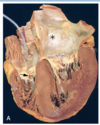
  • Left ventricular hypertrophy
    • thickness of left ventricular wall > 1.5 cm
  • Cardiomegaly
    • heart weight > 450 g, often > 500 g
  • Something will eventually push the heart over the edge where it is no longer able to compensate by thickening the left ventricle
    • this can lead to heart failure
    • wall may become more thin & ventricle may become dilated
    • atrium may become dilated
    • will often have right heart failure due to back up in the pulmonary circulation
      • causes pulmonary hypertension
29
Q

What features differentiate left ventricular hypertrophy from hypertrophic cardiomyopathy?

A
  • Septum is not thicker than outer left ventricular wall
    • more uniform
  • Ventricle is not narrowed
    • nice V shaped cavity rather than banana-shaped in hypertrophic cardiomyopathy
  • Cardiomegaly
30
Q

What pathology is shown in the right image as compared the left?

A

Since myocytes cannot multiply, they undergo hypertrophy

  • Left
    • normal myocardium
  • Right
    • not a higher power view
    • hypertrophied myocardium
    • increase in both cell and nuclear size
31
Q

What the consequence of left-sided hypertensive heart disease?

A
  • may be asymptomatic
  • ischemic heart disease
    • the thicker the ventricle, the harder it is for it to relax & provide blood to the myocardium
  • renal damage
  • cerebrovascular disease
  • progressive heart failure
  • sudden cardiac death due to arrhythmia
    • when have worsening dilation of the atria
  • development of aneurysms, especially aortic
    • & increased risk of aortic dissection
32
Q

Pulmonary (right-sided) hypertensive heart disease is characterized by what features?

What is often the general cause?

A
  • Characterized
    • right ventricular hypertophy
    • ventricular dilation
    • potentially right-sided heart failure
  • Isolated pulmonary hypertenzive heart disease arises from right ventricular pressure overload (lung disease)
33
Q

What are the most common potential causes of right-sided (pulmonary) hypertensive heart disease?

A
  • Disorders of the lung parenchyma
    • most common cause in absence of left-sided heart disease
  • diseases of the pulmonary vessels
  • disorders affecting chest movement
    • ie. severe kyphosis
  • disorders inducing pulmonary arterial constriction
  • left-sided heart disease
    • most common cause overall
34
Q

What pathology is shown in the provided image?

What characteristic features of this pathology are visible in the image?

A

Right-sided (pulmonary) hypertensive heart disease

  • Right ventricular hypertrophy
    • > 0.5 cm thickness
  • right ventricular dilation
    • atrium look dilated as well
  • loss of fat in the wall
  • circumferential orientation of myocytes
    • orderly hypertrophy
35
Q

Which image is an example of systemic hypertensive heart disease & which is an example of pulmonary hypertensive heart disease?

A
36
Q

What is the potential sequelae of pulmonary hypertenzive heart disease?

A
  • tricuspid valve incompetence
  • right-sided heart failure
    • jugulovenous distention
    • anasarca (extreme generalized edema)
    • dyspnea
    • fatigue
    • weakness
    • fainting
    • palpitations and arrhythmias
37
Q

What is the difference between true aneurysms & false aneurysms & dissection?

A
  • Aneurysm
    • widening of the vessel wall
  • False aneurysm
    • extravasation of blood into the blood vessel wall that is contained
    • hematoma that can look from the outside like a hematoma
    • lumen is not dilated
  • Dissection
    • when there is a tear in the intima so blood is able to enter it leading to dissection of the layers of the
      • severe cases can cause closure of the lumen
38
Q

What pathology is shown in the provided image?

A

Abdominal aortic aneurysm

39
Q

What is the most common location for an abdominal aortic aneurysm?

A

just above the bifurcation of the aorta, below the renal arteries

40
Q

What pathology is shown in the provided image?

Identify specfic features

A

Abdominal Aortic Aneurysm

layer of fibrin

atherosclerosis

41
Q

What are risk factors for developing an aortic aneurysm?

A
  • Atherosclerosis
  • Hypertension
  • Trauma
  • Vasculitis
  • Inherited defects
  • Infections
  • Imbalance between synthesis / degeneration of extracellular matrix proteins
42
Q

What is the risk of rupture for aneurysms of the following sizes?

< 4 cm

4 - 4.9 cm

5.0 - 5.9 cm

> 6.0 cm

A
  • < 4 cm : about 0 risk
  • 4 - 4.9 cm : about 1% / year
  • 5.0 - 5.9 cm : about 11% / year
  • > 6.0 cm : about 25% / year
43
Q

Aortic aneurysms can lead to what types of problems?

A
  • obstruction
  • embolism
  • impingement
    • renal arteries or other surrounding structures
44
Q

What pathology is shown in the provided image?

What is the most common cause?

A

Thoracic Aortic aneurysm

Most common cause is chronic hypertension & syphilis

45
Q

What pathology is shown in the provided image?

A

Aortic Dissection

layers of the wall get splayed apart typically by some type of damage or Marfan’s syndrome

double barrel appearance

46
Q

Aortic dissection occures most commonly in what two groups?

Additional risk factors?

A
  1. Middle-aged and elderly
    • most often in setting of hypertension (90% of cases)
  2. Younger patients with generalized or focal abnormalities of connective tissue
    • Marfan or Turner syndrome
  3. Other risk factors
    • atherosclerosis
    • pre-existing aortic aneurysm
    • aortic valve defect
    • coarctation of the aorta
    • iatrogenic
47
Q

What pathology is shown in the provided image?

A
48
Q

Marfan’s syndrom is caused by mutations to what protein?

What are the clinical features of this syndrome?

A
  • Cause
    • mutation to fibrillin gene
    • defects in collagen synthesis
  • Clinical
    • tallness
    • long arms & legs
    • loose-jointed
    • indented or protruding chest bone
    • scoliosis
    • flat feet
    • nearsightedness
    • dislocated lens
    • stretch marks on the skin
49
Q

What pathology is shown by the provided image compared to the normal adjacent image?

A
  • Top image
    • cross section or aortic medial from patient wiht Marfan syndrome
    • asterisks mark the “cystic spaces” of elastic fragmentation (elastic trichrome stain)
  • Bottom image
    • normal aortic medial for comparison
    • regular layed pattern of elastic tissue
50
Q

What are signs & symptoms of aortic dissection?

A
  • Sudden onset of excruciating pain
    • usually beginning in anterior chest
    • radiating to back between scapulae
    • moving downward as dissection progresses