Pediatrics Flashcards

1
Q

What are associated anomalies with this deformity?

A

Klipell Fiel

congential scoliosis

UE abnormalities

diastomeomyelia

renal disease

2
Q

What is the classification of spregnel?

A
3
Q

What are the procedures for spregnel

A

Done for stage 3/4 to improve cosmesis and abduction

Between ages 3-8, >8 is not a good candidate

Woodward with clavicular osteotomy is the preferred procedure

Procedure is done prone, tunnel subfacia anterior to do the clavicle

4
Q

What are risk factors for brachial plexus palsy

A

large babies
difficult presentation
shoulder dystocia
forceps delivery
breech position
prolonged labor

5
Q

What are prognostic factors for brachial plexus palsy?

A

90% will recover without intervention - can occur for up to 2 years

70% of those affected will develop glenoid retroversion

Good - biecps twitch by two months

Bad

lack of biceps function
preganglionic injuries; Horner’s syndrome, loss of rhomboid function

avulsion injuries (worst prognosis)

6
Q

What is Erb’s palsy?

A

Mechanism

results from excessive displacement of head to opposite side and depression of shoulder on same side producing traction on plexus

occurs during difficult delivery in infants or fall onto shoulder in adults

Physical exam

clinically, arm will be adducted, internally rotated, at shoulder; pronated, extended at elbow (“waiter’s tip”)
C5 deficiency

axilllary nerve - weakness in deltoid, teres minor
suprascapular nerve - weakness in supraspinatus, infraspinatus
musculocutaneous nerve - weakness to biceps

C6 deficiency

radial nerve - weakness in brachioradialis, supinator

Prognosis - best prognosis

7
Q

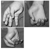
What is klumpke’s palsy?

A

Mechanism

rare in obstetric palsy
usually avulsion injuries caused by excessive abduction (person falling from height clutching on object to save himself)
other causes may include cervical rib, or lung mets in lower deep cervical lymph nodes

Physical exam

deficit of all of the small muscles of the hand (ulnar and median nerves)
clinically, presents as “claw hand”

wrist held in extreme extension because of the unopposed wrist extensors
hyperextension of MCP due to loss of hand intrinsics
flexion of IP joints due to loss of hand intrinsics

Prognosis

poorer
frequently associated with a preganglion injury and Horner’s Syndrome

Ptosis, myosis, anhydrosis

8
Q

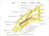
Draw the brachial plexus

A
9
Q

What are indications for microsurgery associated with bracial plexus palsy

A

complete flail arm or horner’s at one month

10
Q

Diagnosis? Incidence? Options for treament (general)

A

Brachial Plexus Palsy - No IR

  • At time of birth
    • PT for ROM and monitor
    • 90% will improve
  • Early micro surgery (graft or repair)
    • fail arm at one month
    • horner’s syndrome at one month
    • root avulsion (only grafting)
  • contracture release (any age)
    • pec, subscap z-lengthening, arthroscopic subscapular release
  • L’Episcopo lat dorsi/teres major transfer with pec major contracture release
    • requires a congruent joint
    • usually children < 4yo
  • Proximal humerus rotational osteotomy
    • incongruent joint
    • >5yo
11
Q

Deformity associated with madelung’s

A

Volar carpal subluxation

proximal radial synostosis

increased radial inclination

volar tilt

volar-ulnar tethering of vickers ligament

12
Q

What is vickers ligament

A

thickening of radiolunate ligament - madelungs

13
Q

What is leri-wiell

A

rare genetic disorder caused by mutation in the SHOX gene
SHOX stands for short-statute homeobox-containing gene
anatomically at the tip of the sex chromosome - pseudo-sex linked disorder
causes mesomelic dwarfism (short stature)
Normal intellegene
associated Madelung’s defomity of the forearm

14
Q

What is the treatment for this deformity

A

watch if they are young, deformity is small; assess with serial XR

Consider OR for pain or functional deformity

Physiolysis or Vickers release - open physis with progression

Radial ostotomy, vickers release, +/- ulnar ostoetomy, +/- ulnar epiphyseodesis

15
Q

What is the evidence for screening DDH

A

No evidence to screen

Imaging prior to 6 months in infants with clinical evidance, fam hx, breech,

16
Q

What factors are associated with increased incidence of LCP?

A

positive family history
low birth weight
abnormal birth presentation
children exposed to second hand smoke
Asian, Eskimo and Central European decent

17
Q

What are the risk factors of LCP?

A

family history of disease in 1.6-20% of cases
has been found to be associated with ADHD in 33% of cases
bone age is delayed in 89% of patients

18
Q

What factors are associated with poor prognosis in this patient?

A

bone > 6 years
female sex
decreased hip range of motion

Lateral pillar, head sphericity

Most patients will do ok until 6th decade

19
Q

What are the caterall risk factors associated with LCP?

A

Gage sign - V-shaped radiolucency in the lateral portion of the epiphysis and/or adjacent metaphysis

calcification lateral to the epiphysis
lateral subluxation of the femoral head
horizontal proximal femoral physis
metaphyseal cyst

20
Q

What are the lateral pillar and stulberg classfications

A

Lateral pillar (fragmentation phase)

A- none

B - >50% no height loss

B/C - >50% height loss

C - < 50% lateral pillar

21
Q

What are treatment options for LCP?

A

Goal : low stulberg classficiation

< 8 = nothing, casting is controversial

> 8, lateral pillar B and B/C in fragmentation phase = femoral osteotomy or pelvic ostetomy containment procedure

C - later treatment

22
Q

Outcomes you should know for LCP

A

no positive effect has been found for containment surgery performed after initial or early fragmentation stage

children with lateral pillar A and those with B under 8 years did well regardless of treatment

large recent studies show improved outcomes with surgery for lateral pillar B and B/C in children > 8 years (bone age >6 years)

poor outcome for lateral pillar C regardless of treatment

23
Q

Risk factors for developing this pathology

A

males (male to female ratio is 3:2)
African Americans
Pacific islanders
obese children - single greatest risk factor

during period of rapid growth
bilateral in 20% (20-40% May go on to develop bilateral)

24
Q

When would you consider endocrine WU for SCFE and what are you looking for?

A

child is < 10 years old
weight is < 50th percentile

  • *hypothyroidism** (labs show elevated TSH)
  • *renal osteodystrophy** (abnormal BUN and creatinine)
  • *growth hormone treatment**
25
What is the risk of AVN associated with the loder classification?
stable - AVN = 10% able to bear weight with or without crutches unstable - AVN = 47% unable to ambulate (not even with crutches)
26
Chondrolysis after SCFE is associated with
unrecognized implant penetration of the articular surface spica cast immobilization Advanced SCFE Intertrochanteric osteotomy
27
AVN following SCFE is associated with
unstable slip (47%) hardware placement in posteriosuperior femoral neck Increased number of pins Cuneiform osteotomy
28
What are options for late deformity in SCFE
arthroscopic vs mini anterior for osteochondroplasty surgical dislocation for more severe deformity Southwick osteotomy
29
Differential? Indications for surgery?
* **Differential for aquired coxa vara** * AVN: (DDH or Perthes) * Fibrous Dysplasia * SCFE * Rickets * Osteomyelitis of Hip and Septic Arthritis * traumatic * Paget's Disease * **Imaging/Indications for valgus osteotomy** * Hilgenreiners angle _\< 45 - nonop_ * _Hilgenreiners angle \> 60_ * _neck shaft angle \< 110_ * **Considerations with osteotomy** * ​might need to tension abductors * may have LLD
30
thought to be a cause of sacral agenesis
maternal diabetes
31
associations with sacral agenesis
gastrointestinal disorders (imperforate anus) genitourinary disorders cardiovascular disorders lower extremity deformities progressive kyphosis
32
what are the clinic findings associated with sacral agenesis
prominent lower lumbar spine extended knees, flexed hips, and equinovarus feet. classic sign of buttock dimping
33
What are leg abnormalities associated with PFFD
**orthopaedic manifestations** fibular hemimelia (50%) ACL deficiency PCL deficiency Hypoplastic lateral condyle = genu valgum coxa vara knee contractures Hypoplastic or dislocated patella **nonorthopaedic manifestations** dysmorphic facies found in rare autosomal dominant type
34
What is the Aitken classficiation for PFFD
35
What are treatment options for PFFD
* **limb lengthening with or without contralateral epiphysiodesis** * _indications_ * predicated limb length discrepancy of \<20 cm at maturity * stable hip and functional foot * femoral length \>50% of opposite side * femoral head present (Aiken classifications A & B) * _contraindications_ * unaddressed varus, proximal pseudoarthrosis, or acetabular dysplasia * can do a containment procedure in A/B * **knee arthrodesis with foot ablation** * _indications_ * ipsilateral foot is at the level of contralateral knee or more proximal * prosthetic knee will not be below the level of the contralateral knee at maturity * need for improved prosthetic fit, function, and appearance * **femoral-pelvic fusion** * _indications_ * femoral head absent (Aiken classifications C & D) * _Fusion_ * fuse the femur to the acetabulum with the femur at 90 degrees * knee flexion and extension becomes hip motion * _Brown's procedure_ * _​_180 degree rotation so the knee becomes the hip and the ankle becomes the knee * function as a BKA * **Van Ness rotationplasty** * _indications_ * ipsilateral foot at level of contralateral knee * ankle with \>60% of motion * absent femoral head (Aiken classifications C & D) * _surgical technique_ * 180 degree rotational turn through the femur * ankle dorsiflexion becomes knee flexion * allows the use of a below-knee prosthesis to improve gait and efficiency * **amputation** * _indications_ * femoral length \<50% of opposite side * _surgical technique_ * preserve as much length as possible * amputate through the joint, if possible, in order to avoid overgrowth which can lead to difficult prosthesis fitting * fit for prosthesis at 6 months for UE and 1 yr for LE
36
What are the goals of pediatric amputation?
(1) Preserve length. (2) Preserve important growth plates. (3) Per- form disarticulation, rather than transosseous amputation, whenev- er possible. (4) Preserve the knee joint whenever possible. (5) Sta- bilize and normalize the proximal portion of the limb. (6) Be pre- pared to deal with other issues in addition to limb deficiency.
37
What is commonly associated with limb hemihypertrophy and what exam do you need to perform?
Wilm's tumour serial US until age 5
38
What are the growth rates of the lower limb?
proximal femur - 3 mm / yr (1/8 in) distal femur - 9 mm / yr (3/8 in) proximal tibia - 6 mm / yr (1/4 in) distal tibia - 5 mm / yr (3/16 in)
39
What are four ways to assess projected limb length
**Green-Anderson tables** uses extremity length for a given age **Moseley straight line graph** improves on Green-Anderson method by reformatting data in a graph form accounts for differences between skeletal and chronologic age minimizes error averages serial measurements **Multiplier method** prediction based on multiplying the current discrepancy by a sex and age specific factor most accurate for congenital LLD **1/2 of final leg length** girls at age 3 boys at age 4
40
What is the most accurate way to predict LLD in congential differences
mutiplier
41
What are the options for LLD
**Shoe lift** \< 2 cm **shortening of long side - epiphysiodesis** 2-5 cm projected LLD **limb lengthening of short side** \> 5 cm projected LLD **lengthening often combined with a shortening procedure (epiphysiodesis, ostectomy) on long side** physeal bar excision indications bony bridge involves \<50% of physis at least 2 years left of growth
42
What are the principles of lengthening
**initiation** perform osteotomy at CORA and place fixator metaphyseal corticotomy to preserve medullary canal and blood supply (too low = risk of nonunion) **distraction** wait 5-7 days then begin distraction distract ~ 1 mm/day following distraction keep fixator on for as many days as you lengthened **concurrent procedures** some ream over a nail so ex-fix can be removed sooner But can put them at risk of infection lengthening often combined with a shortening procedure (epiphysiodesis, ostectomy) on long side **Complications** Incomplete arrest/ angular deformity Pin site infections Fracture Delayed union Premature cessation of lengthening Joint subluxation/dislocation
43
What are the indications for genetic testing or syndromal work-up in PFFD
usually occurs in isolation Do work-up if other associated deficiencies
44
What is the most accurate way to measure LLD
CT scanogram - more accurate in the setting of contractures
45
What are the non-operative treatment options for LLD
\<2cm no treatment 2-5 - shoe lift \>20cm requires extension prosthesis
46
What are the structural components of congential knee dilocation?
quadricep tendon contracture (short, fibrosis) anterior subluxation of hamstring tendon absent suprapatellar pouch tight collateral ligament patella laterally dislocated
47
What syndromes and conditions often occur with congenital knee dislocation?
**Syndromes** myelomeningocele arthrogryposis Larsen's syndrome **Conditions** DDH clubfoot Metatarsus adductus congential vertical talus
48
What is the treatment of congential knee dislocation?
Close reduction, long leg cast - usually in patients with subluxation and most will respond to serial casting to correct hyperextension Takes presedence over DDH and foot deformity
49
What are the operative indications for congential knee dislocation?
usually severe dislocation with associated neuromuscular condition **indications** if failure to gain 30 degrees of flexion after 3 months of casting Or if the knee is not reducible with closed reduction **goal of surgery is to obtain 90 degrees of flexion with** quadriceps tendon lengthening (V-Y quadricepsplasty or Z lengthening) - always anterior joint capsule release hamstring tendon posterior transposition collateral ligaments mobilization **postoperative** cast in 45 to 60 degrees of flexion for 3 to 4 weeks
50
Where is a pediatric popliteal cyst located? How is it different from an adult and what is the treament
semimembranosus medial head of gastrocnemius Not associated with medial meniscal tear Usually resovles without surgery
51
What is the differential for genu valgum
**bilateral genu valgum** ``` physiologic renal osteodystrophy (renal rickets) ``` Because it usually presents when the child is skeletally in valgus, the weakness tends to accentuate this deformity skeletal dysplasia Morquio syndrome spondyloepiphyseal dysplasia chondroctodermal dysplasia **unilateral genu valgum** physeal injury from trauma, infection, or vascular insult proximal metaphyseal tibia fracture (Cozen's fracture) Will see evidence of physeal dysplasia or assymetric growth lines benign tumors fibrous dysplasia osteochondromas Ollier's disease
52
What are the deformities associated with blounts and what are the radiographic features?
genu varum flexion, Internal tibial torsion **Radiographic findings** varus focused at proximal tibia severe deformity asymmetric bowing progressing deformity sharp angular deformity lateral thrust during gait metaphyseal beaking
53
What are two pertinant radiographic measurements to help quantify blounts
**metaphyseal-diaphyseal angle (Drennan)** angle between line connecting metaphyseal beaks and a line perpendicular to the longitudinal axis of the tibia \>16 ° is considered abnormal and has a 95% chance of progression \<10 ° has a 95% chance of natural resolution of the bowing **tibiofemoral angle** angle between the longitudinal axis of the femur and tibia
54
What is your differential for genu varum
blounts osteogenesis imperfecta osteochondromas trauma various dysplasia **Bilateral** Rickets - osteopenia and widened physes Skeletal dysplasia
55
What are the options for treatment of blounts
**KAFO** indications Stages I and II in children \< 3 years (especially if unilateral) bracing must continue for approximately 2 years for resolution of bony changes Outcomes Improved if unilateral Poor results - bilateral, obese Improvement should occur with-in one year **Proximal valgus osteotomy or growth modulation** children \> 3 years risk of recurrence is significantly lessened if performed before 4 years of age stages V and VI failure of brace treatment \*\*\***Make sure you overcorrect by 10 deg if doing an osteotomy**
56
Risk factors for Blounts
A disease with medial physeal interuption, with progression to fibrous tissue and eventually physeal bar ## Footnote Obesity Hispanic/Black Vit D Early walking
57
What are you considerations in the treatment of infantile blounts?
Valgus/flexion/IR - proximal tibial osteotomy Progression - will often need further sx Growth moduation - lateral hemiephyseodesis LLD - unilateral disease Joint line - medial plateau elevation Compartment syndrome - anterior fasciotomy Bar resection - with PMMA or fat, for more severe disease
58
What are the radiographic findings of adolesenct blount
narrowing of the tibial epiphysis widening of the medial tibial growth plate occasional widening of the lateral distal femoral physis metaphyseal beaking less commonly seen with adolescent Blount's
59
How is adolesenct blount different from infantile?
Infantile - bilateral, severe, usually will progress (overcorrect osteotomy by 10-15 deg), \< 3 can be treated with KAFO Adolesenct - unilateral, no progression (do not overcorrect osteotomy), femoral valgus will not resolve,
60
Treatment options for adolescent blounts
* **lateral tibia and fibular epiphysiodesis** * indications * mild to moderate deformity with _growth remaining_ * outcomes * up to 25% may require formal osteotomy due to residual deformity * **proximal tibia/fibula osteotomy** * indications * more severe cases in the _skeletally mature_ * No overcorrection * There should be no further progression of the deformity * outcomes * multiplanar external fixation following osteotomy allows gradual angle and length correction and decreases risk on neurovascular structures * _Will usually appear valgus_ due to big thighs, patient should be warned that their alignment is normal * **distal femoral osteotomy or epiphysiodesis** * indications * for distal femoral _varus deformity of 8_ degrees or greater * Should be done at the same time at the tibia * Will not resolve as it will in infantile blounts
61
What is the diganosis of normal genu valgum
between 3-4 years - 20 degrees after age 7 valgus - 12 degrees after age 7 the intermalleolar distance should be \<8 cm
62
What are the indications for observation of genu valgum
genu valgum \<15 degrees in a child \<6 years of age (there is no role for bracing a pathological deformity because it will often progress, a physiological deformity will correct on it's own; only blount's is an acception to this)
63
What are your indications and options for surgery for genu valgum
* \<10 yo * \> 15-20° of valgus * \>10 yo * if line drawn from center of femoral head to center of ankle falls in lateral quadrant of tibial plateau * Options * Hemiepiphyseodesis/tethering - guided growth * osteotomy (no growth remaining) * TSF if need combined angular and lengthening \> 10% of bone
64
What is the epidemiology of pseudosubluxation of the spine
**demographics** seen in children less than 8 years **location** C2 on C3 is most common C3 on C4 is second most common **Pathophysiology** caused by the horizontal nature of the facet joints at younger ages facet joints become more vertical with age
65
What is a radiographic determinant of pseudosubluxation
**Swischucks line** spinolaminar line drawn from spinolaminar point on C1 to C3 spinolaminar point on C2 should be within 1.5 mm of spinolaminar line helpful to differentiate pseudosubluxation from true injury
66
What factors support pseudosubluxation
reduction of subluxation with neck extension spinolaminar line within 1.5mm of C2 no history or physical findings of significant trauma absence of anterior soft-tissue swelling
67
What are common causes of atlantoaxial rotatory displacement
``` **trauma retropharyngeal irritation (Grisel's disease)​** ``` **other less common causes include** Down's syndrome rheumatoid arthritis tumours congenital anomalies
68
What is the best way to diagnose antlantoaxial rotatory subluxation
## Footnote Dynamic CT gold standard - rotational alignment won't change
69
What is the treatment for atlantoaxial rotatory instability
**Nonoperative** soft collar, anti-inflammatory medications, exercise program indications subluxation present for \< 1 week (traumatic or Grisel's disease) many patients probably reduce spontaneously before seeking medical attention **head halter traction and bracing** indications subluxation persists \> 1 week technique small amount (5 lbs.) usually enough either in hospital or at home muscle relaxants and analgesics may be needed **halo traction and bracing** indications subluxation persists \> 1 mos. **posterior C1-C2 fusion** indications subluxation persists \> 3 mos (or late diagnosis) neurologic deficits present
70
What are the pros and cons for a medial approach for DDH? What are the intervals
* **Approach** * supine, flexed, abducted, ER * incision along adductor longus 3cm from the pubic tubercle * gracilis, adductor longus * adductor brevis, adductor magnus * protect the posterior division of the obturator nerve * Protect the medial circumflex artery, release the psoas * **Pros** * directly addresses block to reduction * can be used in patients under 12 months of age * less blood loss with less soft tissue dissection * less scar formation for future surgery * **Cons** * unable to perform a capsulorrhaphy * higher association of AVN due to disruption of the medial circumflex artery
71
What are the recent AAOS recommendations for DDH
No evdience for screening Should get imaging \< 6 months if family history, breech, clinical evidence No need for U/S , 6 weeks No need to re-examine a hip that was normal at initial imaging No support for bracing an unstable hip with normal radiological markers No evidence to show whether limited or delayed bracing is better No evidence which brace works better No evidence to monitor hip
72
How long should you treat a child in a pavlik?
Age at diagnosis plus 3 months
73
What are blocks to open reduction for DDH
**Extra-articular** iliopsoas contracture - constricts anterior capsule Adductor contracture Adductor tenotomy can improve abduction to maintain reduction capsular constriction - hourglass contracture **Intra-articular** inverted labrum Pulvinar hypertrophied ligamentum teres \*\*Neo-limbus forms around the false acetabulum, it is not recommended to resect this
74
What position do you put a hip spica on for DDH? What is your desired safe zone?
flexion of 100 deg. abduction of 45 deg neutral rotation 3 months 'human position' change cast at 6 weeks for hygiene
75
Physical exam and radiology for DDH
* **Physical** * Overall assessment, assess for torticollis, spina bifida, clubfoot, MA, other dislocations or contractures, LLD * LLD, thigh assymetry * barlow, ortelani (cannot use after 3 months) * ROM - limited abduction * trendeleburg, prominent GT, line from GT to umbelicus \< 45 * **Ultrasound** * \> 6weeks of age, \< 6 month * alpha angle \> 60 * \> 50% coverage * assess psoas, capusule, boney acetaulum * look for subluxation of the femoral head * **Radiography** * **​**Hilginreiners line, perkins line, shentons line * AI - \<25 deg over 6 months * development of teardrop is a good sign for reduction
76
How can you assess if the hip has been reduction for closed reduction in DDH
* Arthrogram * \<5mm dye pool on AP * psoas over the capsule will create the hourglass * Safe zone * \> 60 deg = increased risk of AVN * adductor and psoas release
77
What is the normal alignment of the knee?
Mechanical axis = 87 deg (knee is in 3 deg of valgus) LDFA (lateral distal femoral angle) = 84 deg (accounting for 6 deg of valgus of the femur) Medial proximal tibia angle = 87 deg (accounting for 3 deg of varus)
78
What are special considerations for syndromal kids or kids with poor mobility (MD, CP)
Try to do multi-level surgery at one sitting Trauma/fixation should allow them to WB immediately or they will never walk again
79
Options for tethering the physis in growth modulation
Screws across physis (? Reversisble) Pheimster bone block Drilling Staples - reversible 8 plates - reversible
80
What are important considerations for angular deformity or LLD
Age, amount of growth left Projected growth discrepancy Rotational profile, angular deformity Multilevel surgery should be done at one sitting (Especially in kids with syndromes and slow mobility because they won't be able to rehab several surgeries) Weight bearing (Consider that in some kids with poor mobility, you need to use a strong construct because they won't be able to rehab a long deficit from weight bearing) \*\*Can address angular deformity with modulated growth and then address LLD at maturity - this is an alternative to doing a TSF - Then decide based on LLD Especially with a valgus knee you need to consider the peroneal nerve
81
What are common complications when treating angular deformity
**peroneal nerve injury** (valgus knee) Prevention perform a peroneal nerve release prior to surgery gradually correct the deformity if there is a large defomity utilize a closing wedge technique * *Hardware failure** - staples * *Overcorrection** - monitor every 3 months
82
What are the diagnostic criteria for NF1
83
What are the genetics of NF1
* 50% are spontaneous * autosomal dominant (AD) * mutation in NF1 gene on chromosome 17q21 * codes for neurofibromin protein * negatively regulates Ras signaling pathway * neurofibromin deficiency leads to increased Ras activity * affects Ras-dependent MAPK activity which is essential for osteoclast function and survival * neurofibromatosis is the most common genetic disorder caused by a new mutation of a single gene
84
What are the associated deformities with NF1
**extremity deformities** congenital anterolateral bowing and pseudoarthrosis of tibia/ fibula and forearm hemihypertrophy **spine involvement** scoliosis & kyphosis (dystropic and nondystropic) atlantoaxial instability
85
What is your management plan for scoliosis in NF1
screen early non-dystrophic can become dystrophic MRI and CT to assess dystrophic featrures, soft tissue mass, neuroma that can make approach difficult nondystrophic treated as AIS
86
What are the classes of NF
**NF1 (von Recklinghaussen disease)** most common **NF2** associated with bilateral vestibular schwannomas not associated with scoliosis **Segmental NF** features of NF1 but involving a single body segment
87
Non-orthopedic manifestations of NF1 (other than ocular and neoplasias)
* **Developmental delay** - most common genetic disease to cause learning disorder * **Gross motor, speech, hearing** can all be affected * _Unidentified bright objects on MRI_ in brain with unknown clinical significance * Increased risk for * Stroke * Hypertension * Heart disease * vasculopathy
88
What are the common syndromes of dural ectasia
marfan ehuler danlos NF1 ank spond
89
What are the findings associated with dystropic spine in NF1
90
What is your approach to scolisosis in NF1
Screen early MRI and CT to rule out dystropic monitor every 6 months - can progress to dystrophic non-dystropic can be treated like AIS
91
What is your approach to a dystropic curve in NF1
**a dystrophic curve will progress more rapidly when there are \> 3 features** indications dystrophic scoliosis Monitor \< 20 every 6 months, then fuse \>40 Type 1 - no kyphosis Type 2 - kyphosis \>50 **Indications for anterior (combined anterior and posterior fusion is controversial)** \>90 deg curve Rapid progression \>50 deg kyphosis perform early in young children (\< 7 yrs) with dystrophic curves **3 weeks pre-op traction shown to help** **screen for cervical instability** **use pedical screws, better even with deformation due to poor bone quality** **complications** high rate of pseudoarthrosis with PSF alone (40%) pseudoarthrosis rate still high with ASF&PSF (10%) some recommend augmenting the PSF with repeat iliac crest bone grafting 6 months after the primary surgery
92
What is the most common complication when treated a dystrophic spine in NF1
psuedoarthrosis
93
What percentage of patients with NF1 have anterolateral bowing
50% with anterolateral bowing have NF1 10% of NF1 have anterolateral bowing
94
What is your differential for tibial bowing
**anteromedial** * hemimelia * loss of lateral rays of the foot **posteromedial** * abnormal intrauterine positioning * dorsiflexed foot pressed against anterior tibia * will develop leg length discrepancy * associated with calcaneovalgus deformity **anterolateral** * NF1 * CPT
95
What is the pathology associated with CPT and NF1
The pathology for NF1 and CPT are the same - increased fibrous tissue, decreased functional osteoclasts (RAS pathway) Deficiency of the periosteum to differentiate myofibroblasts and chondrocytes
96
What is your managment for CPT/NF
worse prognosis associated with age and presence of fracture **Bracing** - if no fracture **bone grafting with surgical fixation** in bowing with pseudoarthrosis or fracture - prognosis depends on correction of bowing and stabilization aggressive nonunion debridement, shortening and compression across nonunion IM nail vs ex-fix (no plate on pathological bone) \*osteotomize the fibula and fix with a k-wire as well plan to never take the fixation out **amputation with prosthesis fitting** indications three failed surgical attempts (or when patient asks for it) Syme's often superior to BKA due to atrophic and scarred calf muscle in these patients
97
What are the indcations for amputaiton in CPT/NF1
X3 attempts or when patient asks for it \>5cm LLD unacceptable deformity prolonged hospitalization
98
What are complications associated with CPT after fixation
refracture LLD
99
What are the important concepts when fixing CPT
**resection of pseudarthrosis to grossly normal bone** **correction of deformity bone grafting** With BMP-Rh2 (controversial) Iliac crest graft **stabilization of the remaining segments** IM nail vs ex fix (no plates) Stabilize the fibula also if \<8 yo or for distal fracutre can use a nail crossing the ankle threw the calc
100
What is your differential
CPT/NF1 Fibrous dysplasia OM Fibrosarcoma Fibular hemimelia
101
What is the treatment and expectation of posteromedial bowing of the tib
**Intrauterine positioning** **often associated with calcvalgus foot** **often get LLD later, need to follow and perform epiphyseodesis at appropriate time**
102
What MSK conditions can you get with tibial hemimelia
75% lobster-claw hand preaxial polydactyly ulnar aplasia
103
What is the most common long bone deficiency
fibular hemimelia (linked to sonic hedge hog gene)
104
What are conditions associated with fibular hemimelia
* anteromedial tibial bowing * most common cause is fibular hemimelia * ankle instability * secondary to a ball and socket ankle * equinovalgus foot deformity * tarsal coalition (50%) * absent lateral rays * femoral abnormalities (PFFD, coxa vara) * cruciate ligament deficiency * genu valgum * secondary to lateral femoral condyle hypoplasia * significant leg length discrepancy * shortening of femur and/or tibia
105
What are the key things to look for when assessing tibial hemimelia?
* **Look** * Shortening of the affected limb * ​​anterolateral bowing of the tibia * prominent fibula * **​Feel** * ​flexion contracture * ability to extend knee * stability of knee * **​Foot** * ​rigid equinovarus
106
What are you options for treatment in tibial hemimelia?
* **knee disarticulation followed by prosthetic fitting** * indications * complete absence of the tibia * no active knee extension present (most cases) * **tibiofibular synostosis with modified Syme amputation** * indications * proximal tibia present with intact extensor mechanism and minimal flexion contracture * **Syme/Boyd amputation** * indications * ankle diastasis
107
What is the Achterman and Birch classification system for CFD?
**congenital fibular deficiency** * **Acherman** * Ia - partial deficiency, short fibula * Ib - 30-50% absenct, does not support ankle * II - complete absence * **Birch** * **​**type 1 - functional foot (\>3 rays) * A - \<6% * nothing, orthosis * epiphyseodesis * B - 6-10% * lenghening * epiphyseodesis * C - 11-30% * may require more than one lengthening or orthosis * D - \>30% difference * usually requires amputation * Type 2- non-funcational foot (\<3rays) *
108
What is your work-up and algorythim for treatment of CFD?
* **Necessary Imaging** * **​**AP bilateral lower extremities * look for associated deformities * CT can help assess LLD * MRI if there is functional instability * Echo, renal U/S * **Invovle multi-disciplinary team** * PT, psych, prosthetist * have the families visit clinics to see both reconstructive and amputation options * emphasize amputation is not a failed option * **Principles** * Depends on the functionality of the foot and the LLD * keep in mind ACL def when treating LLD * no role for ACL reconstruction * Not advocated to do tibial osteotomy to correct deformity at the time of amputation * May need a distal femoral osteotomy later to correct valgus * **Lengthening** * contralateral epiphyseodesis * one at 5cm, one at 7cm * may need a final one * **Genu Valgum** * **​**\<15 deg can do epiphyseodesis alone * \>15 deg might need to do an osteotomy * need to correct for amputations as well to ensure proper prosthesis fitting * **Amputation** * Indications * \>30% LLD * \>5cm at birth LLD * functional UE * Boyd or Syme * Done when they start to walk
109
What are the complications associated with each treatment for CFD?
* **_Amputation outcomes_** * higher satisfaction * lower readmission * fewer complications * **Symes amputation** 1. Posterior migration of the heel pad 2. Reformation of the calcaneus (ie, painful stump) 3. Wound infection 4. Heel pad slough 5. Fitting adjustment problems * **Boyd amputation** (maintains more height) 1. Delayed calcaneotibial fusion or nonunion 2. Calcaneal migration and malalignment 3. Wound infection * **Limb lengthening** 1. Pin tract infections 2. Peroneal nerve injury Tibial/femoral fractures 3. Severe depression 4. Nonunion of the osteotomy site Joint stiffness 5. Joint subluxation 6. Persistent or recurrence of knee and foot deformities 7. Residual limb length inequality
110
What are the two most important factors when considering salvage vs amputation for CFD?
1. Functional foot 2. LLD \<30% at maturity
111
What is the difference in etiology for tib hemi and fib hemi?
Tibial hemimelia has a known genetic, AD cause where fibular hemimelia is sporatic
112
What is the normal progression of rotational profile in children?
**40 deg fem anteversion at birth, masked by femoral ER at times; internal tibial torsion** **tibia externally rotates and becomes normal by age 6** **femural anteversion resolves by age 10**
113
What are the 5 components of Staheli's rotational profile?
* **ER Hip/IR hip** * **​**\> 70 IR, \<20 ER = fem anteversion * **thigh-foot-axis (5 IR)** * **​**-10 = internal torsion * **transmalleolar axis (5 IR)** * **heel bisector angle** * **foot progression angle (-5-20)**
114
What are the common pathology by age for intoeing
**0-1 yo - MA** **1-3 yo - internal tibial torsion** **\>3 yo - femoral anteversion**
115
What is the Bleck classification for MA?
* *normal** - heel bisector line through 2nd and 3rd toe webspace * *mild** - heel bisector line through 3rd toe * *moderate** - heel bisector through 3rd and 4th toe webspace * *severe** - heel bisector through 4th and 5th toe webspace
116
What are the normal rotational profiles?
* **Hip Rotation** * **​**Infants - 30 IR, 70 ER * 10yo - 50 IR, 45 ER * at 10yo - IR \> 80 with ER \< 10 is indication for surgery * **Tibia** * ​Infant - IR, resolves by 4yo * 6yo - ER 10 deg * at 80yo - ER \>40 deg is indicatin for surgery
117
What is the berg classfication of MA?
Simple - MTA Complex - MTA, lateral shift of midfoot Skew foot - MTA, valgus hindfoot Complex skew foot (serpentine foot) - MTA, lateral shift, valgus hindfoot
118
What are red flags associated with in-toeing that would make you get further work-up?
pain limb length discrepancy progressive deformity family history positive for rickets/skeletal dysplasias/mucopolysaccharidoses limb rotational profiles 2 standard deviations outside of normal
119
What are the indications for treatment of MA?
**Flexible deformities** that can actively be corrected to midline - No treatment required **Flexible deformities** that _can passively be corrected to midline_ - Serial stretching by parents at home **Rigid deformity with medial crease** - Serial casting with the goal of obtaining a straight lateral border of foot * usually will improve in 6-8 weeks of casting * some will recurr by 2-4 yo
120
What are the indications for surgery and options for MA?
**Indications:** failed non-op severe deformity and difficulty with shoe-wear \> 5yo **Techniques:** \*Soft tissue procedures alone are no longer recommended due to soft tissue necrosis * *lateral column shortening -** done with cuboid closing wedge ostetomy * *medial column lengthening -** includes a cuneiform opening wedge osteotomy with medial capsular release and abductor hallucis longus recession (for atavistic first toe)
121
What is the prognosis of MA?
a benign condition that resolves spontaneously in 90% of cases by age 4 another 5% resolve in the early walking years (age 1-4 years)
122
What pregnancies are associated with MA?
late pregnancy first pregnancies twin pregnancies oligohydramnios
123
Key points of metatarsus primus varus
* **Adduction deformity of the great toe characterized by** * deformity at metatarsophalangeal joint * short thick 1st metatarsal * firm band-like abductor hallucis muscle * **Associated conditions** * _often associated with polydactyly_ – duplication of the great toe * Must be _differentiated from metatarsus adductus_ * Lateral border of the foot is normal * More rigid than MA * Early casting is used, associated with hallux valgus * Severe deformity is indication for osteotomy
124
What is the pathogenesis and treatment for skewfoot?
* Poorly understood why this develops * No good measurement to define skewfoot vs MA * Surgical treatment is only indicated if function is severely limited * **opening wedge and closing wedge osteotomies** * indications * indicated if serpentine deformity is symptomatic and significantly limits function * operative treatment is difficult and often times deformity is accepted and observed * **technique** * _calcaneal osteotomy_ for hindfoot valgus * possible _midfoot osteotomies_ to correct midfoot and forefoot deformities * multiple metatarsal osteotomies with forefoot pinning and _tarsometatarsal capsular release_ (Hamen procedure)
125
What is the indication for a derotational supramalleolar ostoeomty for internal tibial torsion?
\>6yo \>10 deg IR rarely causes issues will not get worse with growth
126
What is the prognosis and associated syndromes with external tibial torsion?
Will increase with age and is associated with malalignment and patellofemoral syndrome * **"miserable malalignment" syndrome** * a condition defined as external tibial torsion with femoral anteversion * **Osgood-Schlatter disease** * **osteochondritis dessicans** * **early degenerative joint disease** * an association between external tibial torsion and early degenerative joint disease has been found * **neuromuscular conditions** * may be found with neuromuscular conditions such as _myelodysplasia_ and _polio_
127
What are indications for surgical treatment of external tibial torsion
* **indications** * ​\>8yo * \>40deg (3 SD above the mean) * **​technique** * **​supramalleolar osteotomy** * ​cross pins for young children * IM nail for skeletally mature * **​Fibular osteotomy for more severe deformity**
128
What is surgery indicated for femoral anteversion?
* **indications** * _\< 10° of external rotation_ on exam in an older child _(\>8-10 yrs)_ * rarely needed - only if there is severe comprimised function - tripping, inefficient gait * Can lead to inefficient abductor function * **Derotational femoral osteotomy** * typically performed at the intertrochanteric level * consider correction of varus if present * amount correction needed can be calculated by _(IR-ER)/2_ * can be done prone or supine depending on what other procedures you will also be performing
129
Differential for cavus foot
* **CNS** * CP * Frederichs ataxia (often can be first diagnosis) * **Spinal Cord** * Myelodysplasia, syrinx/synringomyelia (unilateral), tethered cord (unilateral), tumors, GBS * Polio (more historical) * **Peripheral neuropathy** * CMT/HSMN - most common cause * Result of a number of mutations resulting in dymyelination * Most common is X-linked; Peripheral-myelin-protein 22 (PMP-22) * Aquired - more common in adults * Tibial fracture/compartment syndrome, sciatic nerve injection, peroneal neuropathy * Residual clubfoot
130
What are the key points in evaluation of a cavus foot
131
What imaging is required in the diagnosis of cavus foot
* **Plain radiographs** * ​AP - varus * Lateral * ​meary's angle = 0 * calcaneal pitch = 30 deg * **​​EMG** * ​concerns of CMT or HSMN * may also require genetic testing * **​Spine MRI** * ​unilateral * progressive deformity
132
What is the non-surgical treatment of cavus foot
* **Activity adjustments** * A runner with a cavus foot needs longer time to recover from injury and is more at risk of injury * **Accomodative orthotics** with high shoe box for claw toes * Good running shoes for runners * **PT** * Quads strengthening * Can help decrease the load the ankle sees when running * (2.5X body weight, alleviated by eccentric quads contracure) * **Baclofen, diazepam, Botulinum toxin** * For neuromuscular/CP
133
What are considerations for tendon transfer in cavus?
1. Transfered tendons lose one grade, need at least grade 4 2. should be in straight line of pull 3. try to adhere to bone rather than tendon 4. fixed deformites should be corrected first 5. iatrogenic problems can happen in neuromuscular (ie; CP tib ant transfer; will get a new contracture; better to do a split tendon transfer)
134
What are associations with clubfoot
hand anomalies diastrophic dwarfism arthrogryposis tibial hemimelia Myelomeningocele
135
What are the muscle contractures invovled with clubfoot
``` midfoot Cavus (tight intrinsics, FHL, FDL) forefoot Adductus (tight tibialis posterior) hindfoot Varus (tight tendoachilles, tibialis posterior) hindfoot Equinus (tight tendoachilles) ```
136
what are the boney deformites associated with clubfoot
* talar neck is medially and plantarly deviated * Some believe that this is the primary deformity as it is the most consistent * Talus grows medially toward the navicular * the calcaneus is in varus and rotated medially around talus * Deltoid, spring ligament and PTT keep calcaneous varus * Achilles keeps heel in equinus * the navicular and cuboid are displaced medially * Cuboid is morphologicall normal, just displaced * Navicular is tethered by the above as well as the knot of henry, capsule
137
What are the XR findings associated with clubfoot on the lateral
* talus and calcaneus are parallel; usually talocalcaneal angle is \> 35 deg * dorsiflexion view of turco * flat talar head * talocalcaneal angle \< 35 deg
138
What are the deformities on an AP associated with clubfoot?
``` talocalcaneal angle (Kite's) is \< 20° (normal is 20-40°) talus-first metatarsal angles is negative (normal is 0-20°) ```
139
Treatment
* **CLUBFOOT** * **Month 1-4 - PONSETI CASTING** * Weekly Serial Casting (with knee in 90° of flexion ) * _First correct cavus_ by aligning 1st MT with remaining metatarsals * This requires supination of the forefoot, it is pronated; usually take one week * Then correct _adduction and heel varus_ by rotating calcaneus and forefoot around talus (head of talus acts as fulcrum) * This needs to be done prior to equinus correction, otherwise the calcaneus will be locked under the talus * Usually takes 4 weeks; cast application while feeding can distract the infant * _Achilles tenotomy at week 8_ (required in 80%) * Then correct equinus with TAL (If you don't wait until the navicular is reduced then you will get a rockerbottom foot) * Perform when _foot is 70° abducted_ and _heel is in valgus_ * Ponseti method is complete transverse cut * Can be done in clinic for a small child, or in the OR for a larger/older baby for better analgesia and better case moulding * No stiches need, just a light dressing, then the cast is applied * Cast in maximal dorsiflexion and abduction after TAL, overcorrection will prevent recurrence * This cast is left on 3-4 weeks, more in a larger child * **Month 4-8 - Foot abduction orthosis (FAO)** * full time until ~ 1 year of age * nights only from age 1 to ~4 * With Denis Browne bar in external rotation (_70° in clubfoot and 40° in normal foo_t) * Fit FAO on day of TAL * Normal to be irritable when casts come off, important to explain this to parents and emphasize risk of recurrence * **2-4 years - Anterior tibial tendon transfer** * at 2 yrs of age (10-20% will require) * 10-20% will need TA transfer with or without repeat TAL for recurrent supination, varus, and/or equinus * Occurs due to incomplete reduction of navicular, changes tib ant from a dorsiflexor to a supnator * **4-6 weeks repeat casting** * Keep tib ant under the retinaculum to prevent bowstring * +/- TAL
140
Treatment of failed clubfoot
* **Re-start casting** * **Indications for surgery** * resistant feet * rockerbottom from casting * **​Surgical options** * **​ST release and tendon transfers****​** * **Posteromedial release - plantar fascia, PTT lengthening** * _Deltoid ligament_, _spring ligament_, _knot of henry_ * _Abductor Hallucis_ is released to get access to these structures * Whether to release subtalar or ankle capsule depends on the surgeon * **Tibialis anterior transfer** * To medial cuneiform (only if ossified) * For neuromuscular kids should use a split transfer * **Repeat TAL** * Posterior talo-fib and tib-fib ligaments * Make sure to do your silverskiold * dosiflexion osteotomy * **Medial opening or lateral closing** * ​3-10yo * first ray or cuniform * **​Triple arthrodesis** * ​8-10 yo with refractory * **​Talectomy** * ​8-10 with insensate foot
141
What are the complications associated with nonoperative or operative clubfeet
* **Complications with nonoperative treatment** * _deformity relapse_ * relapse in child \< 2 years * noncompliance with FAO * Inappropraite technique, missed neurological or paralytic foot * treat with repeat casting * relapse in child \> 2 years * treat initially with casting * consider TA tendon transfer * consider repeat TAL * _dynamic supination_ * treat with whole anterior tibial tendon transfer * **Complications with Surgical Treatment** * _residual cavus_ * result of placement of navicular in dorsally subluxated position * _pes planus_ * results from overcorrection * _undercorrection_ * _intoeing gait_ * _osteonecrosis of talus_ * results from vascular insult to talus resulting in osteonecrosis and collapse * _dorsal bunion_ * caused by dorsiflexed first metatarsal and overactivity of anterior tibialis
142
What are the associations and muscle imbalance of equinovarus
* **Associated syndromes** * cerebral palsy (usually spastic hemiplegia) * Duchenne muscular dystrophy * residual clubfoot deformity * tibial deficiency (hemimelia) * **Muscle imbalance include spasticity of** * tibialis posterior _and/or_ tibialis anterior * gastroc-soleus complex * abscense of ligamentous laxity
143
What are two ways to assess with TA or TP is overactive in equinovarus
* **gait analysis** * diagnosis made with gait analysis showing * overactivity of tibialis posterior and/or * overactivity of tibialis anterior * **confusion test** * helps to distinguish AT vs PT as the primarily involved muscle * patient performs active hip flexion against resistance while seated * in a positive confusion test, the tibialis anterior fires with active hip flexion and is typical in patients with CP * if the foot supinates with dorsiflexion, tibialis anterior is most likely contributing to the equinovarus deformity
144
What are the surgical indications for equinovarus
* **TAL with tibialis posterior split transfer to peroneus brevis** * indications * spastic hemiplegia in patient ages 4 to 7 * flexible equinovarus hindfoot * tibialis posterior spastic in both stance and swing phase (continous activity) * technique * reroute half of tendon dorsally and insert into peroneus brevis * **Rancho procedure** * indications * overactive anterior tibialis * flexible equinovarus deformity * technique * split anterior tibialis transfer to cuboid with TAL and intramuscular lengthening of the posterior tibial tendon * **Calcaneal osteotomy** * indications * is required in a rigid hindfoot varus deformity * technique * lateral closing wedge osteotomy to incur valgus to the heel
145
What are the conditions and pathology of equinovalgus
* **Common foot deformity seen with** * spastic diplegic and quadriplegic cerebral palsy * fibular hemimelia * **Deformity consists of** * midfoot abduction * heel valgus * equinus contracture * typically bilateral * **Muscle imbalance includes** (see overview of foot deformity muscle imbalances) * spasticity of * peroneals * gastoc-soleus complex * weakness of * ligamentous laxity * posterior tibialis * anterior tibialis
146
What are the non-operative options for equinovalgus foot
* **bracing and physical therapy** * indications * _flexible deformities_ * technique * ankle foot orthosis or supramalleolar orthosis * **botox injections**
147
What are the surgical options for a rigid foot with equinovalgus
* **soft tissue procedures** * TAL * peroneus brevis lengthening * **calcaneal osteotomy** * medial slide ostetomy or calcaneal lengthening osteotomy * **lateral column lengthening procedure** * performed through calcaneus or cuboid * **Grice procedure** * extra-articular subtalar arthrodesis * place ICBG in lateral subtalar joint to block valgus * does not interfer with tarsal bone growth * **subtalar arthrodesis** * stabalizes subtalar joint in correct alignment without fusion * implant or spacer is placed laterally in the subtalar joint to prop open
148
What are the pros and cons of plaster and fiberglass
149
complications with casting
* Pressure sores * Compartment syndrome * Univalve plaster * 50% release * Then webril is 20% release * Bivalve fiberglass * Atrophy, cartilage degredation, ligament weakness * Don't immobilize for too long * Functional casting when appropraite * Patients with neuromuscular disease are at increased risk of ostetopenia and atrophy * Should immobilize them for less time * Cast saw burn
150
avoid complications with casting
* Complication prevention * Plaster application * Don't apply fiberglass over curing plaster * Don't put plaster on a pillow * Use recommended water temperature * fiberglass * Stretch and relax for fiberglass * Don't leave sharp fiberglass edges * General * Don't go past the MC heads, limits fingers * Don't end at the fibula = peroneal nerve * Use safe position for the hand * Hip spica * Don't put lower limb first and use to traction * Can cause compartment syndrome and popliteal occulsion * Cast application * Appropriate padding * Too much padding can cause loose cast, skin irritation and loss of reduction * Cast should look like and oval * _Optimal width:height = 0.7_ * Don't change position of the cast after applied * Weakens the cast * Puts pressure in the flexion crease
151
What imaging do you need for this patient and what are the imporant angles
* WB AP/lateral, oblique, harris (TC) * **Lateral** * **Meary's angle - 0 deg** * **Calcaneal pitch - 20 deg** * rule out tarsal coalition, verticle talus * assess where break in foot is * **AP** * **Ist MT-talus angle - 20deg** * rule out accessory navicular * **oblique** * **​**best image to see a Cal-nav coalition
152
What is the pes acetabulum?
Navicular, spring ligament, anterior facet calcaneus
153
What is your management of flexible flatfoot?
* **Non-op** * always do this first - counsel parents should resolve as child grows and will not affection function\*\* * supportive shoewear * rigid orthoses * Achilles stretching - if tolerated * **​Operative - only indicated with prolonged resistance to non-op and pain** * Address achilles as per silverskiold * Calcaneal lengthening osteotomy * +/- brevis lengthening (not longus, lengthing longus helps to pronate the foot and encourge arch formation) * If very severe can consider also a calcaneal sliding osteotomy * +/- medial cuneiform osteotomy to address the forefoot supnation ​
154
What are options for osteotomies of the flexible flatfoot
* **calcaneal lengthening osteotomy (Evans)** * with or without a cuneiform osteotomy and peroneal tendon lengthening * **Calcaneal lateral opening wedge osteotomy (Dwyer)** * **Sliding calcaneal osteotomy** * corrects the hindfoot valgus * **plantar base closing wedge osteotomy of the first cuneiform** * used to correct the supination deformity * Can be combined with an opening wedge osteotomy of the cuboid * **"Triple C"** * Sliding calcaneus * Opening cuboid * Closing cuneiform
155
What is your differential for this foot? What imaging do you need?
* Differntial * **Calcaneovalgus** * **posteromedial bowing** * **oblique talus** * **verticle talus** * **paralytic foot deformity** * ​L5 myelomeningocele * **​​imaging** * **​AP/lateral** * **​**CVT will have valgus midfoot and increased Talo-calc angle (\>40 deg) * CVT = meary's angle = 20 deg * **forced plantarflexion** * **​**the navicular will reduce in CV foot (MT will line up with talus) * **Dorsiflexion** * in CVT the talus will remain in equinus
156
What is the prognosis for calcaneovalgus foot?
* Will resovle on it's own * May develop LLD if associated with posteriomedial bowing
157
Painful flatfoot. Corrects with heel rise, suble subtalar joint; with this XR. What's your treatment?
* **Accessory Navicular** * **Convervative** * ​activity restriction, shoe wear modification, NSAIDS, short perior immobilization * _most will become asymptomatic_ at skeletal maturity * **​Resection of accessory navicular** * _​will not correct flatfoot_ * _MRI_ to assess PTT - if needs to be transfered beform _Kindler procedure_ (transfer PTT to inferior navicular) * otherwise straight incision, incise PTT longitudinal, resect with osteotome, then close PTT)
158
Considtions associated with this deformity
**CVT or "Persian slipper" -** 50% will have associated neuromusular conditions * myelodysplasia (common) * arthrogryposis * diastematomyelia * chromosomal abnormalities
159
What is an oblique talus
* similar to vertical talus but reduces with plantarflexion * usually improves with conservative treatment, very few will require CRPP for residual subluxation
160
What age does the navicular ossify?
3 yo
161
What is the treatment of CVT?
* **Deformity** * _fixed hindfoot equinovalgus_ * due to contracture of the Achilles and peroneal tendons * _rigid midfoot dorsiflexion_ * secondary to the dislocated navicular * _forefoot abducted and dorsiflexed_ * due to contractures of the EDL, EHL and tibialis anterior tendons​ * **Casting to stretch lateral tissues** - reverse of clubfoot * supnated forefoot, abducted midfoot, heel valgus * equinus is the same * **Closed or open reduction with pinning** * ​once the foot is stretched, the navicular is reduced to the talus, pinned and casted * **usually require TAL** * +/- tib ant lengthening (limited plantarflexion) * +/- tib ant transfer to talus (\>2yo) * Cast for 8 weeks, remove pin, into boots and bars * **​Talectomy for resistent cases**
162
What are the ages commonly associated with tarsal coalition?
* **demographics** * incidence varies from 1%-6% * most coalitions found are incidental and asymptomatic * **Genetics** * AD, with partial penetrance * **age of onset (when pain develops)** * Calcaneonavicular - 8-12 years old * Talocalcaneal - 12-15 years old
163
What is your differential? What are associated conditions?
* **Tarsal coalition** * **Associated conditions** * fibular deficiency * Apert syndrome * **Differential Diagnosis for Rigid flatfoot** * Verticle talus * Peroneal spastic flatfoot - secondary to another condition such as JRA * Trauma or infection
164
what imaging is necessary to assess for tarsal coalition?
* L**ateral** * ​C sign * anteater sign * **​Internal Oblique (45deg)** * ​best view for CN coalition * **Harris view** * ​TC coalition * **​Saltzman standing view** * ​hindfoot varus * **​CT** * ​best way to assess coalition * assess for other coalition * rule out bilateral * **​MRI only necessary if fibrous lesion**
165
What is your approach to treatment with a patient with rigid flatfoot and this XR
* bilateral CT to assess for other coalition and extent of coalition * **Trial conservative management** * **​**rest, orthotics * many will improve * **Associated Deformities** * _Lateral column lengthening_ (the group that says this - for **\> 16deg valgus**) * If the patient is old enough they will have a more developed flat foot * Some groups won't take down the coalition * _Achilles or gastro lengthening_ * Can do a silverskiold intra-op once you've taken down the coalition * **surgical resection of coalition** with interposition of _fat graft (TC)_ or _extensor digitorum brevis (CN)_ * indications * resistant cases when non-operative management fails to relieve symptoms * outcomes * 80% will experience pain relief * High rates of recurrence, but are lower with tendon interposition (and lower with fat graft than nothing) * _Predictors of poor outcome with resection_ * Coalition \> 50% of the posterior facet (not middle facet * Hindfoot valgus 16 deg * Narrowing of the posterior TC facet * **subtalar arthrodesis** * indications * role has not been well established * Used to be done more often, but now we have good results with surgical resection * **triple arthrodesis** (subtalar, calcaneocuboid, and talonavicular) * indications * advanced coalitions that fail resection
166
Technique to take down of talo-calcaneal tarsal coalition
* Poor prognosis * \>16 deg valgus * \>50% posterior facet * Supine with tourniquette * medial incision underneath FHL centered over the sustentaculum * identify the sheath of FHL and identify the NV structures * Incise the FDL sheath and retract it inferiorly * identify the anterior and posterior facets * use a burr to debride the posterior facet ensuring to not drift superiorly or inferiorly * assess the mobility of the subtalar joint * put bone wax on the bleeding bone and use fat interposition * can use FHL if you are doing a repeat debridement * Repair the FDL sheath with closure * Reasons for failure * \>16 deg valgus * \>50% posterior facet * incomplete resection * lack of interposition * lack of identification of other coalitions
167
Technique to debride a calcaneonavicular coalition
* Incision over the lateral side of the foot between the EDC and the peroneals * Fatty tissue and EDB are elevated to expose the anterior process of the calcaneus * Use small osteotomes to remove a trapezoidal peice of bone * make sure to remove the entire coalition * Fat interposition * taken from a 2cm transverse incision in the buttock crease * Release the tourniquette, get appropriate hemostasis and sew EDB over the interposition
168
What are indicators of poor outcome and complications with coalition resection?
* **Poor prognosis** * Coalition \> 50% of the posterior facet (not middle facet) * Hindfoot valgus 16 deg * Narrowing of the posterior TC facet * **Complications** * **​**incomplete resection * recurrence * wound complications * injury to adjacent cartilage
169
Diagnosis? Prognosis?
**Kholer's disease** * usually 4-7 yo * M\>F * centeral 1/3 is watershed zone prone to stress # and AVN * intermittent symptoms for 1-3 years, treat with NSAIDS, self-limiting * often have deformity but no clinical effect * Can treat with SLC when painful
170
Painful heel with activity. Diagnosis? Differential? Treatment?
**Sever's disease** * sclerosis can be normal as well, as seen below * overuse with running, jumping * often seen during peak growth velocity * **MRI** * _​stress fracture_ * _lytic lesion_ * _infection_ * **​Treatment****​** * activity modificaiton * Achilles tendon stretches (can help decrease recurrence) * ice application before and after athletic endeavors * use of heel cups or heel pads * NSAIDs * short leg cast immobolization of persistent pain
171
Active 10yo, pain lateral foot, improves with rest. Diagnosis?
**Iselin's** * traction apophysis base 5th MT * 8-13yo, girls and boys * self-limited
172
Diagnosis?
**Atavastic great toe (Congential Hallux valgus)** * **Adduction deformity** * deformity at metatarsophalangeal joint * short thick 1st metatarsal * firm band-like abductor hallucis muscle * **Associated conditions** * polydactyly * differentiate from metatarsus adductus * **​**Adductor can be released in rare cases
173
Diagnosis?
* **Polydactyly of the foot** * AD * usually the lateral ray * **Treatment****​** * ​ablation of lateral digit * 9-12 months
174
Diagnosis and Treatment?
**Overlapping 5th toe** * **​congential** * **bilateral** * **problems with shoewear** * **​Anatomy** * ​smaller * adducted, flexed (EDL is contracted) * rotated * MTP dislocated dorsally * **​Butler Procedure****​** * Racket incision * Tenotomy of extensor * dorsal capsulotomy - dorsal medially * Allows toe to flex, reduce and rotate * No tension, no pinning * **McFarland Procedure** * **​**syndactelization of 4th toe
175
**Diagnosis and Treatment**
**Congenital Curly Toe** * flexion and varus deformity of the interphalangeal joints * _flexor digitorum longus (FDL)_ * _flexor digitorum brevis (FDB)_ is the most common cause * True incidence and natural history unknown * usually involves _lateral three toes_ * _usually bilateral_ * **_​_Usually requires no treatment** * **Operative - releases FDL** * ​nailbed deformity * children \> 3yo
176
Associated conditions
**Local Gigantism** * form of gigantism usually isolated to fingers or toes "macrodactyly" but may affect an entire limb * **congenital causes:** * _neurofibromatosis_ * _proteus syndrome_ * _klippel-Trenaunay-Weber_ * **acquired causes:** * amyloidosis * elephantiasis (filariasis) * av malformation * tumor * acromegaly * **Treatment** * ​dependant on case * _epiphyseodesis_ vs _reconstruction_ vs _amputation_
177
Diagnosis and treatment
**Brachymetatarsia** * **Usually 4th** * **​can lead to hallux valgus if 2nd ray** * **Usually bilateral** * **Treatment** * **​Acute lengthening \< 15mm +/- shortening of other MT (see below)** * ​extensor tenotomy, capsulotomy * k-wire to appropraite MT parabola * plate and iliac crest graft * **Gradual distraction \> 15mm** * extensor tenotomy, capsulotomy * ​two mini pins proximal and distal * can use cuneiform or cuboid
178
Options for treatment?
**General rules in the treatment of brachymetatasalgia**
179
What is true about the cervical C-spine in a patient \< 8yo?
* **larger head size relative to the trunk** * _​folcrum is at C2-3_ * **increased physiologic motion** * _horizontally oriented facet joints_ * _shallow facet joints_ * _elevated ligamentous laxity_ * _weak ligaments and capsule_ * _weaker muscles_ * **​​Need a backboard with a head cut-out** * **Injury \>C3 more common (87%)** * ​higher incidence of neuro injury (35-60%) and mortality​ * injuries at C1 lead to a mortality rate of 17% * injuries at C4 lead to a mortality rate of ~4% * The spinal column (but not the cord) can stretch 5cm without rupture
180
What age does the dens ossify and fuse?
Ossify - 3yo Fuse - 6yo
181
What are normal findings in pediatric C-spine imaging?
* **prevertebral swelling \< 2/3** of adjacent vertabral width * **smooth contour lines** of * anterior vertebral bodies * posterior vertebral bodies * spinolaminar line (inside lamina) * tips of spinous process * **parallel facet joints** * normal retropharyngeal space * \< 6 mm at C2 * \< 22 mm at C6 * **retrotracheal space \< 14 mm** * **atlanto-dens interval \< 5 mm in children and \< 3 mm adolescents** * **absent vertebral body wedging** * 7% of normal children have a wedge shaped C3 vertebral body * **absence of of cervical lordosis** * loss of cervical lordosis may be found in 14% of normal children * **C2-3 or C3-4 pseudosubluxation \< 4mm** * considered normal as long as the posterior laminar line is contiguous
182
What are the risk factors that make you suspicous of a pediatric c-spine injury?
183
MVA, 8yo. ATLS clear but having some weakness and reduced sensation. This is your XR as part of the trauma work-up? Diagnosis? Treatment?
**SCIWORA** * 20-30% of pediatric c-spine injuries * Can present in delayed fashion - often more severe (50%) * Soft tissue injury = 40-70% of pediatric injury * Column can stretch 5cm, cord can stretch \< 1cm * **Management** * **​MRI to assess cord** * Complete cord disruption * Major cord hemorrhage * Minor cord hemorrhage * Edema * nothing * **Treatment - no consensus** * _Rest, monitor_ * _Pressure support_ * _Immobilization_ with external collar 12 weeks * Progressive mobilization 12 weeks​ * **Prognosis** * Complete injury worse prognosis (worse at presentation = worse prognosis) * Incomplete injury has good recovery
184
10yo boy in MVA. Comes in with this XR, you are concern of atantoaxial instability. What are your measurements?
BDI \< 12.5mm is normal (best one and easiest to remember)
185
Diagnosis and treatment?
**Atlano-occipital dissociation** * High mortaility rate with neuro injury * MRI or CT to assess boney anatomy * Stabilize initially with halo or minerva * **Posterior Occiput to C2 fusion** * **​****Maintain C-spine precautions** * **​head board cutout \< 8yo** * **fiberoptic intubation** * **sandwich to flip** * **neuromonitoring** * **​Be aware of where you are fusing head** * **Post-op minerva for docile or halo for active kid**
186
Diagnosis and treatment?
**Atlantoaxial instability** * **Anatomy** * _C1-2 transvere ligament_ (runs between lateral mass) is the primary stabilizer * _Alar ligaments_ are secondary * **Imaging** * _ADI \> 5mm = TL rupture_ (or \>3mm in adolescents) * Alar ligaments can prevent cord compression * _ADI \> 10mm = alar ligament rupture_ * **Treatment (with or without fracture)** * Closed reduction in extension * External immobilization * ADI \<10mm * No neurological compromise * C1-C2 fusion * Gallie/brooks in young children (not big enough for screws) * If old enough can treat as adult * Halo to augmnet
187
Risk factors for antaloaxial instability
Downs Larsens Morquio plus many many other dysmorphic syndromes or ligamentous laxity
188
Diagnosis and Differential
**Atlantoaxial Rotatory Displasment (AARD)** * **trauma** * **retropharyngeal irritation (Grisel's disease)** * caused by upper respiratory infection or retropharyngeal abscess * thought to be linked to lymphatic edema in area of cervical spine * other less common causes include * Down's syndrome * rheumatoid arthritis * tumours * congenital anomalies
189
Diagnosis, classification
**Atlantoaxial rotatory displacement** * **Type 1** * Unilateral facet subluxation with intact transverse ligament. * Odontoid acts as a pivot point and there is no anterior subluxation. * Most common and benign type. * **Type II** * Unilateral facet subluxation with 3 to 5 mm of anterior displacement. * Injured Transverse ligament * One facet acts as pivot point and one lateral mass is displaced * **Type III** * Bilateral anterior facet displacement of \> 5 mm. * Rare with higher risk of neurologic involvement or instantaneous death. * Both lateral masses are displaced * **Type IV** * Posterior displacement of atlas (C1). * Rare with higher risk of neurologic involvement or instantaneous death
190
**Diagnosis, Imaging and Treatment**
**Atlantoaxial Rotatory Displacement** * **Radiographs** * recommended views * AP, open-mouth odontoid * look for variation in size and distance from midline of C2 lateral masses (reflects rotation) * lateral * facet joint appears anterior and wedge shaped instead of normal oval shape * cervical flexion & extension views * may be useful to exclude instability * may be difficult due to position of head and resisted neck motion * **Dynamic CT** * is diagnostic gold standard * take CT with head straight forward, and then in maximal rotation to right and left * will see fixed rotation of C1 on C2 which does not change with dynamic rotation * look for anterior displacement, this is more concerning * **Nonoperative** * _soft collar_, anti-inflammatory medications, exercise program * subluxation present for \< 1 week (traumatic or Grisel's disease) * follow-up closely and ensure reduction * can discontinue bracing if reduced * Then one more check to make sure they stay reduced * _halter traction and bracing 4-6 weeks_ * subluxation persists \> 1 week, or if they don't reduced with soft collar * technique - usually admitted * head halter * small amount (5 lbs.) usually enough * muscle relaxants (diazepam) and analgesics may be needed * immobilize for 6 weeks * ensure the reduction is maintained once you take the collar off * anterior displacement more concerning * _halo reduction_ * subluxation persists \> 1 mos (or late diagnosis) * neurologic deficits present * apply halo * start light, will require sedation * can continue traction for up to 3 weeks * prognosis is gaurded, often will not reduce or will re-subluxate following discotinuation of immobilization * _posterior C1-C2 fusion_ - gallie/brooks * unable to reduce at any stage * continued anterior displacement despite reduction * \>3 months
191
What are indications for fusion of a pediatric C-spine
unstable cervical spine with spinal cord injury atlantoaxial instability chronic atlantoaxial rotatory displacement (AARD)
192
Diagnosis? Imaging? Treatment?
**Odontoid fracture** * **Acutely** * Usally seen on lateral * Can get CT to assess * Flex-ex views if appropriate * MRI - static if chronic to assess cord * Then can do dynamic MRI to assess stability of the fragment * **Treatment** * Reduction with Halo or Minerva for 3 months * Acute * Confirm reduction with XR * Confirm healing with fle-ex views * **Posterior C1/2 fusion** * If no decompression necessary * Brooks/Gallie - if young * C1/2 lateral mass screws if older * **Posterior Occiput-C2 fusion** * For chornic injuries that you need to decompress posteriorly * Otherwise hard to get your bone graft in
193
Differential for aquired coxa vara
AVN: (DDH or Perthes) Fibrous Dysplasia SCFE Rickets Osteomyelitis of Hip and Septic Arthritis traumatic Paget's Disease
194
Differential for congential coxa vera?
Cleidocranial Dysplasia Chondrodysplasia Punctata Metaphyseal Chondrodysplasia Gaucher's Disease Multiple Epiphyseal Dysplasia Proximal Focal Femoral Deficiency Spondyloepiphyseal Dysplasia Congenita
195
Diagnosis? How can you confirm this with imaging?
**Pseudosubluxation** * **Up to 40%/4mm subuluxation** * ​C2/3 most common * then C3/4 * **Children \<8yo** * **​**due to physiological changes * **​Imaging** * **​Swischuk's line****​** * spinolaminar line drawn from spinolaminar point on C1 to C3 * spinolaminar point on _C2 should be within 1.5 mm of spinolaminar line_ * **​​Confirmation****​** * _reduction of subluxation_ with neck extension * spinolaminar line _within 1.5mm of C2_ * _no history_ or physical findings of significant trauma * _absence of anterior soft-tissue swel_ling
196
What are the orthopedic and non-orthopedic association with this syndrome?
* **orthopaedic** * generalized ligamentous laxity and hypotonia * C1-2 instability * hip subluxation and dislocation * patellofemoral instability and dislocation * scoliosis & spondylolithesis * pes planus * primus varus * SCFE * **Phenotypic presentation** * Facial features * Flat nasal bridge Epicanthal folds Upward-slanting palpebral * fissures Open mouth * Hand abnormalities * Small finger hypoplasia Small finger clinodactyly Single, deep palmar crease (simian crease) * Characteristic pelvis with lateral flare of iliac wings * Joint hypermobility Ligamentous laxity Hypotonia * Short stature * Mental impairment * **Medical Issues** * _Cardiac abnormalities Congenital heart disease (50)_ * Atrioventricular septal defect (45) * Ventricular septal defect (35) * Patent ductus arteriosis (7) * Tetralogy of Fallot (4) * _Leukemia_ * Leukemia (1) * Acute lymphoblastic leukemia (0.33) * Acute myeloid leukemia (0.33) * _Otolaryngologic abnormalities_ * Hearing loss (75) * Otitis media (50 to 75) * _Obstructive sleep apnea (50 to 75)_ * _Ophthalmologic disorders_ * Refractive errors (35 to 75) * Strabismus (27 to 57) * Congenital cataracts (NA) * _Gastrointestinal disorders_ * Gastrointestinal atresias (12) * Celiac disease (7) * Hirschsprung disease (1) * _Skin disorders (87)_ * _Neurologic and psychiatric disorders_ * Mental impairment (NA) * Seizures (8) * Alzheimer’s disease (in adulthood) (75) * Disruptive behavior disorders (17) * Depression (in adulthood) (6) * _Endocrine disorders_ * Hypothyroidism (15) * Diabetes mellitus (1)
197
12yo female with downs. Diagnosis? Measurements? Approach?
**C-spine instability in down syndrome** * Take a complete history; common symptoms * neck pain * sensory deficiet * bladder control * Always get **supervised flex-ex views**, **static MRI, get dynamic MRI** if difficulty with visualizaiton or concerns of cord * especially if young and not yet ossified spine * ADI \> 10mm = C1/2 pathology * Powers \> 1.0 = occiput pathology * In multi-level, need to consider more the SAC and cord encroachment (MRI) * SAC \< 14mm * **Screening** - highly controversial * prior to high risk activity (olympics) * surgery * **Asymptomatic, ADI \< 10mm on flex-ex** * family education on what to expect if it develops * full activity * **​Asymptomatic, ADI \> 10mm on flex-ex** * MRI to confirm no cord pathology * family education * ​restrict activities * annual imaging * **​Symptomatic, ADI \> 10mm** * ​posterior fusion depending on pathology * decompression as necessary (occ-C2 fusion required for graft) * Halo 3 months post-op * **​Complications** * ​pin site infection * 60% pseudoarthrosis * Wound dehisence * Junctional instability * Loss of reduction * Neurological deterioration * Resorption of bone graft ​
198
C-Spine Pathology in downs
Higher than sex match, healthy children * **C1-2 Pathology** (assessed with observed flex-ex and ADI measurement) * Atlantoaxial instability * Os odontoideum * persistent dentocentral synchondrosis of C2 * spina bifida occulta of C1 * ossiculum terminale * **Atlano-occipitial instability** (assessed with powers ratio)
199
This patient has down syndrome. How is the pathology different from DDH? Management options (general)
* usually have normal, function hips at birth and develop issues later at mobilization * Multiple presentations * Reducible and stable * Some will reduce spontaneously, might resolve with age * Reducible and unstable * fixed * Subluxation or habitual painful dislocation can be progressive and lead to degenerative arthritis and pain, so it is advocated to do surgery in childhood if boney pathology is present * **Treatment** * _Bracing or prolonged immobilization_ * indicated in younger child without bony changes * _capsulorrhaphy and pelvic and femoral osteotomies_ * indicated in the symptomatic older child * surgery associated with high complication rate
200
Diagnosis? Associated Conditions?
**Klipell-Fiel** * Rare disease with congenital fusion of C2-C7 * **Classic Triad****​** * low posterior hair line * short webbed neck * limited cervical ROM * **​Associated conditions****​** * congenital scoliosis * Sprengel's deformity (33%) * renal disease (aplasia in 33%) * synkinesis (mirror motions) * congenital heart disease * brainstem abnormalities * congenital cervical stenosis * basilar invagination * atlantoaxial instability (~50%) * adjacent level disease (100%)
201
Diagnosis? Differential? Treatment?
**Torticollis** * **Pathophysiology** * some studies suggest association with _intrauterine compartment syndrome of SCM_ * non-SCM causes include * ophthalmologic * vestibular * congenital * traumatic * tumors * **Associated conditions** * Packing disorders * DDH (5 - 20% association) * metatarsus adductus * traumatic delivery * plagiocephaly (head assymetry) * congenital atlanto-occipital abnormalities * **Differential** * Rotatory atlanto-axial instability / Grisel's disease * Klippel-Feil syndrome * **Imaging** * _​XR/CT_ * if no palpable mass * to rule out AARD/klippel-fiel * _US_ can show lesion in SCM * **​Treatment** * _passive stretching_ * condition present for less than 1 year * limitation less than 30° * should include **lateral head tilt away from the affected side and chin rotation toward the affected side** (opposite of the deformity) * 90% response with-in one year * _Z plasty lengthening or distal bipolar release of SCM_ * \> 1 year * \> 30°
202
Diagnosis? Classification?
**Sponylolisthesis** * **Myerding** * **​** Grade I - \< 25% * Grade II - 25-50% * Grade III - 50-75% * Grade IV - 75-100% * Grade V - Spondyloptosis * **Wiltse-Newman** * I. _Dysplastic_ * II. _Isthmic_ * IIA, Disruption of pars as a result of stress fracture * IIB, Elongation of pars without disruption related to repeated, healed microfractures * IIC, Acute fracture through pars * III. _Degenerative_ * IV. _Traumatic_ * V. _Pathologic_
203
Diagnosis? Presentation? Exam?
**Spondylolisthesis** * **epidemiology** * ​47% incidence in hyperextension athletes * 15% of pars lesion progress to spondylolisthesis * most common at L5-S1 (90%) in pediatric population (L4-5 most common in adult population) * **risks of progression** * \>50% * dysplasic slips (Wiltse Type I) * **History​** * _neurologic symptoms include_ * hamstring tightness (most common) and knee contracture * radicular pain (L5 nerve root) * bowel and bladder symptoms * cauda equina syndrome (rare) * _listhetic crisis_ * severe back pain aggravated by extension and relieved by rest * neurologic deficit * hamstring spasm - walk with a crouched gait * **Physical Exam** * **Look** * flattened lumbar lordosis * Phalen-dixon gait * Scoliosis - Sciatic, olisthetic * **Feel** * palpable step off of spinous process * _popliteal angle \> 50 = hamstring tightness_ * **Move** * limitation of lumbar flexion and extension * pain with single-limb standing lumbar extension * **neurologic exam** * ASIA * straight leg raise may be positive * rectal exam if bowel and bladder symptoms present
204
Appropriate imaging for sponylolisthesis
* **AP** * **​**Napolenons hat sign * Spina bifida * **Lateral** * **​**dysplastic vs isthmic * Myerding slip grade * **sacral slope - \>50 deg = progression** * ​line along the sacrum and then perpendicular along the superior endplate * line along superior endplate L5 * **Pelvic indidence** * ​doesn't change but correlates to severity of disease * center of S1 endplate to center of femoral head * **Sacral slope =** pelvic incidence - pelvic tilt * **Other Imaging** * **​Bone Scan -** may be hot * **CT -** assess boney morphology * **MRI -** if neuro symptoms
205
Options for treatment of spondylolisthesis
* **Best indication for non-op** * Low grade isthmic slips * Less likely that dysplastic will respond to non-op, but can always try it with close follow-up * **observation with no activity limitations** * asymptomatic patients with low-grade spondylolithesis or spondylolysis * may participate in contact sports * **physical therapy and activity restriction** * symptomatic isthmic spondylolysis * symptomatic low grade spondylolithesis * technique * physical therapy should be done for 6 months and include * hamstring stretching * pelvic tilts * abdominal strengthening * watch low grade dysplastic carefully as there is a higher chance of progression * **TLSO bracing for 6 to 12 weeks** * acute pars stress reaction spondylolysis * isthmic spondylolysis that has failed to improve with physical therapy * low grade spondylolithesis that has failed to improve with physical therapy * outcomes * _brace immobilization is superior to activity restriction alone for acute stress reaction spondylolysis_ * **Operative** * for low grade slips requires minimum 6 months trial non-op * **par interarticularis repair** * L1 to L4 isthmic defect that has failed nonoperative management * multiple pars defects * **L5-S1 in-situ posterolateral fusion with bone grafting** * L5, low grade spondylolithesis (Myerding Grade I and II) that * has failed nonoperative treatment * is progressive * has neurologic deficits * is dysplastic due to high propensity for progression * **L4-S1 posterolateral fusion, +/- reduction, (+/- ALIF)** * high grade isthmic spondlylithesis (Meyerding Grade III, IV, V) * reduction is extremely controversial with no accepted guidelines
206
What are your considerations for reduction of this patient? What is your plan for treatment?
* **Benefits of reduction** * Improves saggital balance * Reduces stenosis (better in patients with cauda eqina, radiulopathy) * Reduces risk of pseudoarthrosis * Sacral slope \> 45 deg * Hypermobile L5/S1 * Decompression * Sacral dysplasia * Spina bifida * Secondary changes of S1 from slip * Cons of reduction * risk of significant complications (8-30%) including * **L5** and S1 nerve root injuries * sexual dysfunction * catastrophic neurologic injury * **Options for reduction** * Pre-op halo * Pantaloon cast in hyperextension with uninstrumented fusion * Anterior fusion with ALIF * Intra-op reduction with posterior fusion * **Your plan for treatment - Wide decompression with instrumented partial reduction and posterolateral fusion** * Prone positioning, neuromonitoring * Posterior approach from L4-S2 * L5 laminectomy with wide decompression of L5/S1 nerve roots * Partial excision of the disc with posterosuperior aspect of S1 * Pedicle screws L4, L5, S1 with iliac wing screws * Gradual reduction with cantilever flexion of the pelvis with downward force on the sacrum * Over 1 hour * Goal to reduce to grade 2 * Visualize neural elements, neuro check * If concerns, reduce correction * Improve decompression * ​Post-op TLSO for 3 months * **Complications** * Always rule out infection * _​Pseudoarthrosis (most common)_ * ​if asx with mild slip progression then no treatment * ​Pain, gait disurbance or neurological injury = revision instrumentation, or decompression * _​Neurological injury_ * **​**L5 weakness, bowel/bladder * best is prevention * ​neuromonitoring * decompression before reduction * gradual, partial reduction * ​Post-op cauda equina = exploration with wide decompression of sacral roots
207
This patient in non-amulatory. What is your concern? Risk Factors? Differential?
* **demographics** * most common in children \< 3 years old * **social risk factors** * low income, unemployed * single parent homes * parents were abused * drug abuse * recent job loss of parent * children with disabilities (cerebral palsy, premature) * step children * **Prognosis** * If unreported, 30-50% chance of repeat abuse and 5-10% chance of death from abuse * **Differential Diagnosis** * true accidental injury * osteogenesis imperfecta * metabolic bone disease
208
What are red flags and injuries that make you concerned for child abuse?
* **Common injuries associated with abuse** * long bone fxs in infant that is not yet walking * multiple bruises * retinal hemorrhage * multiple fxs in various stages of healing * corner fxs * primary spongiosa (metaphyseal) * high specificity for child abuse * posterior rib fractures * bucket handle fractures * same as corner fractures * avulsed bone fragment is seen en face as a bucket handle * transphyseal separation of the distal humerus * **Symptoms** * pain related to fractures * fractures are the second most common lesion in abused children (skin is the first) * frequency of fractures * humerus \> tibia \> femur * diaphyseal fractures 4 times more common than metaphyseal * **Physical exam** * skin lesions * most common presenting lesion
209
Managment of this fracture?
**Corner fracture associated with abuse** * **Differential** * **​**abuse * osteogenesis imperfecta * keep in mind that kids with OI are also at risk of abuse * metabolic bone disease * **Radiographs** * AP and lateral of bone or joint of suspicion * initial evaluation should include skeletal survey * **Bone scan** * used as an alternative or adjunctive study for patients 5 years of age or older as skeletal surveys not as reliable in older children * **Head CT** - shaken baby * subdural hematoma * retinal hemorrhage * skull fracture * **Report abuse to appropriate agency** * failure to report = 50% risk of repeat abuse and 15% mortality * early involvement of social workers and pediatricians is essential * reporting suspected child abuse is mandatory * **Hospital admission** * admit infants with fractures to the hospital and consult child protective services * **Obtain social service consult** * **Best to involve pediatric medical team or any other teams necessary** * ​neuro * optho * derm * gyne
210
What are the most common types of abuse
* Neglect (52% of victims in 1995) * physical abuse (25%) * sexual abuse (13%) * emotional maltreatment (5%) * medical neglect (3%)
211
Clinical findings of child abuse
212
Differential Diagnosis of child abuse
213
This patient had an ankle fracture 2 years ago. What is your approach?
* **overview** * complete arrest leads to shortening * see Leg Length Discrepancy * partial arrest leads to angulation * **CT to assess location of bar** * **​**Peterson classification * **treatment** * bar resection with interposition * \< 50% physeal involvement * \> 2 years or 2cm growth remaining * approach: straight lateral vs metaphyseal * drill out area with burr, ensure no residual bar * fat or PMMA * ipsilateral completion of arrest * \> 50% physeal involvement * can combine with contralateral epiphysiodesis and/or ipsilateral lengthening * osteotomy +/- lengthening * skeletally mature
214
Classification? Other Imaging? Plan for treatment?
* **Salter-Harris classification** * SH-I is most frequent in \<5 year olds * SH-II is most frequent in \>12 year olds * **Neer-Horowitz classification** * type I is minimally displaced (\< 5mm) * type II is displaced less than 1/3 of the shaft width * type III is displaced greater than 1/3 and less than 2/3 the shaft width * type IV is displaced greater than 2/3 the shaft width * **Non-operative Indications** * _​\< 6yo with any displacement_ * proximal humerus has 80% growth of humerus with lots of ability to remodel * **Indiations for reduction and pinnning** * severly displaced fractures in adolescents * \<50% apposition or \>45° angulation * Neer-Horowitz III-IV * open fractures in any age * fractures associated with vascular injuries * intra-articular displacement * **closed reduction** * performed with the arm in 90° abduction and 90° external rotation * stab incision, be aware of axillary nerve 5cm below acromion * blocks to reduction can include * _long head the biceps tendon_ * _joint capsule_ * _Periosteum_ * **open reduction** * required if block is present or acceptable reduction is unable to be obtained * deltopec appraoch * **fracture stabilization** * stabilized with immobilization * threaded percutaneous pins * non-threaded pins migrate * screw fixation or retrograde flexible nail fixation are other options * **Complications** * _Nerve injuries_ * occur in less than 1% of cases * typically are neuropraxias * associated with a medially displaced shaft * close proximity to brachial plexus * functional recovery by 9 months without treatment * _Malunion_ * deformities are well tolerated due to range of shoulder motion * _Growth arrest_ * rare​
215
Diagnosis? Concerns? Treatment?
**transphyseal distal humerus fracture/distal humerus physeal seperation** * Typically seen in children _under the age of 3_ * Associated with abuse or _battered child syndrome (up to 50%)_ * **Imaging** * ​difficult to diagnosis - humerus is _posterior and medial_ * can confirm with _MRI or arthrography_ * **Nonoperative** * posterior molded splint then long arm casting * indicated in nondisplaced fractures * **Operative** * CRPP vs ORIF * indicated in displaced fractures * **Complications** * Cubitus varus * Medial condyle AVN
216
Classification? Concerns of nerve damage? Imaging?
* **Common nerve injuries** * _anterior interosseous_ * the most common * extension * _radial nerve palsy_ * second most common neurapraxia * _ulnar nerve palsy_ * seen with flexion-type injury patterns * usually resolve with time, median nerve injury is a reason for urgent fixation with open exploration * **Gartland Classification** * Type I Nondisplaced, beware of subtle medial comminution leading to cubitus varus * Type II Displaced, posterior cortex intact * IIa - no rotation, translation * IIb - rotation and translated * Type III Completely displaced * \*Type IV Complete periosteal disruption with instability in flexion and extension * **Imaging** * **​Anterior humeral line** * ​bisects middle 1/3 capitellum * **​Bauman angle** * ​line along diaphysis, line
217
Slip angle calculation
218
Abnormalities associated with congenital sponylolisthesis
hypoplastic facets maloriented facets sacral deficieny abnormal pars development (leads to elongation and slippage)
219
Diagnosis? Treatment principles?
* **posterior molded splint then long arm casting at at 90° or less** * indications * Type I (non-displaced) fractures which are very rare * Type II fractures that meet the following criteria * anterior humeral line intersects capitellum * minimal swelling present * no medial comminution * typically used for 3 weeks * **Treatment in emerg****​** * Splint in 20-40 deg of flexion * Admit to monitor compartment Q2H by nursing * Non-sedating anaglesia * **closed reduction and percutanous pinning** * Flexion - still do closed technique * closed reduction (extension-type) * patient supine * gentle traction, milk the fracture out of the brachialis * Corrent translation and varus/valgus * posteromedial fragments: forearm pronated with hyperflexion * posterolateral fragments: forearm supinated with hyperflexion * ​Flex the arm with pressure on the olecranon to translate the fracture anterior * Hyperflex the elbow to lock the reduction * _two lateral pins_ * one in each column * add a third if concerned about stability * flex to 90, confirm stability * crossed pins * biomechanically strongest to torsional stress * higher risk of ulnar nerve injury (3-8%) * highest risk if placed with elbow in hyperflexion * ​**Post-op****​** * Cast at 60-80 deg flexion in posterior splint in neutral rotation, montior for compartment syndrome * Follow-up with XR one week, re-inforce cast with fiberglass * Cast off, pins out at 3 weeks with start ROM and splint for protection * Other considerations * **open reduction with percutaneous pinning** * reduction cannot be obtained closed * more frequently required with flexion type fractures * technique * anterior approach typically utilized * **immediate closed reduction and percutanous pinning** * vascular compromise is present (e.g, pale, cool hand) * "floating elbow" - most commonly radius * ​increased risk of compartment syn * ​Patient who cannot be monitored for compartment syndrome (young, disability) * TIII is NOT an indication
220
This patient comes in with a pale, cool, pulseless hand. Managment?
* **Managment in emerg** * full NV exam * Best way to assess vascular status * colour * cap refill * temp * splint in position of comfort * call vascular and make them aware * plan for urgent closed, possible open CRPP * angiography is not used, slows time to assessment * **Urgent reduction and pinning** * Sterile doppler, drap the leg * Attempt a closed reduction as outlined * don't have very good parameters for closed reduction * If acceptable * reassess pulse * if no pulse but pink * splint at 60-80 deg, admit and monitor for 48 hours * If not acceptable - open * anterior approach * if vessel requires repair call vascular
221
Factors that indicate emergent open reduction SCH#
* Open fracture * Dysvascular limb * Skin puckering * Floating elbow * Median nerve palsy * Evolving compartment syndrome * Young age * Cognitive disability
222
This patient comes in one year following SCH#. Diagnosis? Treatment? Other complications associated with SCH?
**Cubitus Varus/Gunstock demformity/malunion** * **Cubitus varus (gunstock deformity)** * caused by fracture malunion * usually a cosmetic issue with little functional limitations - this is changing * Can lead to attenuation of the LUCL and eventually PLRI * Changes the force of the triceps (assymetric) * Corrective osteotomy is recommended if substantial deformity is present * Explain to parent risk of PLRI * Opening lateral wedge * Timing * \>1 year after no improvement in ROM * When old enough to comply with PT * **Compartment syndrome** * TIII, admit and monitor pre-op and post-op with Q2H compartment checks * Increased risk with worse deformity and vascular injury (even after repair) * Treatment - forearm fasciotomy * **Pin migration** * _most common complication (~2%)_ * **Infection** * occurs in 1-2.4% * typically superficial and treated with oral antibiotics * **Recurvatum** * common with non-operative treatement of Type II and Type III fractures * **Nerve palsy** * usually resolve with no need for treatment or investigation * median nerve injury is the only one that is an indication for urgent treatment and open exploration * **Vascular Injury** (as discussed) * Volkmann ischemic contracture * rare, but dreaded complication associated with supracondylar humerus fractures * more often as a result of brachial artery compression with treatment utilizing elbow hyperflexion and casting than true arterial injury * increase in forearm compartment pressures and loss of radial pulse with elbow flexed greater than 90° * rarely seen with CRPP and postoperative immobilization in less than 90° * Stiffness * Rare after casting or pinning * Will resolve by 6 months * Literature does not support use of PT
223
Most common complication in SCH#?
pin migration
224
Order ossification center of elbow
C - captiellum (2) R - radial head (4) T - trochlea I - internal/medial epicondyle (6) O - olecranon (8) L - lateral epiconyle (10)
225
Diagnosis? General principles of treament?
**Medial Epicondyle fracture** * 9-14 yo * chronic apophysitis can occur in pitchers * Injury mechanism * _Elbow dislocation_ - often incacerated * Direct injury * **Physical** * _**​**block to extension_ can indicate incarceration * **Imaging** * ​_AP, lateral, internal oblique_ * Evidence of incarceration * ​incongruent joint line * irregular physeal line * fragment at the level of the joint should be consider encarcerated ​ * **Treatment** * _brief immobilization (1 to 2 weeks) in a long arm cast or splint_ * \< 5mm displacement * 5-15 mm remains controversial * _open reduction internal fixation_ * indications * displaced fx with entrapment of medial epicondyle fragment in joint * Open injury * relative * ulnar nerve dysfunction * \> 5-15mm displacement * high level athletes * **posteromedial approach to elbow** * incision is made directly anterior to medial epicondyle * Identify and protect the ulnar nerve * patient supine on table with arm abducted to 90 degrees and externally rotate * **reduce fracture** * Flex the wrist, supinate the arm, flex the elbow * Apply eshmarch distally to proximally * Preliminary fixation with k-wire superolaterally, avoid olecranon fossa * Use c-arm to confirm position * **Fracture fixation** * Unicortical cannulated 4-0 screw for fixation * Bicortical associated with complications * K-wires indicated for smaller fragments or in younger children * Bulky dressing, sling for comfort * ROM to start in 2 weeks * Splint for a non-compliant patient
226
Complications associated with medial epicondyle fracture
* Complications are low * Infection, cubitus varus * Nerve injury * ulnar nerve can become entrapped * neuropathy with dislocation which usually resolves * Missed incarceration * missed incarceration of fragment in elbow joint * Elbow stiffness * loss of elbow extension, avoid prolonged immobilization * Non-union
227
Diagnosis? Classification? Helpful imaging?
**Lateral Condyle Fracture** * **Milch Classification** * Type I - Fracture line is lateral to trochlear groove (can be very hard to tell on XR) * SH4 * Type II- Fracture line into trochlear groove * SH2 * **Fracture Displacement Classification** * _Type 1_ - Displacement \<2mm, indicating intact cartilaginous hinge * non-operative if cartilagenous hinge is intact * _Type 2_ - Displacement 2-4mm, displaced joint surface * _Type 3_ - Displacement \>4mm, joint displaced and rotated * **Radiographs** * ap, lateral, and _internal oblique views_ * internal oblique view most accurately shows maximum displacement and fracture pattern * **Ultrasound** * can identify fracture as well, and can accurately assess whether the cartilage hinge is intact * needs an experienced technician * **MRI** - good, but expensive and need to sedate child * Good to assess integrity of cartialge * **Arthrogram** * Usually done in OR so often pinned at the same time * can assess if cartilage hinge is intact; if intact can do CRPP * Good to differentiate SCH from lateral condyle in young children ​
228
Diagnosis? Treatment principles?
**Displaced lateral condyle fracture (type 2)** * **Nonoperative** - long arm casting * only indicated if \< 2 mm of displacement, which indicates the _cartilaginous hinge is most likely intact_ * Keep in mind that the fragment is mostly cartilaginous and that the amount of displacement may be more than perceived on the XR * If concerns get an _US or MRI_ * technique * _weekly follow up_ * radiographs out of cast may be useful * _5-10% risk of late displacement_ * _6 weeks of immobilization_ * You can get stress views to access for fracture stability, evidence of instability of the fracture with delayed union may lead you to ORIF * **CRPP** * indications * no evidence of intra-articular incongruity * technique * _athrogram_ to confirm intact articular surface * lateral soft spot * straight posterior if too much swelling * _divergent pin configuration_ most stable * **open reduction and percutaneous pinning** * indications * if \> 2mm of displacement * any joint incongruity * technique * direct lateral approach * Between _brachioradialis and triceps_ (or can use kocher interval) * Extend the wrist to relax the extensors, reduce the fragment * a_void dissection of posterior_ aspect of lateral condyle (source of vascularization) * _Two lateral pins_ versus screw fixation, may need a third for stability * Screw fixation may be more stable and prevent overgrowth * If there is a large metaphyseal segment, try to pin the metaphysis and the epiphysis sepearately * If there is just a small metaphyseal segment, you may have to pin the physis to the metaphysis * Pins are left in place until evidence of fracture healing (6 weeks) * **Open reduction with Grafting** * Can be used for delayed union of \> 12 weeks * A small wedge of bone is placed in the fracture site, trying to _avoid displacing the fibrous union of the articular surface_ - this could result in growth disturbance * no good awnswer of when to take down the fibrous union and it is up to the descrepancy of the surgeron to some degree
229
Complications of lateral condyle fractures
* **AVN** * posterior dissection can result in lateral condyle osteonecrosis * may also occur in the trochlea * **Nonunion** * fracture is intra-articular and bathed in synovial fluid * caused from delay in diagnosis and improper treatment * If a nonunion develops, the fragment will migrate proximally and this is where the valgus deformity comes from * Fix this only if they are apprehensive - bone graft and screw, don't reduce the fibrous union * Rarely results following surgery * 15-30% with nonop treatment * **Malunion** * More common get _cubitus varus (20%)_, poorly understood why * may result in _cubitus valgus (10%_)_and tardy ulnar nerve palsy_ * Treatment * Osteosynthesis with ulnar nerve transposition * **Lateral overgrowth/prominence (spurring)** * in up to 50% of cases regardless of treatment, families should be counselled in advance, usually resovled with time and remodelling * lateral periosteal alignment will prevent this from occurring * **Growth arrest with or without angular deformity** * Several osteotomies are available to correct this deformity if necessary * Care must be taken not to malalign the forearm * **Unsatisfactory appearance of surgical scar**
230
Diagnosis? Management?
​**Radial head fracture with 90 deg angulation** * Discuss the reduction ladder with the family * **Physical** * **​**assess joint above and below, NV exam * compartments * assess ROM - specifically looking at pro/sup * elbow stability * **More imaging** * Greenspan view - can be helpful * 45 deg oblique with elbow in neutral rotation * **immobilization +/- closed reduction** * most fractures can be treated closed * if _\< 30° angulation_ immobilize without closed reduction * if \>30° angulation perform closed reduction and immobilize if angulation reduced to \< 30° * followup * _begin early ROM at 3-7 days_ to prevent stiffness * **operative percutaneous reduction** * indications * _\> 30° of residual angulation_ * _3-4 mm of translation_ * _\< 45° of pronation and supination_ * reduction techniques * _Patterson maneuver_ * hold the elbow in extension and apply distal traction with the forearm supinated and pull the forearm into varus while applying direct pressure over the radial head * _Israeli technique_ * pronate the supinated forearm while the elbow is flexed to 90° and direct pressure stabilizes the radial head * _elastic bandage technique_ * tight application of an elastic bandage beginning at the wrist continuing over the forearm and elbow may lead to spontaneous reduction * _K-wire joystick technique_ * _​_be careful of the PIN * pronate the forearm * use a posterolateral pin placement, if it remains unstable after reduction pin the fracture * _Metaizeau technique_ * involves retrograde insertion of a pin/nail across the fracture site * fracture is reduced by rotating the pin/nail * Check the stability of your fracture and ROM once you have your reduction * Posterior splint, change at 1 week * Assess healnig at 3 weeks, if not healed then 2 more weeks * ROM starts when healing and pins are out * **open reduction** * fracture that cannot be adequately reduced with closed or percutaneous methods * outcomes * open reduction has been associated with a greater _loss of motion_, increased rates of _osteonecrosis_ and _synostosis_ compared with closed reduction. * _lateral approach_ - anconeus/ECU * be mindful of the pin 2.5cm distal to radial head * repair the annular ligament if it's disrupted * fixation * avoid transcapitellar pins * internal fixation only used for fractures that are grossly unstable
231
Compliations associated with radial head fractures
* **Decreased range of motion** * loss of pronation more common than supination * **Radial head overgrowth** * 20-40% of fractures * usually does not affect function * **Osteonecrosis** * 10-20% of fractures * up to 70% of cases occur with open reduction * **Synostosis** * most serious complication * occurs in cases of open reduction with extensive dissection or delayed treatment
232
What is the normal rotational alignment of the forearm
* relationship of _bicipital tuberosity and radial styloid_ should be 180 degrees from each other on the AP radiograph * relationship of _coronoid process and ulnar styloid_ should be 180 degrees from each other on the lateral radiograph
233
What are the forces acting on a forearm fracture?
234
What are acceptable angulations when treating this fracture
235
What is your approach to this patient? 8yo boy, fell off the monkey bars. NV intact, compartments soft.
* **complete neurovascular assessment, check for open wounds and compartment syndrome** * **closed reduction and immobilization** * most pediatric forearm fractures can be treated without surgery * _Concious sedation_ * _Reduction_ _with flouro__​_ * apex volar fractures (supination injuries) * forearm pronation * apex dorsal fractures (pronation injuries) * forearm supination * _Long arm cast in stable position_ * _3 ways of remodelling_ * Bone will straighten as adjacent physes grow * Physeal growth will adjust as per heuter-volkman law * True shaft remodelling via wolf's law * _Followup_ * close monitoring in cast with weekly radiographs for first 3-4 weeks following reduction * change to munster at 4 weeks * casting for 6-12 weeks total typical * **percutaneous vs open reduction and nancy nailing** * absolute indications * \> 10 yo = \>15 degrees, rotation \>45 * \<10 = \>10 degrees, rotation \>30 degrees * bayonet apposition in children older than 10 years * both bone forearm fractures in children \> 13 * relative indications * highly displaced fractures * allows smaller dissection and advantage of a load-sharing device allowing rapid healing * _fixation of one bone often sufficient stability_ * considerations * shorter surgical time than ORIF * less blood loss than ORIF * equal union rates, radial bow and rotation as ORIF * techniques * radius is done first, retrograde * ulna is second * one may suffice * Post-op care * Splint that allows and and elbow motion * See at 4-6 weeks to assess * Pins are removed at 6 moths​ * **open reduction and internal fixation** * absolute indications * unacceptable alignment following closed reduction (as above) * open fractures * refractures * same technique as an adult
236
Complications of both bone forearm fractures
* **Refracture** * occurs in 5-10% following both bone fractures * is an indication for ORIF * **Malunion** * loss of pronation and supination is common but mild * **Compartment syndrome** * may occur due to high energy injuries * may occur due to multiple attempts at reduction and rod passage
237
Diagnosis? Injury mechanism? Concerns? Classfication?
**Tibial Tuberosity Fracture** * **Mechanism** * ​_active quads, flexed knee (basketball)_ * ​​extensor mechanism pulls off secondary ossification center * **​Compartment syndrome** * ​due to _recurrent anterior tibial artery_ * **​Ogden Classification****​** * _Type I_ - fracture of the secondary ossification center near the insertion of the patellar tendon * _Type II_ - fracture propagates to proximal to the junction with the primary ossification center * _Type III_ - fracture extend posteriorly to cross the primary ossification center * Modifier: A (nondisplaced), B (displaced) * Newer descriptions have been added to the original system * Type 4 - is a fracture through the entire proximal tibial physis * Type 5 - is a periosteal avulsion of the extensor mechanism from the secondary ossification center * **can get an US to discen patellar sleeve vs tibial tubercle**
238
Diagnosis? Treatment?
**Displaced, Comminuted, Tibial Tuberosity Fracutre** * **Nonoperative - long leg cast in extension for 4-6 weeks** * usually Type 1 and 2 injuries * minimal displacement (\< 2 mm) * acceptable displacement after closed reduction * **Closed reduction and percutaneous fixation** * approach * closed reduction often done under anesthesia * percutaneous clamping * internal fixation with _4.0 cancellous partially threaded screws_ * larger screws can be used but may cause soft tissue irritation in the long-term * **pros** * no open reduction * excellent healing potential * **cons** * _inability to clean fracture_ site or removal soft tissue interposition * _hardware irritation_ can necessitate implant removal * **Open reduction and internal fixation** * approach * midline incision to the fracture site * technique * evaluate and clean fracture site * remove any soft tissue interposition (periosteum) * anatomic reduction of fracture fragments * internal fixation with _4.0 cancellous partially threaded screws_ * **pros** * _anatomic reduction_ and stable fixation * excellent healing potential * may allow for earlier range of motion * **cons** * hardware irritation can necessitate implant removal * **Open reduction with arthrotomy and internal fixation** * approach * midline approach or median parapatellar arthrotomy * joint surface must be visualized to assure anatomic reduction * alternatively, arthroscopy can be used to directly assess the articular reduction * technique * same as above * evaluate for meniscal tears and repair or debride as appropriate * evacuate intraarticular hematoma * visualize joint surface to achieve anatomic reduction * **pros** * addresses intraarticular extension and soft tissue injuries * **cons** * arthrotomy may require longer immobilization and/or rehabilitation * **Open reduction and soft tissue repair** * approach * midline incision to the soft tissue injury site * technique * evaluate soft tissue injury * remove any soft tissue interposition (periosteum) * heavy suture repair of periosteum back to the secondary ossification center * postoperative care * immobilization * **long leg cast for 8-10 weeks** * **cons** * _prolonged healing time_ due to soft tissue healing * **post-op care** * immobilization * long leg cast for 4-6 weeks * prolonged immobilization needed in Type 2 and 3 injuries * non-weight bearing * rehabilitation * _progressive extensor mechanism_ strengthening * _return to sports no sooner than 3 months_
239
Complications associated with tibial tuberosity fixation?
* **Recurvatum deformity** * more common than leg length discrecancy * growth arrest anteriorly as posterior growth continues leading to decrease in tibial slope * **Compartment syndrome** * related to injury of anterior tibial recurrent artery * **Loss of range of motion** * **Bursitis** * due to prominence of screws and hardware about the knee
240
Syndromes associated with dural ectasia
Marfans Ankylosing Spondylitis Euler's Danlos OI Neurofibromatosis
241
MRI findings associated with scoliosis and neurofibromatosis
Paraspinal mass (helps to distinguish from AIS) Dumbell lesions Dural ectasia Vertebral body scalloping
242
Diagnosis? Associated conditions
**Marfans** * **Orthopaedic conditions** * Arachnodactyly * scoliosis (50%) * protrusio acetabuli (15-25%) * ligamentous laxity * recurrent dislocations (patella, shoulder, fingers) * pes planovalgus * **Nonorthopaedic conditions** * cardiac abnormalities * aortic root dilatation * possible aortic dissection in future * Most lethal consequence * May require prophylatic surgery to repair if \> 5cm * mitral valve prolapse * superior lens dislocations (60%) (ectopia lensitis) * Mypoia * Glaucoma * cataracts * pectus excavatum * spontaneous pneumonthoraces * dural ectasia (\>60%) * Meningocele
243
Genetics and recommendations for sports for marfans
* **autosomal dominant** * **mutation in fibrillin-1 (FBN1) gene** * located on chromosome 15 (locus CH 15q21) * multiple mutations identified * **Recommendations for sports** * Low energy activity allowed * Contact sports, rapid accel/deccl contra-indicated (weight lifting, martial arts) * Lens dissection * Aortic dislocation * Maintain aerobic level \< 50%
244
Assessment and necessary pre-op tests for patient with marfans
* **History** * scoliosis is usually the first to be diagnosed * may be a history of ankle sprains secondary to ligamentous laxity * possible history of dislocations * Chest pain, family history * **Symptoms** * asymptomatic in most cases * **Physical exam** * _dolichostenomelia_ (arm span greater than height (\>1.05 ratio) * _arachnodactyly_ (long, thin toes and fingers) * Wrist sign * ligamentous hyperlaxity * pes planus * scoliosis, pectus excavum * listen to lungs, heart​​ * **Radiographs** * scoliosis series of spine * CXR - pneumothorax, heart * findings * scoliosis * kyphosis * **CT** * Narrow pedicles * Wide TP * Vertebral scalloping * Thin laminae * **MRI** * MRI of spine prior to surgery * look for dural ectasia * Commonly between L5-S2 * Thinning of the pedicel cortices * Widening of the foramen * Anterior myelomeningocele * **Cardiac studies** * cardiac evaluation prior to surgery * cardiac consult * echocardiogram ​
245
Approach to scoliosis in marfans
* Often progresses more rapidly than AIS * Often have double and triple major curves * 40% have kyphosis \> 50deg * **bracing** * Most are small and will not require operative treament * _Indications_ * early treatment of mild curve * \<45 deg * Risser 0-2 * _outcomes_ * ineffective in most cases - need to discuss this with family * **ASF +/- PSF with instrumentation** * _indications_ * \>45 deg * rapidly progressing curve in a skeletally immature patient * large curve in a skeletally mature patient * _Technique_ * May need to use growing rods if young * Often need to go to iliac * Selective thoracic fusion is less successfu * Avoid short fusion * _Outcomes_ - higher complication rate than idiopathic scoliosis surgery * Blood loss * infection * pseudoarthrosis * Failure of fixation is common due to thin bones * May develop deformity around instrumentation = 'adding on'
246
Approach to acetabular protrusio in marfans
* **Diagnosis** * center edge angle of Wiberg \>40° * crossing of the ilioischial line by the acetabular line medially by * \>3 mm in men * \>1 mm in boys * \>6 mm in women * \>3 mm in girls * crossing of the iliopectineal line by the acetabular line * **Nonoperative** * observation unless severe symptoms develop * If \<8yo, progressive can consier epiphyseodesis * Will be stable after 20 yo and do not adversly affect function
247
Four differentiating features of perthes and MED
* MED is bilateral at the same stage * MED has no metaphyseal cysts * acetabular changes associated with MED * MED will have changes in other joints
248
3 ways to tell perfusion in a pulseless hand in SCH#
* capillary refill * skin temperature * skin colour
249
Factors that predict failure of pavlik
* bilateral * irreducible hip * \> 7 weeks old (controversial and debated)
250
differnetial diagnosis for neonatal septic hip
* Developmental dysplasia of the hip * Transient synovitis of the hip * Osteomyelitis of the proximal femur/ pelvis * Henoch-Schönlein purpura * Pyomyositis of the surrounding musculature * Traumatic synovitis * Fracture * Intra-abdominal pathology * Sacral agenesis * Superficial cellulitis * Superficial abscess * Psoas abscess * Pyogenic sacroiliitis * Acute leukemia * Various rheumatologic disorders * Nonspecific arthritides * Proximal focal femoral deficiency * Acute rheumatic fever
251
4 mechanical risk factors of SCFE
* vertical physis * coxa varum * femoral retroversion * obesity
252
Why do lateral condyle fractures progress to nonunion
* synovial fluid * pull extensor mechanism * poor blood supply
253
Advantages of U/S in children
radiation inexpensive dynamic no need for sedation or fasting can be done before ossification
254
Pathology that U/S can diagnose in children (12)
* **DDH** - diagnose and follow treatment * can assess acetabulum, femoral head and dynamic stability * **SCFE** - can't see pre-slip * **FAI** - only to assess CAM lesion * **Psoas** - dynamic evaluation of snapping hip * guided injection can rule out pathology * **Clubfoot** - can monitor realignment of the TN joint * assess the adequacy of complete reduction; predict failure * **Spine** * _Tethered cord_ - assess for location of conus * conus below L2/3 with limited motion = tether cord * _Spinal dysraphysm_ * closed - lipomyelocele, lipomyelomeningocele, posterior meningocele, and myelocystocele * **Peripheral nerves** * _subluxation_ * _extent of damage_ (endo/epi/perineruium vs scar) * **Neonatal brachial plexus palsy** - assess glenoid, position of head, stability * **Infection** * periosteal abcess from OM * hard to reach joint aspiration * abcess diagnosis and culture * **Biposy** (obviously) or **assessment of cyst/tumor**
255
Indications for spine U/S in the neonate
subcutanoeus mass bowel/bladder anorectal or urogenitary abormalities foot abormalities from neuro dysfunction VATER syndrome
256
List uses of US in treatment of pediatric pathology (3)
* U/S guided nerve blocks - last longer, less medication, fewer complicaitons * U/S guided botulism injection - imporved needle placement, less time, improved results * Removal of foreign body
257
Associated conditions with this deformity
* ***Note that these are not mutally exclusive and you should always screen for all of them; see baby below with thumb hypoplasia, fanconi's anemia; also has several features of VACTERL*** * ***​CBC*** * ***echo*** * ***Renal U/S*** * ***full head-toe exam*** * ***These patients should be treated with a team based approach - invovle genetics, pediatrics and all necessary specialties*** * **TAR** * autosomal recessive condition with thrombocytopenia and absent radius * thrombocytopenia fluctuants over time * exaccerbated by infection, stress * presents in first week of life * purpura, petechiae * intracranial hemmorhage - fatigues, lethary, difficulty feeding * low platlets, normal chormosomes * Basic first aid, platelet transfusion * different in that _thumb is typically present_ * **Fanconi's anemia** * autosomal recessive condition with aplastic anemia * can get hemmorhage, infection, neoplasia * Work-up * CBC - pancytopenia or complete aplasia * _Diagnosis - Fanconi screen and chromosomal breakage test to screen_ * treatment is bone marrow transplant * **Holt-Oram syndrome** * autosomal dominant condition characterized by cardiac defects * ASD most common * diagnoses with blood test and genetics * **VACTERL Syndrome** * vertebral anomalies * anal atresia * cardiac abnormalities * tracheoesophageal fistula * renal agenesis * limb defects * **VATER Syndrome** * vertebral anomalies, anal atresia, tracheoesophageal fistula, esophageal atresia, renal agenesis)
258
Classification of radial clubhand
**Bayne and Klung** * **Type I** * Deficient distal radial epiphysis * **Type II** * Deficient distal and proximal radial epiphyses * **Type III** * Present proximally (partial aplasia) * **Type IV** * Completely absent (total aplasia - most common)
259
What are the principles of treament
* Rule out associated disorders and life threatening conditions * Team based approach with peds/genetics * Assess function, estabilish goals; invovled senior colleague * **Nonoperative** * _Indications_ * (1) functional compensation established * 2) mild deformities with good function and cosmesis * (3) associated medical anomalies * (4) severe, bilateral elbow extension contractures in patients who rely on wrist flexion and radial deviation of the hand for movement of the hand to the face. * _passive stretching_ * target tight radial-sided structures * should also be done pre-op * **hand centralization** * _indications_ * good elbow motion and biceps function intact * done at 6-12 months of age * Stage the forearm first, and then the thumb at 18 month * followed by tendon transfers * technique - centralization of the wrist with overcorrection * involves resection of varying amount of carpus, shortening of ECU, and, if needed, an angular osteotomy of the ulna (\> 30 deg bowing; be sure to spare ulnar distal physis) * _options_ * may do as two stage procedure in combination with a distration external fixator * vascularized free fibular with lateral rays are experimental
260
Diagnosis? How can you tell?
**Congenital radial head dislocation** * can bedifferentiated from a traumatic dislocation by * bilateral involvement * hypoplastic capitellum * convex radial head * other congenital anomalies * lack of history of trauma * difficult to reduce * **Pathoanatomy** * almost always posterior dislocation of radial head * often combined with bowing and shortening of radius
261
Classification and treament options
**Bluth Classification Thumb Hypoplasia** * **Type I** * Minor hypoplasia * All musculoskeletal and neurovascular components of the digit are present, just small in size * _No surgical treatment required_ * **Type II** * All of the osseous structures are present (may be small) * MCP joint ulnar collateral ligament instability * Thenar hypoplasia * _Treatment_ * Stabilization of MCP joint * Release of first web space * Opponensplasty * **Type IIIA** * Musculotendinous and osseous deficiencies * CMC joint intact * Absence of active motion at the MCP or IP joint * **Type IIIB** * Musculotendinous and osseous deficiencies. * Basal metacarpal aplasia with deficient CMC joint * Absence of active motion at the MCP or IP joint. * _Thumb amputation & pollicization_ * **Type IV** * Floating thumb * Attachment to the hand by the skin and digital neurovascular structures * **Type V** * Complete absence of the thumb
262
Treatment of thumb hypoplasia \<3a
​ 1. **First web space deepening** * usually performed with Z-plasty * Excision of redudant FPL if causing contracture 2. **Opponensplasty (opposition transfer)** * flexor digitorum superficialis - 2 * abductor digiti minimi (subsidized with palmaris) - 3b 3. **Stabilization of MCP joint** * fusion * reconstruction of UCL with FDS * reconstruction of UCL with free tendon graft
263
Treatment of thumb hypoplasia \>3b
* Complete thumb ablation if still present * Other option is 2nd toe transfer * Pollicization * plan skin incision to avoid skin grafts * isolate index finger on its neurovascular bundles * detach first dorsal and palmar interosseous muscles * shorten digit by removing index finger metacarpal and epiphyseal plate * stabilize index MCP joint * reattach and balance musculotendinous units * reconstruct long extensor tendons * rebalance flexor tendons
264
Presentation and treatment of congential trigger thumb
* **get an increase FPL thickening with triggering at A1** * 25% are bilateral * presents with a fixed deformity * \> 2yo = resolution unlikley * Usually not painful * Notta's nodule = flexor tendon prominence * **passive extension exercises and observation** * _indications_ * usually considered first line of treatment * not recommended for fixed deformities in older children * _technique_ * passive thumb extension exercises * duration based on clinical response * _outcomes_ * 30-60% will resolve spontaneously before the age of 2 years old * \<10% will resolve spontaneously after 2 years old * **extension splinting** * _indications_ * consider alongside stretching regime * not recommended with fixed deformities in older children * _technique_ * splints maintain IP joint hyperextension and prevent MCP joint hyperextension * duration for 6-12 weeks * _outcomes_ * 50-60% resolution in all age groups * high drop out rate from therapy * **A1 pulley release** * _indications_ * fixed deformity beyond age of 12 months of age * failed conservative treatment * _outcomes_ * 65-95% resolution in all age groups
265
Complications of release of A1 pulley in trigger thumb
* **Radial Digital nerve injury** * caution must be performed during release as digital nerves at high risk due to proximity to flexor tendon and A1 pulley * **Wound complications** * scar contracture * **abscess** * **infection** * **IP flexion deficit** * **Bow-stringing of flexor tendon** * usually related to release of the _oblique pulley_
266
Pathoanatomy and conditions associated with congential clasp thumb
* Congenital flexion-adduction deformity of the thumb that persists beyond the 3rd or 4th month of life * male-to-female ratio is approx 2.5:1 * **risk factors** * consanguinity * family history * **Pathophysiology** * autosomal dominance inheritance of variable expressivity * may be sporadic * _attenuation or deficiency of extensor pollicis longus or brevis, or both_ * _contracture of the 1st web space_ * _contracture of adductor pollicis_ or first dorsal interosseous muscle * _global instability of first MP joint_ * _abnormal articular cartilage of first MP joint_ * **Orthopedic considerations** * _lower limb anomalies_ * congenital vertical talus * congenital talipes equinovarus (bilateral) * _upper limb anomalies_ * flexion deformities of the four fingers * **Associated conditions** * arthrogryphosis (congenital joint contractures affect tow or more areas in the body) * digitotalar dysmorphisms * Freeman-Sheldon syndrome * X-linked MASA syndrome
267
Anatomical pathoanatomy of congenital clasp thumb
* attenuation or deficiency of extensor pollicis longus or brevis, or both * contracture of the 1st web space * contracture of adductor pollicis or first dorsal interosseous muscle * global instability of first MP joint * abnormal articular cartilage of first MP joint
268
Options for treatment of congential clasp thumb
* **serial splinting and stretching for 3-6 months** * first-line treatment over a trial period of 3-6 months for all types * begin treatment around the age of 6 month old * _outcome_ * good definitive results with Type I congenital deformities when one of the EPL or EPB tendons are present * poor results with Type I deformities when both EPL/EPB tendons are absent * poor results with Type II or III deformities * **tendon transfer to EPL** * _indications_ * Type I or II with residual deficiency in active extension * _technique_ * extensor indicis transfer to remnant of extensor tendon * **thumb reconstruction** * _indications_ * failed conservative treatment * soft-tissue deficiency in the thumb-index finger webspace (Type III) * Type II or III deformity with significant MCP joint contractures * technique * usually delayed until the age of 3 to 5 years old * _widening the first webspace_ * transposition flap of skin (dorsal rotational advancement flap) * four-flap or five-flap Z plasty of skin * _deepening the first webspace by releasing soft-tissue_ * releasing origins of thenar musculature from transverse carpal ligmant * releasing joint capsule of first MP joint * _tendon transfer_ * FPL Z-lengthening in the forearm * isolated EPB tendon will not usually require tendon transfer * EPB and EPL absence is best reconstructed with tendon transfer * **arthrodesis** * severe deformities when skin release and tendon trasnfer cannot overcome joint deformity.
269
Classification of congential clasp thumb
* **Type I (Supple clasped thumb)** * Thumb able to be passively abducted and extended against resistance of thumb flexors. No other digital anomly present. * **Type II (Clasped thumb with contracture)** * Thumb cannot be passively extended and abducted. This may occur with or without other digital anomaly. * **Type III (Rigid clasped thumb)** * Clasped thumb that is associated with arthrogryposis and marked soft-tissue deficits.
270
Conditions associated with syndactyly
* acrosyndactyly * digits fuse distally and proximal digit has fenestrations (e.g., constriction ring syndrome) * Poland Syndrome * Apert Syndrome * Carpenter syndrome * acrocephalopolysyndactyly
271
Classification of syndactyly
* **Simple** * Only soft tissue involvement, no bony connections * **Complex** * Side to side fusion of adjacent phalanges * **Complicated** * Accessory phalanges or abnormal bones involved in fusion * Complete vs. Incomplete * **Complete:** syndactyly the skin extends to finger tips * **incomplete:** skin does not extend to fingertips
272
Goals and indication for reconstruction of syndactyly
* **Goal is to have as many independant and functional digits as possible** * **​**deep and wide web * cover deficits with soft tissue * **Indications** * **​**skin bridge that hinders use * **Contraindications** * **​**super digit * complex digit that functions in unison * no active muscle control with functional digits
273
What do you need to consider in the treatment of syndactyly of the hand
* **Timing** * _syndactyly_ * perform at ~ 1 year of age * controversial * _acrosyndactyly_ * perform in neonatal period * if multiple digits are involved perform procedure in two stages to avoid compromising vasculature * early surgery allows this * thumb and index at \< 6months * release digits with significant length differences first to avoid growth disturbances * **technique** * zigzag flaps are created during release to avoid longitudinal scarring * a nailbed can be re-created in complete syndactyly * **complications** * _Web creep_ * most common complication of surgical treatment * reconstruct web space with local skin flaps * _Nail deformities_ * _Scar_ * _Avascular digit_ * _​_never operate threw both sides of the digit
274
What is poland syndrome? What are the associated deformities
* unilateral chest wall hypoplasia * due to absence of sternocostal head of pectoralis major * hypoplasia of the hand and forearm * symbrachydactyly and shortening of middle fingers * result of absence or shortening of the middle phalanx * simple complete syndactyly of the short digits * hypoplasia or absence of the pectoralis major, pectoralis minor, deltoid, serratus anterior, external oblique, and latissimus dorsi * Sprengel’s deformity * scoliosis * dextrocardia * absence or underdevelopment of the breast * carpal coalition or hypoplasia * radioulnar synostosis * nail agenesis
275
What are the deformities associated with Apert Syndrome
* FGFR2 gene * **Rosebud Hands** * bilateral complex syndactyly of hands and feet * index, middle, and ring fingers most affected * symphalangism * **Dysmorphic Facies** * premature fusion of cranial sutures (craniosynostosis) results in flattened skull and broad forehead (_acrocephaly_) * _hypertelorism_ (increased distance between paired body parts, as in wide set eyes) * **normal to moderately disabled cognitive function** * **glenoid hypoplasia** * **radioulnar synostosis**
276
What are the forms of polydactyly
* preaxial polydactlyly * thumb duplication * postaxial polydactlyly * small finger duplication * central polydactlyly
277
What is the Wassel classifiation of pre-axial polydactyly
Type I Bifid distal phalanx Type II Duplicated distal phalanx Type III Bifid proximal phalanx _Type IV Duplicated proximal phalanx (most common)_ Type V Bifid metacarpal Type VI Duplicated metacarpal Type VII Triphalangia
278
What are the goals of treatment in pre-axial polysyndactyly
* **goals of treatment** * to construct a thumb that is _80% of the size_ of the contralateral thumb * resect smaller thumb (usually radial component) * preserve / reconstruct medial collateral structures in order to preserve pinch function * reconstruction of all components typically done in one procedure * **type 1 combination procedure (Bilhaut-Cloquet)** * indications * type I, II, or III * technique * involves removing central tissue and combining both digits into one * outcomes * approximately 20% have late deformity * problems include stiffness, angular and size deformity, growth arrest, and nail deformities * **type 2 combination procedure** * _indications_ * usually favored approach for type III and IV * type V and VI usually require more complex transfer of intrinsics and collateral ligaments * technique * preserve skeleton and nail of one component and augment with soft tissue from other digit and ablation of lesser digit (radial digit most commonly) * **type 3 combination procedure** * indications * when one digit has superior proximal component and one digit has superior distal fragment (type V, VI, and VII) * _technique_ * a segmental distal transfer (on-top plasty)
279
Classification and treatment of post-axial polydactyly
* **Classification** * Type A - well formed digit * Type B - rudimentary skin tag (vestigial digits) * **formal reconstruction with a Type 2 combination** * indications * Type A * technique * preserve radial digit * preserve or reconstruct collateral ligaments from ulnar digit remnant * preserve muscles * **tie off in nursery or amputate before 1 year of age** * Type B
280
Prognosis and prediction of AIS curve progression
* **natural history** * increased incidence of acute and chronic pain in adults if left untreated * curves \> 90° are associated with cardiopulmonary dysfunction, early death, pain, and decreased self image * **risk factors for progression (at presentation)** * _curve magnitude_ * _before skeletal maturity_ * \> 25° before skeletal maturity will continue to progress * _after skeletal maturity_ * \> 50° thoracic curve will progress 1-2° / year * \> 40° lumbar curve will progress 1-2° / year * _remaining skeletal growth_ * \< 12 years at presentation * Tanner stage (\< 3 for females) * Risser Stage (0-1) * open triradiate cartilage * **peak growth velocity is the best predictor of curve progression** * in females it occurs _just before menarche_ and before _Risser 1_ (girls usually reach skeletal maturity 1.5 yrs after menarche) * most closely correlates with the Tanner-Whitehouse III RUS method of skeletal maturity determination * **if curve is \>30° before peak height velocity there is a strong likelihood of the need for surgery** * _curve type_ * thoracic more likely to progress than lumber * double curves more likely to progress than single curves
281
What classifies a structural curve in AIS
* Side bending cobb \> 25 deg * Kyphosis \> 20 deg * Largest curve is always structural
282
What are the components of the Lenke classification
* Anatomic definition * thoracic T2-T11 * thoracolumbar T2-L1 * Lumbar L1-L4 * **Identifiy the primary structural curve (1-6)** * proximal thoracic * main thoracic * thoacolumbar/lumbar * **Lumbar modifier** * relationship of central sacral vertical line to apical lumbar vertebrae * A - CSVL between pedicles * B - CSVL hits pedicles * C - CSVL medial to pedicles * **Saggital thoracic modifier****​** * Measure sagital Cobb from T5 to T12 * Assign modifier * hypokyphotic (-) if \< 10° * normal if 10-40° * hyperkyphotic (+) if \>40° *
283
What are the goals of fusion in AIS
* prevent progression * maintain/improve coronal & sagittal alignment * level shoulders * correct deformity * save motion segments
284
What are important definitions and concepts when considering levels to fuse in AIS
* **End vertebra:** * **​**vertebra that define ends of curve, are tilted maximally toward the concavity * **Neutral vertebra:** * the most cephalad & caudal vertebrae in a curve without axial rotation * **Stable vertebra:** * **​** the thoracic or lumbar vertebra cephalad to a lumbar scoli that is most closely bisected by the CSVL assuming that the pelvis is level (SRS) * **Selecting upper instrumented vertebra (UIV):** ​ * Goals * prevent shoulder imbalance * prevent prox curve progression * prevent prox junctional kyphosis * Levels * T2 for pre-op L shoulder elevation * T3 for pre-op level shoulders * T4 for pre-op L shoulder depression * **Selecting lower instrumented vertebra (LIV):** * Try to preserve motions and levels * Goals for inclusion * all structural curves * lumbar non-structural curves \>45° on PA XR * significant rotation/wedging * Levels * LIV Lenke 1, 2: modifier A → vert touching CSVL; mod B/C → prox stable vert * LIV Lenke 3-6: distal end vertebra [many exceptions; no scoli surgeons agree..]
285
Indications for an MRI in AIS
* atypical curve pattern (left thoracic curve, short angular curve, apical kyphosis) * rapid progression * excessive kyphosis * structural abnormalities * neurologic symptoms or pain * foot deformities * asymmetric abdominal reflexes * a syrinx is associated with abnormal abdominal reflexes and a curve without significant rotation
286
Things to note on plain radiographs in AIS
* Standing AP, lateral, traction films, bending films * **Cobb angle** - \>10deg = scoliosis * **coronal balance** * C7 plumb line to central sacral vertical line * **sagittal balance** * C7 plumb from center of C7 to the posterior-superior corner of S1 * **Stable vertebrae, end vertebrae, apical vertebrae, neutral vertebrea** * **clavicle angle** * best predictor of postoperative shoulder balance * AC-AC
287
Treatment of AIS
* **observation alone** * cobb angle \< 25° * obtain serial radiographs to monitor for progression * **bracing** * cobb angle from 25° to 45° * only effective for _flexible deformity_ in skeletally immature patient _(Risser 0, 1, 2)_ * goal is to stop progression, not to correct deformity * **posterior spinal fusion** * cobb angle \> 45° * can be used for all types of idiopathic scoliosis * remains gold standard for thoracic and double major curves (most cases) * **anterior spinal fusion** * best for thoracolumbar and lumbar cases with a normal sagittal profile * **anterior / posterior spinal fusion** * larges curves (\> 75°) * stiff curves * young age * Risser grade 0 * girls \<10 yrs, boys \< 13 yrs) * in order to prevent crankshaft phenomenon
288
Recommendations for bracing in AIS
* Indications * Risser 0-2 * flexible curve * 25-45 * recommended for **16-23 hours/day** until skeletal maturity or surgical intervention deemed necessary * **brace types** * curves with apex above T7 * Milwaukee brace (cervicothoracolumbosacral orthosis) * extends to neck for apex above T7 * apex at T7 or below * TLSO * Boston-style brace (under arm) * Charleston Bending brace is a curved night brace
289
Poor prognostic factors associated with bracing AIS
poor in-brace correction hypokyphosis (relative contraindication) male obese noncompliant (effectiveness is dose related)
290
Approach to monitoring and neurologic comprimise intra-operatively in AIS surgical correction
* **Monitoring** * somatosensory-evoked potentials (SSEPs) * upper exteremity peripheral nerve from positioning (most common reversible neuro injury) * motor-evoked potentials (MEPs) * impending spinal cord dysfunction * need to use propofol for intubation * Ankle clonus * Stagnaras wake-up test * **Causes** * **​**direct injury - screws; wires * expanding hematoma * distraction of the cord * prolonged hypotension * decreased HbB * ligation of segmental vessels * **Algorythm for neurological injury (\>50% amp, \>10% latency)** * check hemoglobin and transfuse as necessary * elevate blood pressure (MAP \>80) * improve oxygenation and temperature * reverse correction * remove instrumentation if the spine is stable * administer Stagnaras wake up test * Steroids and admit to ICU if still no movement * MRI to assess cord
291
Management of delayed neurological injury in AIS correction
* Assess HbB * Assess MAP \> 80 * fluids * transfuse * pressors * Temperature * MRI to assess hematoma * Go to OR * if dealy to get MRI * decompress hematoma * release correction
292
Complications with AIS correction
* **Neurologic injury** * paraplegia is 1:1000 * increased risk with kyphosis, excessive correction, and sublaminar wires * **Pseudoarthrosis (1-2%)** * presents as late pain, deformity progression, and hardware failure * an asymptomatic pseudarthrosis with no pain and no loss of correction should be observed * **Infection (1-2%)** * presents as late pain * incision often looks clean * _Propionibacterium acnes_ most common organism for delayed infection (requires 2 weeks for culture incubation) * attempt I&D with maintainence of hardware if not loose and withing 6 months * **Flat back syndrome** * early fatigability and back pain due to loss of lumbar lordosis * rare now that segmental instrumentation addresses sagittal plane deformities * decreased incidence with rod contouring in the sagittal plane and compression/distraction techniques * treat with revision surgery utilizing posterior closing wedge osteotomies * anterior releases prior to osteotomies aid in maintenance of correction * **Crankshaft phenomenon** * rotational deformity of the spine created by continued anterior spinal growth in the setting of a posterior spinal fusion * can occur in very young patients when PSF is performed alone and the anterior column is allowed continued growth * avoided by performing anterior diskectomy and fusion with posterior fusion in very young patients * **SMA syndrome** (superior mesenteric artery syndrome) * presents with symptoms of bowel obstruction * treat with NG tube and IV fluids * **Hardware failure** * late rod breakage can signify a pseudarthrosis
293
Risk factors for SMA syndrome in AIS correction
* **Risk factors** * BMI \< 25th percentile for age * Stiffer thoracic curve, less than 60% correction on lateral bending films. * Laterally displaced lumbar curve (Lenke B or C) * Two stage procedure * Anterior surgery * Thoracotomy * **Presentation** * **​**bowel obstruction at 3rd part of duodenum * nausea, vomiting, bilious vomiting, hyperactive bowel sounds, post prandial fullness, epigastric pain or discomfort, abdominal distension * **Treatment** * **​**NPO * NG tube * fluids
294
SIADH following scoliosis correction
* Drowsiness, irritability, muscle cramps - hyponatremia
295
neural axis anomalis associated with juvinille scoliosis
* high incidence of neural axis abnormalities (18-25%) * **syringomyelia** * cyst or tubular cavity within spinal cord * can be seen in a scoliotic curve without rotation * can manifest as an asymmetric umbilicus reflex * **Arnold-Chiari syndrome** * cerebellar tonsil are elongated and protruding through the opening of the base of the skull and blocking CSF flow) * **tethered cord** * **dysraphism** * **spinal cord tumor**
296
Good prognostic factors in infantile scoliosis
Cobb \< 25 degrees RVAD \< 20 degrees (rib-vert angle diff; mheta) Phase 1 ribs
297
Indications for treatment of infantile scoliosis
* **observation alone** * _indications_ * Cobb \< 25 degrees * RVAD \< 20 degrees * Phase 1 ribs * 90% will resolve spontaneously * **casting and bracing** * Cobb \< 35 * RVAD \> 20 degrees * **surgical deformity correction (growing rod construct vs ASF&PSF)** * Cobb \> 35 to 40 degrees
298
Causes of congential scoliosis
* most cases occur spontaneously * maternal exposures * diabetes * alcohol * valproic acid * hyperthermia * genetic * knowlege evolving * notch gene
299
Strategies to preserve blood loss in scoliosis surgery
* VItamin supplementation (e.g. Iron, folate, B12) * Erythropoietin * bipolar sealant ( no clear benefit) * Argon-bean coagulation ( no clear benefit) * Topical hemostatic agents ( eg. collagen agents, fibrin sealants) * Antifibrinolytics (TXA - topical or IV) * Reinfusion systems (cell-saver) * Transfusion protocols ( lower transfusion trigger - HGb 80)
300
Associated malformations and symdromes with congenital scoliosis
* with associated systemic anomalies, up to 61% * cardiac defects - 10% * genitourinary defects - 25% * spinal cord malformations * Most commonly associated with other MSK issues * Vertical talus * clubfoot * spregnels * limb hypoplasia * with underlying syndrome or chromosomal abnormality * **VACTERL syndrome** * in 38% to 55% * **Goldenhar/OculoAuricularVertebral Syndrome** * hemifacial microsomia and epibulbar dermoids * **Jarcho-Levin Syndrome/Spondylocostal dysostosis** * short trunk dwarfism, multiple vertebral and rib defects and fusion * most commonly autosommal recessive * often associated with thoracic insufficiency syndrome (TIS) * caused by shortening of the thorax and rib fusions * result is thorax is unable to support lung growth and respiratory decompensation * **Klippel-Feil syndrome** * short neck, low posterior hairline, and fusion of cervical vertebrae * **Alagille syndrome** * peripheral pulmonic stenosis, cholestasis, facial dysmorphism
301
Predictors of progression of congential scoliosis
* most rapid in the **first 3 years of life** * Also with growth spurts, namely puberty * **Thoracolumbar \> upper thoracic** * **presence of fused ribs increases risk of progression** * Rate of progression from _greatest to least_ is: * unilateral unsegmented bar with contralateral hemivertebra * greatest potential for rapid progression (5 to10 degrees/year) * unilateral unsegmented bar \> * fully segmented hemivertebra \> * unsegmented hemivertebra \> * incarcerated hemivertebra \> * unincarcerated hemivertebra \> * block vertebrae * little chance for progression (\<2 degrees/year)
302
Indications for surgery in congential scoliosis
* **observation and bracing** * some partially segmented hemivertebrae * nonsegmental hemivertebrae * incarcerated hemivertebrae * Block vertebrae * absence of documented progression * **bracing** * not indicated in primary treatment of congenital scoliosis (no effectiveness shown) * may be used to control supple compensatory curves * **in situ posterior fusion** * indications * older patients * girls \> 10 yrs * boys \> 12 yrs * significant progression * neurologic deficits * declining respiratory function * drawback is stunted growth of the thorax and abdomen * **anterior/posterior spinal fusion +/- vertebrectomy** * _indications_ * In young patients anterior fusion needed to prevent progression or crankshaft * young patients with significant progression, neurologic deficits, or declining respiratory function * girls \< 10 yrs * boys \< 12 yrs * ***patients with failure of formation with contralateral failure of segmentation at any age*** * _technique_ * nutritional status of patient must be optimized prior to surgery * **growing rod construct** * _indications_ * may be used in an attempt to control deformity during spinal growth and delay arthrodesis * _outcomes_ * need to be lengthened approximately every 6 months for best results * **VEPTR (vertical expanding prosthetic titanium rib)** * _indications_ * thoracic insufficiency syndrome * _outcomes_ * long-term follow up is needed to determine efficacy
303
Options for surgery in congenital scoliosis
* **in situ arthrodesis, anterior/posterior or posterior alone** * indications * unlateral unsegmented bars with minimal deformity * As risk for crackshaft and may not prevent progression in children \<10 * **hemiepiphysiodesis** * indications * intact growth plates on the concave side of the deformity * Cannot use in a patient with unsegmented bars * patients less than _5 yrs. with \< 40-50 degree_ curve * mixed results * **osteotomy** * osteotomy of bar * for more severe; rigid deformities * **hemivertebrectomy** * hemivertebrae with progressive curve causing truncal imbalance and oblique takeoff * often caused by a lumbosacral hemivertebrae * _patients \< 6 yrs_ * _flexible curve \> 40 degrees_ * Difficulty procedure requiring both anterior and posterior exposure * **spinal column shortening resection** * indications * deformities that present late and have severe decompensation * rigid, severe deformities * pelvic obliquity, fixed
304
Options and indications for arthrodesis/vertebrectomy in congential scoliosis
* **in situ arthrodesis, anterior/posterior or posterior alone** * unlateral unsegmented bars with minimal deformity * **hemiepiphysiodesis** * _intact growth plates_ on the concave side * _patients less than 5 yrs_ * _\< 40-50 degree curve_ * mixed results * **osteotomy** * osteotomy of bar * **hemivertebrectomy** * hemivertebrae with progressive curve * truncal imbalance * oblique take-off * often caused by a lumbosacral hemivertebrae * Best candidate * _patients \< 6 yrs_ * _flexible curve_ * _\< 40 degrees_ * **spinal column shortening resection** * deformities that present late and have severe decompensation * rigid, severe deformities * pelvic obliquity, fixed
305
Implants that can be used for guided growth in young patients with scoliosis
* **Distraction Based** * **​**_growing rods_ * ​significant remaining axial growth * progressive deformity \>50° * flexible spinal deformity * _Verticle Expandable Prosthetic Titanium Rib_ * **_​_Guided growth implants** * **​**Luque trolley - historical * **Compression based** * **​**tethers the concave side * Heuter-volkaman law
306
What is the definition of scheurmans' kyphosis
* **Definition** * \> 45 degrees * anterior wedging of \>5 degrees across three consecutive vertebrae * *\>75 will cause thoracic pain* * *throacic \> thoracolumbar* differentiated from postural kyphosis (20-45 deg) by rigidity of curve
307
XR findings associated with schuerman's scoliosis
* **findings** * anterior wedging across three consecutive vertebrae * disc narrowing * endplate irregularities * Schmorl's nodes (herniation of disc into vertebral endplate) * scoliosis * compensatory hyperlordosis * important to look for spondylolysis on lumbar films * hyperextension lateral xrays * can help differentiate from postural kyphosis * Scheuermann's kyphosis remains inflexible on bending xray
308
Treatment indications for schuermans' kyphosis
* **observation alone** * indications * _kyphosis \< 60°_ * _asymptomatic_ (or pain is mild) * most patients fall in this group and can be treated with observation alone * **bracing with extension-type orthosis (CTLSO - modified Milwaukee brace)** * _kyphosis 60°-80°_ * _\< Risser stage 3_ * _asymptomatic_ * outcomes * patient compliance is often an issue * usually does not lead to correction but can stop progression * **PSF with dual rod instrumentation +/- anterior release and interbody fusion** * kyphosis \> 75 degrees * rigid * skeletally mature patient * neurologic deficit * spinal cord compression * severe pain in adults * **anterior release** * improved pedicle screw posterior constructs are often sufficient and use of anterior procedures is decreasing * indicated in larger curves that are more rigid in nature * outcomes * studies show 60-90% improvement of pain with surgery (no correlation with amount of correction)
309
Approach to posterior fusion for schuerman's kyphosis
* **approach** * posterior midline to thoracic spine * **arthrodesis** * current recommendation is to include entire kyphotic Cobb angle and stop distally to include the _first lordotic disc_ * in thoracolumbar Scheuermanns's spine fusion _usually extends to L3_ * posterior bone grafting from iliac crest is recommended to enhance fusion * **fixation technique** * usually a combination of pedicle screws and hooks * hooks may be dangerous at apex of curve as they can potentially compress spinal cord * _do not always have to instrument at apex_ * **correction technique** * in situ bending * may be used to obtain final correction * goal is to obtain correction to _final kyphosis from 40-50°_f * posterior spine shortening technique of Ponte * indicated in stiff curves where correction is needed * done by removing spinous processes at apex, ligamentum flavum, and performing complete superior and inferior facet resection * **it is important not to overcorrect as it may lead to junctional kyphosis** * **​do not exceed 50% of original correction**
310
Complications following surgical correction of schuerman's kyphosis
* **Neurologic complications** * higher than idiopathic scoliosis corrective surgeries * must monitor with motor and somatosensory evoked potentials * **Junctional kyphosis** * occurs in 20-30% of patient * avoid by * making proper selection of fusion levels * avoid overcorrection (correction should not exceed 50% of original curve)
311
Natural history and diagnosis of transient synovitis
* **natural history of disease** * usually benign * marked improvements in 24-48 hours * complete resolution \< 1 week * **Kocher Criteria** * **​**WBC \>12 * _unable to WB_ * Fever \> 38.5 * ESR \> 40 * _CRP \>20_
312
What should your history and physical include when examining the newborn
* **History** * Pregnancy History * diabetes, valproic acid * infection * screening * Birth * presentation, delivery * ​Trauma * Family history * **Physical Exam** * Skin - nevi/café au lait * Neck ROM - klippel-feil, torticollis * Clavicles * Hips - Ortalani/Barlow/Galleazi * Foot and lower limb alignment * ROM - arthrgryposis * Spine - gluteal cleft, hairy patch, dimples * Reflexes * Grasp reflex * Present until 2-4 months * Plantar response * Assess for assymetry (CP) * Moro Reflex * Should be gone by 6 months * Consider developmental delay (CP) * Walking reflex * Should be gone by 6 months
313
Differnetial for a newborn who is not moving their upper extremity
* **Brachial Plexus Injury** * No pain * See topic * **Fracture** * Crepitus * Can be difficult due to large ossification centers * US can be very useful * Clavicle # common, femur less common * After discharge must consider child abuse * If multiple fractures need to consider rickets or OI * Treatment * Upper extremity - pin the arm to the shirt * Femur - Hip spica * Prognosis * Only need to immobilize for one week * LLD or deformity is very rare * **Infection** * Difficult because of their immature immune system * Joint may or may not be red * 76% of OM in infants will become septic arthritis * Leukopenia may be more suggestive of infection * Staph Aureus \> GBS * Blood cultures positive 30-40% of the time
314
Differential diagnosis for periosteal new bone in the newborn
* **Some reaction is normal** * Between 1-4 months and bilaterally is likely normal * \> 2mm is likely pathological * **Differential** * Rickets (multiple) * Osetogenesis imperfecta (multiple) * Caffey disease * Congential syphilis * Infection * Malignancy * Child Abuse * Hypervitamniosis A * Prostaglandin infusion * Extracorporeal membrane oxygenation * Scurvy
315
What can present as mild contractures in the newborn
* **Mild Contractures** * Some contractures are normal and will usually resolve with-in a few months * **Early DDH** * See topic * _Klisic Test_ * Place a middle finger over the GT and an index finger over the ASIS * This should point to the umblicus * In a dislocation the line will be below the umbelicus because the hip is sitting superiorly * **Congenital Knee Dislocation** * Likley due to contracture of the quads femoris * Associated with clubfoot, arthrogryposis, larsen syndrome and myelodysplasia * Treatment * Traction and closed reduction followed by progressive increased flexion with casting * Need to treat this before the DDH so you can get them in a pavlik * **Congenital Muscular Torticollis** * Etiology is controversial - possible intrauterine compartment syndrome and fibrosis of the sternocleidomastoid muscle * Often seen with MA and DDH * Can often feel an olive sized mass in the SCM muscle that is replaced by fibrous tissue * **Risk Factors** * Breech * Difficult delivery * **Treatment** * Massage and stretching * Rule out klippel-Feil syndrome or C-spine anomalies in non-responders * SCM muscle release at 5 yo for sever cases
316
Risk factors and presentation for septic OA of the neonate hip
* **Exposure** * **​**GBS - transvaginal delivery * NICU - MRSA * **Risk Factors** * invasive procedures * catheterization * heel puncture * NICU treatment * thin skin * access lines * prematurity/immunocompromise * breech * Presentation * limited movement * edema of buttock or leg * different resting position of hip * anorexia irritability, lethargy * **often afebrile, labs often unreliable**
317
Differential diagnosis of acute (or chronic) pain and swelling in the irritable neonate
**Dislocated septic hip** * DDH * Transient synovitis of the hip * Osteomyelitis of the proximal femur/ pelvis * Henoch-Schönlein purpura * Pyomyositis * Traumatic synovitis * Fracture * Intra-abdominal pathology * Sacral agenesis * Superficial cellulitis * Superficial abscess * Psoas abscess * Pyogenic sacroiliitis * Acute leukemia * Various rheumatologic disorders * Nonspecific arthritides * Proximal focal femoral deficiency * Acute rheumatic fever
318
What investigations should you perform for diagnosis of neonatal septic hip
* **aspiration & inspection of jt fluid** * cell ct, gm stain, Cx (an/aero, fungal) * + gm stain or Cx, WCC \>30,000, PMNs \>75% = septic arthritis [Cx often negative] * Orgs: s. aureus, GBS, strep pneumo, Klebsiella, and Proteus * **Imaging: XR** * capsular swelling * widening of the jt space * subluxation or dislocation * radiolucency in the proximal femoral metaphysis, or periosteal elevation * **Imaging: US** * **​**effusion; _difference \>5 mm_ compared with the contralateral side is considered an indication for aspiration because the larger measurement represents an additional 5 mL of fluid
319
Deformities and treatment associated with bone damage following septic hip of the newborn
* **Type 1: almost nml** * 1B is coxa magna * _treatment_ * address like LCP as needed * may require abduction bracing and NWB for extended period of time (until reossification) * +/- pelvic osteotomy * **Type 2: coxa breva, vara or valga** * GT overgrowth * LLD * _treatment_ * Pavlik/brace * prox fem redirectional osteotomy * **Type 3: fem neck involved** * angular deformity * ante/retroversion * pseudarthrosis * _treatment_ * ​varus or valgus DRO * ± bone graft * (& address LLD) * **Type 4: severe; fem neck stump** * (4A) or absent neck / no hip artic * (4B), LLD, acetab dysplasia, prox migration of femur * _treatment_ * ​GT arthroplasty w/ abductor advancement (+FVO & pelvic ost) * pelvic support procedures to prevent pistoning * arthrodesis * do nothing and allow to migrate superiorly (also has had ok reported outcomes)
320
Joints with intra-articular metaphysis
* **hip** * **​**more at risk in neonates because of epiphyseal artery, resolves by 18 months * shoulder * elbow * ankle * (not the knee)
321
Poor prognostic factors of septic OA
age \< 6 months associated osteomyelitis hip joint (versus knee) delay \>4 days until presentation MRSA
322
Complications of septic OA
* **Femoral head destruction** * complete destruction of the femoral head and neck, easily visible on x-ray * salvage operations exist including varus/valgus proximal femoral osteotomies * **Deformity** * physeal damage leads to late angular deformity and leg length discrepancy * **Joint contracture** * **Hip dislocation** * **Growth disturbance** * **Gait abnormalities** * **Osteonecrosis** * **Ankylosis or fibrous union of the joint**
323
Treatment of septic hip in a neonate
* ID consult * Emperic antibiotics - IV because often become septic * vancomycin * gentamycin * Urgent open irrigation through an anterior approach * some studies will suggest that repeat aspiration can also lead to good outcomes without the need for anesthetic and scarring
324
Risk factors for DVT following OM
CRP \> 6 surgical treatment age \> 8-years-old MRSA Meningitis
325
Compare involucrum to sequestrum
* **involucrum** * a layer of new bone growth outside existing bone seen in osteomyelitis * **sequestrum** * necrotic bone which has become walled off from its blood supply and can present as a nidus for chronic osteomyelitis
326
Common organisms in pediatrics
* **Staph aureus is the most common organism in all children** * recent strains of community acquired (CA) MRSA have genes encoding for Panton-Valentine leukocidin (PVL) * PVL-positive strains are more associated with complex infections * MRSA is associated with increased risk of DVT and septic emboli * **Group B strep** * **​**neonates * **Kingella kingae** * younger age groups * **Pseudomonas** * direct puncture wounds to the foot * H. influenza has become much less common with the advent of the haemophilus influenzae vaccine * **Mycobacteria tuberculosis** * children are more likely to have extrapulmonary involvement * biopsy with stains and culture for acid-fast bacilli is diagnostic * **Salmonella** * **​**sickle cell patients
327
Radiological classification of subacute osteomyelitis
types IA and IB show lucency type II is a metaphyseal lesion with cortical bone loss type III is a diaphyseal lesion type IV shows onion skinning type V is an epiphyseal lesion type VI is a spinal lesion
328
What is the diagnosis and treatment for chronic multifocal osteomyelitis
* **Criteria** * multiple sites of apparent osteomyelitis * pathology and cx’s are negative * no response to antibiotics * **responds well to NSAIDS** * traditionally it has been through of as having relatively benign sequelae. * however, several case reports of growth disturbance have been reported.
329
What is Pustulosis palmoplantaris syndrome
* **Definition** * a rare chronic relapsing condition causing red patches and pustules on the soles of the feet and palms of the hands * considered a rheumatologic condition and no infectious agents have been identified * following the outbreak o the lesions, desquamation occurs several days later * associated with CRMO * **Treatment** * responds well to NSAIDS * oral steroids may be indicated if NSAIDS are not effective
330
What are the current recommendations for pediatric seal belt safety?
*Up to 99% of kids are not belted appropraitely* * Rear-facing = 10Kg (22lbs) * Forward-facing = 10-22kg (22-48lbs) * Booster seats = 18-27Kg (40-60lbs) * Seatbelts = 27Kg (\>60lbs)
331
What is the seatbelt syndrome
hip & abd contusions intra-abd injuries pelvic & L-spine fxs during deceleration
332
What is the correct seat belt restraint
shoulder strap mid-clavicle & sternum; strap low on pelvis; knees bent 90°
333
What are the types are arthrogryposis
* Type I * Single localized deformity (e.g., forearm pronation) * Type II * Full expression (absence of shoulder muscles, thin limbs, elbows extended, wrists flexed and ulnarly deviated, intrinsic plus deformity of hands, adducted thumbs, no flexion creases) * Type III * Full expression (type II) with polydactyly and involvement of non-neuromuscular systems
334
What are the typical findings associated with this condition
* shoulders adducted and internally rotated (absense of shoulder muscles) * elbows extended (no flexion creases) * wrists flexed and ulnarly deviated * hands with intrinsic plus defomity topic * thumb adducted * hips flexed, abducted, and externally rotated * subluxation or teratologic dislocation common * knees extended (classical), most of the time flexed * clubfeet * normal intelligence, facies, sensation, and viscera
335
What are the options for upper extremity contracture associated with arthrogryposis
* allow optimal functioning in wheelchair or at computer * Elbow extension * Triceps V-Y lengthening and posterior capsulectomy at 1.5 to 3 years (4 yrs and older?) * Wrist palmar flexion and ulnar deviation * Flexor carpi ulnaris release, lengthening and/or transfer to wrist extensors * dorsal carpal closing wedge osteotomy * Thumb in palm contracture and syndactyly * Z-plasty * syndactly release * Finger defomity * PIP arthrodesis
336
Treatment for hip dislocation associated with arthrogryposis
* **nonoperative** * _indications_ * bilateral dislocations (controversial) * unilateral dislocation in older child (controversial) * Pavlik harness is contraindicated * **closed reduction** * _indications_ * rarely successful * **medial open reduction with possible femoral shortening** * _indications_ * unilateral dislocation * may lead to worse function if it leads to a hip flexion contracture because flexion deformities worsen the patient's gait
337
What operative procedures are available for knees contractures in arthrogryposis
* **soft tissue releases (especially hamstrings)** * _indications_ * flexion contracture \>30 degrees * best performed early (6-9 months of age) * perform before hip reduction to assist in maintenance of reduction * **femoral shortening through guided growth (epiphysiodesis)** * _indications_ * useful in conjunction with osteotomies * _outcomes_ * may not effectively correct chronic poor quadriceps function * **supracondylar femoral osteotomy** * _indications_ * may be needed to correct residual deformity at skeletal maturity
338
Two foot conditions associated with arthrogryposis and their treatment
* **Clubfoot** * _Ponseti casting_ * useful in many patients * _soft tissue release_ * first line of treatment in rigid clubfoot * failed Ponseti casting in more flexible types * _talectomy vs. triple arthorodesis_ * failed soft tissue releases * triple arthrodesis in adolescence * **Vertical Talus [topic]** * _soft tissue releases_ * first line of treatment * _talectomy_ * if deformities recur despite soft tissue releases
339
What is the pathophysiology of muchopolysacharoidosis and treatment
GAGs accumulate in lysosomes of chondrocytes, synovium → disorganized physeal architecture → 2° inflammatory response → irregularly shaped metaphyses → skel dysplasia * Treatment * IV enzyme replacement * bone marrow transplant * Improved cardiac/resp, improved hearing and resolution of hydrocephalus * no effect on orthopedic manifestations
340
Clinical findings associated with muchopolysaccharoidosis
* typically none at birth. After a period of normal growth * coarse facial features * fullness of skin/brows * enlarged tongue * retained epicanthal folds * short neck * hydrocephalus * corneal clouding * inguinal/umbilical hernias * hepatosplenomegaly * otitis media, conductive hearing loss * chronic resp tract infections * cardiac d/o, carpal tunnel syn.
341
What will a skeletal survery show in a child who has sanfilipino
* hip dysplasia * coarse long bones * Madelung deform, ulnar shortening * short metacarpals w/prox tapering * platyspondyly (vert body flattening) * broad clavicles, oar-shaped ribs, skull enlargement * gibbus * L1-2 beaking * Progression of skeletal abnormalities is monitored with regularly scheduled cervical spine MRI, AP and lateral scoliosis radiographs (preferably standing), AP pelvic XR, NCS of hands * lower ext XRs as clinical deformity demands (genu valgum)
342
What are the operative treatments available for a patient with hunters
* **C-spine** * indications * myelopathy * instability (\>8mm) * cord signal change * isolated occipitocerv stenosis → decompression * MPS IV (Morquio) instability → decompression w/fusion O-C2 * (augment w/halo if tenuous fixation) * **Gibbus** * Indications * progression \>70° or myelopathy * Ant + Post fusion w/instrumentation (augment with brace x3 months) * **Hip Dysplasia/Osteonecrosis** * **​**indications * prevent progressive subluxation & arthrosis * San Diego / Dega + FVO * THA for young adult pts with MPS IV (Morquio) and disabling hip arthrosis * **Genu Valgum** * **​**growth modulation (8-plates) can be used * growth slows after age 5 & may end after age 8 in this population (NOT A NORMAL PHYSIS) * Prox tib or distal fem osteotomy * TKA for disabling arthrosis * **Carpal Tunnel Syndrome** * carpal tunnel release (TCL)
343
What is the classification of muchopolysaccaroidosis
* **Hurler** * most severe * α-L-iduronidase * Dermatan sulfate, heparan sulfate * **Hunter** * Iduronate-2-sulfatase * Dermatan sulfate, heparan sulfate * **San Filipino** * most common * Heparan sulfate, chondroitin sulfate * **Morqouio** * Keratan sulfate, chondroitin sulfate
344
What is the dalbet classification of pediatric femoral neck fractures?
345
What is the appropriate wrist position for an unstable DRUJ for galeazzi fracture?
Overall - supnation is the most stable position for the DRUJ dorsal - supnation volar - pronation LAC - 6 weeks
346
Treatment of a pediatric galeazzi fracture
* **Closed reduction in LAC for 6 weeks** * **ORIF with immoblization** * **​**loss of reduction at follow-up * unable to get a reduction at time of injury
347
What is the silence classification for OI
* **Type I:** * **​**mildest form * lifelong, blue sclera * AD * **Type II:** lethal * blue slcera, AR * **Type III:** severe * normal sclera, AR * **Type IV:** intermediate * normal sclera, AD
348
What are the non-orthopedic manifestations of OI
blue sclera hearing loss brownish opalescent teeth (dentinogenesis imperfecta) wormian skull bones (puzzle piece intrasutural skull bones) Thin skin, prone to bruising Cardiac manifestations **increased risk of malignant hyperthermia**
349
What are the orthopedic manifestations of OI
* **Defect in Type 1 Collagen** * **​**90% have genetic mutations * COL 1A1, COL 1A2 * causes abnormal collagen cross-linking via a **glycine substitution in the procollagen molecule** * _bone fragility and fractures_ * fractures heal in normal fashion initially but the bone is does not remodel * Usually decrease with age and stop after puberty * can lead to _progressive bowing_ * _ligamentous laxity_ * _short stature_ * _scoliosis_ * _codfish vertebrae_ (compression fx) * _basilar invagination_ * _olecranon apophyseal avulsion fx_ * On imaging * _​_saber shins (bowing) * generalized osteopenia * thin corices
350
What is the treatment of OI
* **Fracture prevention** * early bracing * indicated to decrease deformity and lessen fractures * bisphosphonates * indicated in most cases of OI to reduces fracture rate and pain * improves amulation, vertebral height improves * functions to increase cortical thickness by inhibiting osteoclasts * does not affect development of scoliosis * chronic use causes horizontal metaphyseal bands seen on radiographs * growth hormone * bone marrow transplantation * has been used with some success * **Fracture treatment** * observation * indicated if child is \<2 years (treat as child without OI) * fixation with telescoping rods * consider in patients \> 2 years * allow continued growth
351
Histology associated with OI
increased diameter of haversion canals and osteocyte lacunae replicated cement lines increased number of osteoblasts and osteoclasts decreased number of trabeculae decreased cortical thickness
352
Side effects of bisphosphanates in children
* Delay in healing after osteotomy * transient hypocalcemia/hypophosphatemia * GI upset * NO growth inhibition * NO delay in fracture healing * NO AVN jaw * associated with mets/chemo
353
What is the etiology of DMD
* **Epiemiology** * prevalence is 2-3/10,000 * affects young males only * age of onset is between 2-6 years of age * **Pathophysiology** * _dystrophin absence_ leads to * poor muscle fiber regeneration * progressive replacement of muscle tissue with fibrous and fatty tissue * skeletal and cardiac muscle lose elasticity and strength * **Genetics** * X-linked recessive * _Xp21.2 dystrophin gene defect_ due to point deletion and nonsense mutation * 30% result from spontaneous mutations * **Presentation** * **​**typically presents with waddling gait and toe walking
354
What is the prognosis of DMD
most are unable to ambulate independently by age 10 most are wheelchair dependent by age 15 most die of cardiorespiratory problems by age 20
355
What are the outcomes and side effects associated with steroid treatment in DMD
* **outcomes** * significant positive effect on disease progression * acutely improves strength * slows progressive weakening * prevents scoliosis formation * prolongs ambulation * delays deterioration of pulmonary function * **side effects** * osteonecrosis * weight gain * cushingoid appearance * GI symptoms * mood lability * headaches * short stature * cataracts
356
What are soft tissue releases that can prolong ambulation in DMD
* **hip abductor** * **hamstring releases** * **Achilles tendon** * **posterior tibialis lengthenings** * postoperative care * early mobilization and ambulation to prevent deconditioning
357
Presentation and diagnosis of DMD
* **Symptoms** * progressive weakness affecting proximal muscles first (begins with gluteal muscle weakness) * gait abnormalities * delayed walking * toe walking * clumsy, waddling gait * difficulty climbing stairs, hopping, or jumping * decreased motor skills * **Physical exam** * calf pseudohypertrophy (infiltration of normal muscle with connective tissue) * deep tendon reflexes present (unlike spinal muscular atrophy) * lumbar lordosis * compensates for gluteal weakness * Gower's sign * rises by walking hands up legs to compensate for gluteus maximus and quadriceps weakness * Trendelenburg sign * Labs * markedly elevated CPK levels (10-200x normal) * CPK leaks across defective cell membrane * Muscle biopsy * will show connective tissue infiltration and foci of necrosis * will show absent dystrophin with staining * DNA testing * shows absent dystrophin protein * EMG * myopathic * decreased amplitude, short duration, polyphasic motor
358
What is the treatment of scoliosis in DMD
* **early PSF with instrumentation** * _indications_ * curve \> 20° in nonambulatory patient (treat early, \< 30° curve, before pulmonary function declines) * *can wait slightly longer (Cobb ~ 40°) if patient is responding well to corticosteroids* * FVC drops below 35% * rapidly progressive curve * **techniques** * extension to pelvis is controversial * **Complications** * _malignant hyperthermia_ is common intraoperatively * pretreat with _dantrolene_ * intraoperative cardiac events
359
What is the treatment of the foot deformity associated with DMD
Equinovarus * **Muscle imbalance includes** * tibialis posterior persistent function or overactivity * gastrocnemius-soleus complex contracture * absence of ligamentous laxity * **Diagnosis** * made with gait analysis showing overactivity of tibialis posterior * **stretching, physical therapy, AFO use, and botulinum toxin injections** * **TAL with tibialis posterior split transfer** * reroute half of tendon dorsally and insert into peroneus brevis * _indicated when affected muscle is spastic in both stance and swing phases_ * **Rancho procedure** * TAL * tibialis posterior lengthening * reroute half of tibialis anterior laterally and insert into cuboid
360
Causes of toe-walking
* Cerebral palsy * Congenital muscular dystrophy * Tethered cord syndrome * Diastematomyelia * Autism * Schizophrenia * Global developmental delay * Charcot-Marie-Tooth disease * Spina bifida * Transient dystonic reaction * Venous malformation of the posterior calf muscle * Ankylosing spondylitis * Congenital or posttraumatic limb-length discrepancy
361
Options for achilles lengthening
362
What is the GMFCS scale for CP
* Level I * Near normal gross motor function, independent ambulator * Level II * Walks independently, but with limitations * Level III * Dependent ambulator * Level IV * Minimal walking ability, uses adult assisted or powered devices for community ambulation * Level V * Nonambulator with global involvment, dependent in all aspects of care
363
What are common UE deformities in CP
Shoulder internal rotation contracture forearm-pronation / elbow flexion deformity wrist-flexion deformity thumb-in-palm deformity finger-flexion deformity
364
Treatment options for a pediatric femur fracture
* **\< 6 months** * Pavlik * Early Spica if needs more control * **7m-5 years** * \< 2-3 cm short * spica * \> 3 cm short or polytrauma * Traction with spica * ORIF with bridge plate * Flexible nails * Ex-fix (open or polytrauma) * 6-11 years * Length stable * Flexible IM Nail * Length unstable (comminuted) or Proximal or distal * ORIF with plate * Ex-fix (damage control) * \> 11 years * Length stable and \< 100 lbs * Flexible IM Nail * Length unstable or \> 100 lbs * IM nail with lateral start point * Length unstable or Proximal or distal * ORIF * Ex-fix in open/polytrauma/damage control
365
Goals of reduction of a pediatric femur fracture
\< 10° of coronal plane \< 20° of sagittal plane deformity no more than 2cm of shortening \< 10° of rotational malalignment
366
Complications of femoral IM nails
* most common complication is pain at insertion site near the knee * reported in up to 40% of patients * recommended that less than 25mm of nail protrusion and minimal bend of the nail outside the femur are present * increased rate of complications in patients \>11 - 12 years of age or \> 45 kg * increased rates of malunion and shortening in very proximal and distal fractures, as well as significantly comminuted fractures
367
Complications of antegrade rigid nailing in the pediatric population
* ON risk is 1-2% with piriformis start in a patient with open proximal physes * exact risk of ON with greater trochanter and lateral entry nails is unknown * secondary deformities of the proximal femur can occur after greater trochanteric insertions * narrowing of the femoral neck * premature fusion of greater trochanter apophysis * coxa valga * hip subluxation
368
Complications associated with any treatment of a pediatric femur fracture
* **Leg-Length Discrepancy** * most common complication * overgrowth * 0.7 - 2 cm is common in patients between the ages of 2 - 10 years at time of fracture * typically presents within 2 years of injury * shortening * is acceptable if less than 2 - 3 cm because of anticipated overgrowth * can be symptomatic if greater than 2 - 3 cm * temporary traction or internal fixation used to prevent persistent shortening * **Osteonecrosis (ON) of femoral head** * has been reported with piriformis and greater trochanter entry nails * **Nonunion** * higher risk with load bearing devices * external fixator or submuscular plates * can occur after flexible intramedullary nailing in patients * aged over 11 years old * who weigh \>49 kg (\>108 lb) * **Malunion** * typical deformity is varus and flexion of the distal fragment * remodeling is greatest in sagittal plane (ie flexion/extension deformity) * remodeling does not occur with rotational malalignment and therefore must be corrected at the initial surgery * rarely symptomatic * **Refracture** * most commonly seen after external fixator removal * highest risk in transverse and short oblique fractures * less likelihood of secondary callus formation
369
Medical treatment of spasticity in CP
* Benzodiazepine * works systemically to reduce spasms and anxeity * used with caution due to effects of sedation * Bacolfen * works centrally to decrease GABA receptors * works to inhibit Ca influx from pre-synapic vessels * drug of choice for spinal cord pathology in adults * Dantrolene * works peripherally on the muscles to decrease the release of Ca from the sarcoplasmic reticulum * Botox - not given centrally * injected into the muscle * works locally to block release of Ach from pre-synaptic vessels
370
Indications and contraindications to rotationplasty
* **Indications** * _Congenital_ * Proximal femoral focal deficiency * _Acquired_ * Sarcomas of the hip, femur, knee, and proximal tibia * Failed limb salvage following tumor resection * Traumatic bone loss * Severe burns with an intact distal limb * Failed hip or knee arthroplasty * **Contraindications** * _Absolute_ * clinical sciatic nerve dysfunction * en-casement of the sciatic nerve * ankle stiffness * anticipated motor loss due to expected muscle resection * (insensate foot, unstable pelvis) * _Relative_ * _​_bilateral * psychosocial issues
371
Options for amputation sites for rotationplasty. What is required pre-operativley
* Discussion of all possible complications, risks, outcomes * Expected concerns of parents/patient * Meet with physiotherapist and prothetist * maybe other patients with ampuations * psychologist * Pre-op planning * bone age, tanner stage * expected growth of the child * Plastic surgery * vascular anatstomosis - if vessels are invovled * skin incision and closure
372
Surigcal prinicples of knee rotationplasty
* Careful skin incision, incorperate biopsy tract * protect sural nerves and vessles inervating gastroc, disect muscle at the pre-planned level of the femoral ostotomy ensureing adequate normal soft tissue margins, the femur is cut and remove * Soleus is elevated off the tibia, anterior and lateral compartments are disected at the osteotomy site * The tibia is turned 180deg, ensuring the NV structures move medially without tension * Fix with 4.5 hole plate, 4 above, 4 below * can use a nail * can also fuse the knee and rotate through the knee * anterior structures and posterior structures are tenodesed with the ankle in neutral dorsiflexion * the vessels are coiled medially and protected in soft tissue * should be able to close with tension-free closure
373
Principles of hip rotationplasty
* protect sciatic nerve * femoral vessels may need to be cut and re-anastomized * cut at least 5 cm of fibula * have plastics available for help with closure and anastomosis * children \<10 can remodel a tibial plateau into a femoral head * If \>10 can use a prosthesis for hip stability
374
What is the post-operative care and expected complications with rotationplasty
* **Rehabilitation** * **​**hip spica in young patients * mobilize with crutches POD1, gentle ROM of the ankle * start chemo 2-3 weeks after surgery * 3 months fitted for prosthesis * use ishial bearing prosthesis if inadequate callus * **Outcomes** * **​**better function than endoprosthesis or other amputations * no increase in recurrence * minimal amounts of depression compared to healthy peers * **Early Complications****​** * **​**ischemia secondary to kinking * reperfusion injury * compartment syndrome * wound or skin necrosis * deep or superficial infection * transient peroneal nerve palsy * **Late Complications** * **​**nonunion, malunion * infection * nerve plasy * PFFD - derotation * may require further rotation procedures * not recommended in patients less than 12 (patients less than 6 often need more than 2 procedures) * doesn't happen in patients with tumors
375
Compare hip dysplaisa in DDH and CP/neuromuscular
* **DDH** * hip dysplastic at birth * recognized \< 1yo * physical examination is diagnosis * mechanical factors * pain in fourth/fifth decade * no progressive subluxation * anterior * pavlik harness * Salvage - THA * **Neuromusulcar** * not present at birth * progressive subuluxation with later diagnosis * posterior-superior * closed reduction not adequate if diagnosed later * Salvage - castle procedure, interpositional osteotomy
376
Management of a subluxed/dislocated hip in CP
* Principles * identify goals of treatment with parents * Prevention * monitor * botox * antispasmatics - diazepam, baclofen, dantrolene, tziandine * PT * If surgery is performed, should be bilateral, multi-level surgery * **Hip at risk** * Hip abduction of \<45° with partial uncovering of the femoral head on radiographs * Reimers index \<33% * _Treatment_ * Attempt to prevent dislocation with adductor release + psoas release, avoid obturator neurectomy * **Hip subluxation** * Reimers index \>33% * Disrupted Shenton's line * _Treatment_ * Treat with adductor tenotomy if abduction is restricted. * Consider proximal femur and pelvic osteotomies if significant dysplasia is present * **Spastic dislocation** * Frankly dislocated hip * Reimers index \>100% * _Treatment_ * Open reduction with varus derotational osteotomy, + femoral shortening, and pelvic osteotomies * can do releases but femoral shortening is better * **Salvage for failed surgery** * chiari * **​**Interposition arthroplasty - Castle procedure * femoral head resection distal to LT * oversew rectus and vastus lateralis * Interpose gluteal muscles into acetabulum * arthrodesis * **Windswept hips** * Abduction of one hip with adduction of the contralateral hip * Brace adducted hip with or without tenotomy and release abduction contracture of abducted hip
377
Principles of surgical management of hip dislocation in CP
* Bilateral, multi-level, single stage surgery * Need abduction to 45deg or VDRO will result in abduction * Shortening and varus angulation has the most effect on function * in nonambulators can perform ostotomy at the LT and resect it as your psoas release * open soft tissue lengthening of psoas and adductors and distal hamstrings * lateral approach for femoral osteotomy * osteotomize the femur and assess if it will reduce * 100 deg nonambulators * 120 deg in ambulators * if not then will need to perform a capsulotomy * Anterior approach * to address the capsule if you need to * do the periacetabular dega osteotomy * extends from the ASIS to the triradiate and hinges through the intact triradiate * Then decide how much femoral shortening to do * needs to be rigid fixation so ambulation can start right away * assess with hips at 90 deg, usually 1-3 cm * can address any LLD * 0-15 deg derotation, don't be overzealous * Rehab * sitting POD1 * walking with-in one week *
378
Indications that support congential radial head dislocation
* posterolateral more common * bowed radius does not point at capitellum * abnormal radial head * dysplastic capitellum * bilateral * other skeletal dysmorphism * family history
379
Nancy Nailing
* Supine on a radiolucent table with c-arm * fixation of one bone often sufficient stability * Radial pin * Usually do this first, because passage of the first nail is easier * The ulna is easiest to access if you need to open to pass the nail * Small incision over first compartment * Protect the superficial radial nerve, retract the tendons * Use an awl to get your start point with flouro * Precontour the nail, feel it hit the cortex and scrape as it is advanced, don't use a power tool * Reduce the fracture and rotate the nail to pass it * If you are malreduced and having trouble might need to switch to a smaller nail * Ulnar pin * Start on the lateral subcutaneous border of the ulna * Avoids the olecranon physis * Get your point with an awl, make a small hole and then advance the nail * Assess the whole construct, ensure reduction, cut the pins * Post-op care * Splint that allows and and elbow motion * See at 4-6 weeks to assess * Pins are removed at 6 moths
380
What level myelomeningocoele causes increased risk of hip dislocation?
L3 +adductors, absent abductors (L4/5) = increased risk of dislocation