Hand Flashcards
(174 cards)
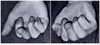
What is the blood supply to the lunate?
- Y-pattern
- X-pattern
- I-pattern -31% of patients
- postulated to be at the highest risk for avascular necrosis

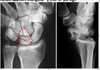
Diagnosis? Etiology?

Keinbochs
- Not well defined, multifactoria
-
biomechanical factors
-
ulnar negative variance
- leads to increased radial-lunate contact stress
- repetitive trauma
-
ulnar negative variance
-
anatomic factors
- geometry of lunate
-
vascular supply to lunate
- “I” pattern
- disruption of venous outflow
-
Patient factors - presumptive but not proven
- Sickle cell
- Steroids
- Septic emboli
- Gout
- Carpal coalition
Diagnosis? Classification

Keinboch’s - Litchman Classification
-
Stage I
- No visible changes on xray, changes seen on MRI
- Immobilization
- NSAIDS
- No visible changes on xray, changes seen on MRI
-
Stage II
-
Sclerosis of lunate
- Ulnar negative - ulnar shortening
- Ulnar positive - Radial wedge, STT fusion
- Distal radius core decompression
- Revascularization procedures
-
Sclerosis of lunate
-
Stage IIIA
- Lunate collapse, no scaphoid rotation
- Same as Stage II above
- Lunate collapse, no scaphoid rotation
-
Stage IIIB
- Lunate collapse, fixed scaphoid rotation
- Proximal row carpectomy
- STT fusion
- Lunate collapse, fixed scaphoid rotation
-
Stage IV
- Degenerated adjacent intercarpal joints
- Wrist fusion
- proximal row carpectomy
- limited intercarpal fusion
- Degenerated adjacent intercarpal joints

Imaging to help define collapse of lunate
-
AP, lateral, oblique views of wrist
- findings (see table above)
- Can help define collapse and adjacent scelorsis
-
CT
- most useful once lunate collapse has already occurred
- best for showing
- extent of necrosis
- trabecular destruction
- lunate geometry
-
MRI
- best for diagnosing early disease
- findings
- decreased T1 signal intensity
- reduced vascularity of lunate
options in the treament

-
immobilization, NSAIDS
- initial management for Stage 1 disease
- Immobilize for up to 3 months
-
Outcomes
- a majority of these patients will undergo further degeneration
- BUT worth is to try because there are a small number of patient who will improve
-
temporary scaphotrapeziotrapezoidal pinning
- adolescent with radiographic evidence of Kienbock’s and progressive wrist pain
-
Ulnar Negative Variance - Stage I, II, IIIa
-
joint leveling procedure
- Stage I, II, IIIA disease with ulnar negative variance
- technique
- can be radial shortening osteotomy or ulnar lengthening
- Goal is to have 1mm ulanr postive variance
- Otherwise you risk ulnocarpal abutment
- Outcomes
- Changing ulnar variance by 4mm can offload lunate 45%
-
joint leveling procedure
-
Ulnar positive variance - Stage I, II, IIIa
- radial wedge osteotomy
- Capitate shortening +/- hamate fusion
-
vascularized bone grafts
- 2nd dorsal intermetacarpal artery is commonly used
- early results promising, but long-term data lacking
- greatest success in Stage II patients
-
distal radius core decompression
- creates a local vascular healing response
-
Stage IIIb disease
- Capitate lengthening with lunate excision
-
STT fusion
- must address internal collapse pattern (DISI)
-
proximal row carpectomy (PRC)
- some studies have shown superior results of STT fusion over PRC for stage IIIB disease
- Others show no difference in outcome, grip strength, pain
-
Stage IV disease
-
Proximal row Carpectomy
- Benefits
- Preserved motion
- Can do a wrist fusion later
- Resect 1cm of the posterior interosseous nerve (which innervates the joint capsule in the floor of the 4th extensor compartment)
- Benefits
- Contraindications
- Severe arthritis of the head of the capitate
-
wrist fusion
- must remove arthritic part of joint
-
total wrist arthroplasty
- long-term results not available
-
Proximal row Carpectomy
What are the components of the TFCC
-
dorsal and volar radioulnar ligaments
- deep ligaments known as ligamentum subcruatum
- central articular disc
- meniscus homolog
- ulno-lunate; ulno-triquetral (UCL)
- ECU subsheath
- origin of ulnolunate and ulnotriquetral ligaments

What are the primary stabilizers of the DRUJ
-
Primary
-
volar radioulnar ligaments
- volar translation
- supination
-
dorsal radioulnar ligaments
- dorsal translation
- pronation
- trigangular fibrocartilage
-
volar radioulnar ligaments
-
Secondary
- ulnar head
- sigmoid notch
- interosseous membrane
- pronator quadratus
Presentation of TFCC tears
-
History
- ulnar sided wrist pain
- turning a door key often painful
- Get a full and complete history of pain, may be associated with instability
- Swelling
- History of trauma
- ulnar sided wrist pain
-
Physical exam
- Examine for areas of fullness or swelling
- ROM and instability
- Assess stability in neutral, sup/pro
- Hypersupnation will stress the DRUJ
- Provacative test
-
Ulnocarpal stress test
- Ulnar deviation, axial load, extension
-
positive “fovea” sign
- tenderness in the soft spot between the ulnar styloid and flexor carpi ulnaris tendon, between the volar surface of the ulnar head and the pisiform
- 95% sensitivity and 87% specificity for foveal disruptions of TFCC or ulnotriquetral ligament injuries
- pain elicited with ulnar deviation (TFCC compression) or radial deviation (TFCC tension)
-
Ulnocarpal stress test

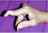
What is the most sensitive test for TFCC tear
foveal sign
- tenderness in the soft spot between the ulnar styloid and flexor carpi ulnaris tendon, between the volar surface of the ulnar head and the pisiform
- 95% sensitivity and 87% specificity for foveal disruptions of TFCC or ulnotriquetral ligament injuries

Palmar Classficiation of TFCC tears
-
Class 1 - Traumatic TFCC Injuries
- 1A - Central perforation or tear
- 1B - Ulnar avulsion (without ulnar styloid fx)
- 1C - Volar avulsion (origin of UL, UT, UC ligaments)
- usually results in dislocation
- 1D - Radial avulsion
- only time you might try to fix this is in a young patient
-
Class 2 - Degenerative TFCC Injuries
- 2A - TFCC wear and thinning
- 2B - Lunate and/or ulnar chondromalacia + 2A
- 2C - TFCC perforation + 2B
- 2D - Ligament disruption + 2C
- 2E - Ulnocarpal and DRUJ arthritis + 2D

How is the load distribution in the wrist affected by ulnar variance?
-
+2 mm ulnar variance approximately
- 40% of the load goes to the ulna
- 60% to the radius
-
normal neutral wrist approximately
- 20%: ulna
- 80%: radius
-
-2 ulnar variance
- 5%: ulna
- 95% radius
What conditions are associated with the pathological finding of this XR? What would you expect to find on physical exam?

Ulnar Positive Variance
- Load 60% radius, 40% ulna
-
Associated conditions
- ulnar abutment syndrome
- Scapho-lunate instability
- TFCC tears
- arthrosis
- ulnar head
- lunate
- triquetrum
- lunotriquetral ligament tears
-
Physical Exam
- ulnar sided wrist pain from increased impact stress on the lunate and triquetrum

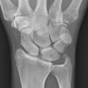
What conditions are associated with the pathological finding of this XR?

Ulnar Negative Variance
- Kienbock’s disease
-
ulnar impingement syndrome
- ulna impinges on the radius proximal to the sigmoid notch
How does position of the forearm affect ulnar variance?
- Ulnar variance increases in pronation
- decreases in supination
- ulnar variane increases during grip
A pronated grip view is the best to determine your ulnar varience
Pathophysiology of altered ulnar variance
-
congenital
- Madelung deformity (positive UV)
- reverse Madelung deformity (negative UV)
-
trauma/mechanical
- distal radius/ulnar fracture with shortening
- growth arrest (previous Salter-Harris fracture)
- DRUJ injuries (Galeazzi and Essex-Lopresti)
-
iatrogenic
- joint leveling procedures (radial or ulnar shortening/lengthening)
- radial head resection (positive UV)
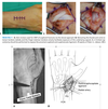
Landmarks for wrist scope portals
-
Radiocarpar Portals
-
3-4
- Located just distal to Lister tubercle, between EPL and EDC
- Established first, primary viewing portal
-
4-5
- Located in line with ring finger metacarpal, between EDC and EDM;
- Portal for instrumentation, visualization of TFCC
-
6R
- Located just radial to ECU tendon;
- Primary adjunct for visualization and instrumentation, ulnar-sided TFCC repairs
- Dorsal sensory branch of ulnar nerve
- 6U
- Located just ulnar to ECU tendon;
- Primary adjuct for visualization and instrumentation, ulnar-sided TFCC repairs
- Dorsal sensory branch of ulnar nerve
-
1-2
- Located between APL and ECRB, along dorsal aspect of snuffbox;
- Not often utilized, provides access to radial styloid and radial aspect of joint, sometimes used for inflow
- Superficial branch of radial nerve; Radial artery
-
3-4
-
Midcarpal Portals
- MCR
- Located 1 cm distal to 3-4 portal along axis of radial border of middle finger metacarpal, between ECRB and EDC.
- Allows visualization of scapholunate, scaphocapitate, and scaphotrapezoid joints.
- MCU
- Located 1 cm distal to 4-5 portal along axis of ring finger metacarpal, between
- Allows visualization of lunocapitate, lunotriquetral, and triquetrohamate joints.
- STT
- Located along axis of index finger metacarpal just ulnar to EPL at level of STT joint
- Allows visualization of scaphotrapezial and scaphotrapezoid joints.
- MCR
-
First CMC Joint
-
1U
- Located on ulnar aspect of EPL at level of first CMC joint (basal joint)
- Allows diagnosis of DJD of first CMC joint and arthroscopic debridement.
- Superficial sensory branch of radial nerve
-
1R
- Located on radial aspect of EPL at level of thumb CMC joint, just volar to APL tendon
- Allows diagnosis of DJD of first CMC joint and arthroscopic debridement.
- Superficial sensory branch of radial nerve
-
1U

Indications for wrist arthroscopy
- TFCC injuries
- interosseous ligament injuries
- anatomic reduction assistance (distal radius, scaphoid fxs)
- ulnocarpal impaction
- debridement of chondral lesions
- removal of loose bodies
- synovectomy
- excision of dorsal wrist ganglia
- assistance in treatment of SNAC and or SLAC wrist
- septic wrist irrigation and debridement
- diagnosis in unexplained mechanical wrist pain
Complications associated with wrist arthroscopy
overall complication rate is 1-2%
-
Dorsal sensory branch of ulnar nerve
- averages 8mm from 6R portal
- at risk with establishment of 6U and 6R portals
- to a lesser extent main ulnar nerve and artery also at risk
- when performing a TFCC repair, small open incision is typically made prior to knot tying to prevent injury to this nerve.
-
Superficial sensory branch of radial nerve
- averages 16mm from 3-4 portal
- at risk during arthroscopy of basal joint, as 1U and 1R portals are on either side of the first branch of this nerve
- at risk during placement of 1-2 portal
-
Radial artery Injury
- associated with establishment 1-2 portal, used for arthroscopic radial styloidectomy.
-
Extensor tendon injury
- most commonly EPL and EDM due to improper portal placement
-
Chondral injuries
- iatrogenic from scope or instrument placement
- Portal site infection
- Stiffness
-
MCPJ pain
- typically caused by over-distraction
What are the common wrist extensor tendons that get injured during wrist athroscopy
most commonly EPL and EDM due to improper portal placement
What portals do you need to be most concerned about neurovascular injury?
-
Dorsal sensory branch of ulnar nerve
- averages 8mm from 6R portal
- at risk with establishment of 6U and 6R portals
- to a lesser extent main ulnar nerve and artery also at risk
- when performing a TFCC repair, small open incision is typically made prior to knot tying to prevent injury to this nerve.
-
Superficial sensory branch of radial nerve
- averages 16mm from 3-4 portal
- at risk during arthroscopy of basal joint, as 1U and 1R portals are on either side of the first branch of this nerve
- at risk during placement of 1-2 portal
-
Radial artery Injury
- associated with establishment 1-2 portal, used for arthroscopic radial styloidectomy.

Differential for DRUJ pain
DRUJ instability or arthritis
TFCC tear
LT ligament tear
pisotriquetral arthritis
ECU tendonitis or instability
Ulnocarpal impaction
History and physical for a patient with ulnar sided wrist pain?
-
Differential for DRUJ Pain
- DRUJ instability or arthritis
- TFCC tear
- LT ligament tear
- pisotriquetral arthritis
- ECU tendonitis or instability
- Ulnocarpal impaction
-
History
- Associations with unlocarpal abutment
- pain on dorsal side of DRUJ
- increased pain with ulnar deviation of wrist
- pain with axial loading
- ulna sided wrist pain
- Painful snapping or instability
- History of trauma or instability; gymnastics or repetative loading as a child
- madelungs, distal radius malunion, TFCC
- PMHx - RA, JRA
- Associations with unlocarpal abutment
-
Physical exam
-
Examine
- Assess the wrist for ulnar prominence
- Irritation over the DRUJ
-
Full ROM
- Especially sup/pro
- DRUJ strained in hypersupnation
- Assess for ECU subluxation
- Assess for crepitus (OA)
- Especially sup/pro
-
Ballottement test
- dorsal and palmar displacement of ulna with wrist in ulnar deviation
- positive test produces pain
-
Ulnar stress test
- ulnar deviation of pronated wrist while axially loading, flexing and extending the wrist
- positive test produces pain
-
Fovea test
- used to evaluate for TFCC tear or ulnotriquetral ligament tear (most sensitive for TFCC pathology)
- palpation of the ulnar wrist between the styloid and FCU tendon to
-
Examine

Radiographic work-up for ulnar sided wrist pain
-
Radiographs
- AP radiograph with wrist in neutral supination/pronation and zero rotation
- required to evaluate ulnar variance
- Assess for congruence of the joint
- Evidence of OA or ulnocarpal impaction
- Assess for space between the proximal row carpi
-
pronated grip view
- increases radiographic impaction
- Findings assoicated with ulnocarpal abutment
- ulna positive variance
- sclerosis of lunate and ulnar head
- AP radiograph with wrist in neutral supination/pronation and zero rotation
-
Arthography
- joint injection shows extravasation
- can help determine
-
MRI
- Important to determine presence of TFCC tear
- Helps to determien procedure
- has largely replaced arthrography
- tear at ulnar part of lunate indicates ulnocarpal impaction
- sensitivity = 74-100%
- Not felt to be a good assessement tool for TFCC tear
- Accuracy 55%
- Important to determine presence of TFCC tear
-
Arthroscopy
- most accurate method of diagnosis
- indicated in symptomatic patients after failing several months of splinting and activity modification
- Benefit is that you can treat the pathology at the time of diagnosis
- Components of the DRUJ you can see with a scope
- Triangular fibrocartilage
- Volar extrinsic ligaments
- Components you can only see with a tear
- Radial attachment of radial-ulnar ligaments
- Can test indirectly with ballottement of the DRUJ
- Ulnar attachement of the RU ligaments
- Radial attachment of radial-ulnar ligaments

Patient with ulnar sided wrist pain. Young. Failed non-op. Options for treatment?

Ulnocarpal Abutment
-
Nonoperative
-
indications
- may attempt supportive measures as first line of treatment
- ECU strengthening, NSAID, Splint
-
indications
-
Arthroscopy
- any procedure can be combined with a diagnostic arthroscopy for further treatment determination, or to address isolated TFCC pathology
-
Ulnar shortening osteotomy
-
indications
- most cases of ulnar positive variance
- most cases of DRUJ incongruity
-
indications
-
Wafer procedure
-
technique
- 2 to 4mm of cartilage and bone removed from under TFCC arthroscopically
-
technique
-
Darrach procedure (ulnar head resection)
-
indications
- reserved for lower demand patients
-
technique
- 40 deg oblique cut just proximal to the DRUJ
- Try to preserve the R-U ligaments, TFCC and periosteium, ECU sheath
- Some will leave the ulnar styloid to maintain staibilty
-
indications
-
Sauvé-Kapandji procedure
-
indications
- good option for manual laborers
- Can be used in patients with a torn TFCC
-
technique
- Dorsal-ulnar approach
- Resect the distal ulna 10-15mm from the joint
- Stabilize the DRUJ with k-wires and 3.5mm cannulated screws
- creates a distal radioulnar fusion and a ulnar psuedoarthrosis proximal to the fusion site through which rotation can occur
- ECU/FCU can be tenodesed in the ulnar stump for additional stability (to help reduce issues with post-op residual instability)
- Proximal ECU slip
- Distal FCU slip
- Woven to create a stable stump
-
indications
-
Ulnar hemiresection arthroplasty (HIT)
-
indications
- usually requires an intact TFCC
- appropriate treatment option in the presence of post-traumatic DRUJ with concomitant distal ulnar degenerative changes
-
indications
-
ulnar head replacement
- indications
- Severe OA
- salvage for failed Darrach
- Isolated instability
- Occasionally after trauma
- Options
- Partial ulnar head arthroplasty
- There is also a hemiarthroplasty available, but can only be done after hit
- Severe OA in an otherwise stable joint
- Total ulnar head arthroplasty
- Wider indications but can still have instability; often asymptomatic
- Partial ulnar head arthroplasty
-
Total DRUJ arthroplasty
- Experimental, most often used in the setting of madelungs
- This is commonly used by surgeons as a salvage technique
- indications