Pathoma Ch. 2 (Inflammation/ Wound Healing) Flashcards
What 2 things characterize acute inflammation?
edema + neutrophils
Where are toll-like receptors found? What do they do? Give a specific example.
TLRs are on macrophages and dendritic cells
they recognize PAMPs (pathogen associated molecular patterns)
ex: TLR 4 on macrophages recognizes LPS on gram (-) bacteria, which activates NF-kappa-B (transcription factor that leads to making of pro-inflammatory cytokines)

What 4 things attract neutrophils?
- Leukotriene B4
- C5a
- IL-8 (Remember 8 looks like infinity and there are “infinity neutrophils” bc they’re so abundant)
- Bacterial products
What are the arachadonic acid metabolites? (What’s the pathway?) What does each thing cause?

What do leukotrienes cause?
LT B4–> attracts neutrophils
LT C4, D4, E4–> smooth muscle contraction—> (1) vasoconstriction, (2) bronchospasm, (3) inc vascular permeability (by contraction of pericytes—> creates opening between endothelial cells—> fluid leaks into tissue space) *they also increase mucus secretion

What things do prostaglandins cause?
Vasodilation, inc vascular permeability, E2 also does pain + fever
also: protective to GI mucosa and dilate the afferent arteriole (—> inc GFR and RPF)

What 2 things does TXA2 (thromboxane A2) cause?
- Vasoconstriction
- Platelet aggregation
(within platelets: COX-1–> TXA2–> aggregates platelets, which is why we give aspirin in MI…it blocks COX—> blocks TXA2–> prevents platelet aggregation to limit thrombosis/ clotting from platelets sticking together)
What 2 things do prostacyclins cause?
- Vasodilation
- Stop platelet aggregation

What 3 things activate mast cells? When activated, what gets released from the mast cells themselves and what does it cause?
These 3 things activate mast cells:
- Tissue trauma
- C3a, C5a (complement)
- Pathogen cross-linking of IgE antibodies
Once active—> degranulation/ release of histamine—> vasodilation and increased vascular permeability

What is complement?
pro-inflammatory serum proteins that “complement inflammation”
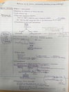
Describe/ draw out the complement cascade. What are the 3 pathways that feed into it? What activates them?…
C3—(C3 convertase)—> C3b——-> bacteria killed
- Mannose binding lectin (MB-lectin) pathway- MBL binds mannose on pathogen surfaces to activate complement cascade
- Classic pathway- C1q binds IgG or IgM (antibody-antigen complexes activate complement cascade) (“General Motors makes classic cars”)
-
Alternate pathway- spontaneous/ pathogen surfaces directly activate complement
* How does the cascade kill bacteria?* C3b is a key opsonizer—> C3a, C5a (anaphylaxins)—> mast cell degranulation (vasodilation, inc vascular permeability). MAC attack formation punches holes in cell membrane of bacteria to kill it.

What is the Hageman factor (factor XII/ factor 12)? When does it get activated and what 3 things does it do?
pro-inflammatory protein made by the liver that gets activated when subendothelial collagen is exposed (response to endothelial damage)
- Activation of the coagulation/ fibrinolytic cascade
- Activation of the kinin cascade (—> bradykinin—> vasodilation, inc vascular permeability, pain)
- Activation of complement

What are the 4 signs of inflammation. State the inflammatory mediators involved and what they do also.
- Redness (rubor) and warmth (calor)- histamine, PGs, and bradykinin—> vasodilation (inc blood flow)
- Swelling (tumor)- histamine (inc vascular permeability) and tissue/ endothelial damage—> leakage of exudate
- Pain (dolor)- bradykinin and PG E2–> sensory nerve endings
- Fever- pyrogens (compounds from bacteria, like LPS, that lead to fever)—> macrophages release IL-1 and TNF-alpha—> gets into perivascular cells of hypothalamus—> inc COX—> makes PGE2–> inc temp set-point= fever

What are the names of them 7 steps of neutrophil arrival and function? What happens in each step? What are the key players?
- Margination- vasodilation slows blood flow and moves neutrophils to the wall of the blood vessel
- Rolling- neutrophils slow down due to P-selectin and E-selectin “speed bumps” (unregulated by IL-1 and TNF)
- Adhesion- neutrophils adhere to endothelial cells—the neutrophils have integrans like LFA-1, etc (unregulated by C5a and LTB4, which are neutrophil attractants) that stick to cellular adhesion molecules like I-CAM and V-CAM (unregulated by IL-1 and TNF) on the surface of endothelial cells
- Transmigration and chemotaxis- neutrophils migrate across the wall of the blood vessel/ endothelium of postcapillary venules with the help of PECAM-1 and they move toward chemical attractants (chemokines)
- Phagocytosis- IgG and C3b are key opsonins that tag for phagocytosis. The bacteria gets phagocytsoed by the neutrophil into a phagosome and fuses with a lysosomes that holds the digestive enzymes—> phagolysosome
- Destruction of phagocytosed material- once in the phagolysosome, a series of reactions called “oxidative burst” does the killing
- Resolution- once neutrophils successfully got rid of the problem, they undergo apoptosis (no longer needed)
Baby with delayed separation of the umbilical cord. What immuno disorder should come to mind?

What immuno disorder is due to a defect in CD18? What’s the presentation?
Leukocyte adhesion deficiency
-AR defect of integrans (CD18 subunit)—> (1) delayed separation of the umbilical cord (no neutrophils to clean debris), (2) increased neutrophils in circulation (they can’t stick to vessel wall), and (3) recurrent bacterial infections (w/o pus)
*CD18 is needed to form integrans like LFA-1

What immuno disorder is due to an autosomal recessive mutation in the LYST lysosomal trafficking gene? What does it present with?
Chediak-Higashi syndrome
AR mutation in LYST (lysosomal trafficking gene)—> microtubule dysfunction—> failure of lysosomes to fuse with phagolysosomes (to form phagolysosomes)
- recurrent bacterial infections (since phagolysosome didn’t form to kill bacteria by respiratory burst)
- albinism (no “railroad system” to bring melanin to cell surfaces)
- neuro issues (no protein trafficking to keep peripheral nerves alive)
- decreased neutrophils + giant granules in leukocytes (no “railroad system” for granules to disperse so they pile up)

What immuno disorder involves failure of the phagolysosome to form? How does it present?
Chediak-Higashi syndrome
AR mutation in LYST (lysosomal trafficking gene)—> microtubule dysfunction—> failure of lysosomes to fuse with phagolysosomes (to form phagolysosomes)
- recurrent bacterial infections (since phagolysosome didn’t form to kill bacteria by respiratory burst)
- albinism (no “railroad system” to bring melanin to cell surfaces)
- neuro issues (no protein trafficking to keep peripheral nerves alive)
- -*decreased neutrophils + giant granules in leukocytes (no “railroad system” for granules to disperse so they pile up)

Albino kid presents with recurrent infections. His neutrophil count is low and WBCs have large granules on blood smear. Diagnosis?
Chediak-Higashi syndrome
AR mutation in LYST (lysosomal trafficking gene)—> microtubule dysfunction—> failure of lysosomes to fuse with phagolysosomes (to form phagolysosomes)
- recurrent bacterial infections (since phagolysosome didn’t form to kill bacteria by respiratory burst)
- albinism (no “railroad system” to bring melanin to cell surfaces)
- neuro issues (no protein trafficking to keep peripheral nerves alive)
- decreased neutrophils + giant granules in leukocytes (no “railroad system” for granules to disperse so they pile up)

What’s Chronic Granulomatous Disease (CGD)? What 6 infections are you at high risk for? What result do you get doing a nitroblue tetrazolium test?
CGD= defect in NADPH Oxidase (X-linked or AR)
leads to recurrent catalase (+) infections (catalase negative can steal H2O2 from the bacteria themselves to mediate bacteria killing, but have no way of killing catalase positive bugs with the respiratory burst reactions):
- Staph A.
- Pseudomonas
- Serratia
- Nocardia
- Aspergillus
- Burkholderia Cepacia
Nitroblue tetrazolium test—> neutrophils fail to turn blue when exposed to dye (“negative nitroblue tetrazolium test”)

MPO deficiency will lead to an increase in what type of infections?
candida infections

Neutrophils vs. macrophages. O2-depending and O2-independing killing of pathogens?
neutrophils—> mainly do O2-dependent killing (“respiratory burst”) *can also do O2-independent killing (lysozymes in macrophages, major basic protein in eosinophils), but it’s less effective
macrophages—> mainly do O2-independent killing
What are the special names for macrophages in: liver, CNS, bone?
liver- Kupffer cells
CNS- microglia
bone- osteoClasts
If the infection is acute, what immune cells are definitely involved in the response?
neutrophils
(these guys are the 1st responders!)

Neutrophils= 1st responders. Then macrophages come into the area and decide to do one of four things. Explain them:
- Resolution and healing
- Continued acute inflammation
- Abscess
- Chronic inflammation
- Resolution and healing- neutrophils took care of it. Let’s release IL-10 and TGF-beta to wrap it up/ shut down inflammation (“IL-10 and TGF-Beta Both aTENuate the Immune response”)
- Continued acute inflammation- release IL-8 to recruit additional neutrophils to help out
- Abscess- release fibrogenic growth factors/ cytokines to wall off the infection w/ fibrosis
- Chronic inflammation- eat up the pathogen—> express antigen on their MHC II—> activate CD4+ T-cells to secrete cytokines (get adaptive immune system involved to handle the situation)

What cells characterize chronic inflammation?
lymphocytes (T-cells, B-cells) and plasma cells (mature B-cells that produce antibodies)
*the adaptive immune system, a response to persistent infection, viruses/ mycobacterium/ parasites/ fungi, autoimmune dz, foreign material, etc.

What are all the steps and enzymes of respiratory burst?

What are the 3 antigen-presenting cells (APCs)?
- Dendritic cells
- Macrophages
- B-cells
When an APC presents to a CD4+ T-cell, what 2 signals/ connections are going on between them?
B7 (APC)——CD28 (T-cell)
(*this^ is the 2nd activation signal, can be remembered by 28/7= 4 for CD4)
MHC II (APC)——TCR (T-cell)

Th1 and Th2 cells are both subtypes of CD4+ helper T-cells.
- Role of Th1 and 1 major cytokine it releases?
- Role of Th2 and 3 major cytokines it releases?
- (Also say what the cytokines do)*
Th1 helps CD8+ Cytotoxic T-cells
-releases IFN-gamma (which activates macrophages, does class switching IgM—> IgG, and activates Th1/ blocks Th2)
Th2 helps B-cells
-releases IL-4 (class switching to IgE), IL-5 (class switching to IgG), and IL-13
What does IL-2 do?
activates CD8+ T-cells (“two” cells)
(secreted by Th1 cells, which are the helpers of CD8+)

2 ways that CD8+ cytotoxic T-cells do killing?
- Secrete perforin and granzyme (activates apoptosis via the cytotoxic CD8+ pathway)
- Expresses FAS ligand which binds to the FAS death receptor on target cells (activates apoptosis via the extrinsic receptor-ligand pathway)
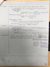
When a B-cell serves as an APC and presents antigen to a CD4+ (Th2) T-cell, what 2 signals/ connections are going on between them? What happens?
CD40 Ligand (Th2)——CD40 receptor (B-cell)
(*this^ is the 2nd activation signal)
TCR (Th2)——MHC II (B-cell/ APC)
Once the B-cell shows the antigen to CD4+ Th2 cells, the Th2 cells secrete cytokines (IL-4, IL5) that enable Ig class-switching

What are the steps and cytokines involved in granuloma formation?
- APC—> Th1 (by IL-12)
- Th1–> macrophage (by INF-alpha)
- Macrophage—> recruits more macrophages/ became epitheliod histiocytes and giant cells that wall off infection (by TNF-alpha)

Caseating vs. non-caseating granulomas?
caseating- central necrosis, infectious cause (TB/ fungi)
non-caseating- non central necrosis, non-infectious (sarcoidosis, Crohn’s, cat scratch dz, reaction to breast implants, etc.)
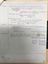
Kid has recurrent infections. No thymic shadow seen. Labs show hypocalcemia. Diagnosis? State the cause and other things a patient with this immuno disorder may present with.
DiGeorge syndrome
22q11 microdeletion—> failure of the 3rd and 4th pharyngeal pouch to form—> no parathyroid glands and no thymus
- recurrent infections (no thymus—> no mature T-cells)
- hypocalcemia, seizures (no parathyroid to secrete PTH to raise calcium levels—> low serum calcium)
- facial abnormalities
- heart defects
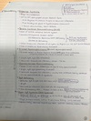
What structures are formed from the 4 pharyngeal pouches?
1st—> ear
2nd—> tonsils
3rd—> inferior parathyroid + thymus
4th—> superior parathyroid
(“ear, tonsils, bottom to top”)

3 causes of SCID?
- Cytokine receptor defect
- Adenosine deaminase (ADA) deficiency
- MHC II deficiency
(all potential causes lead to T-cell dysfunction—> B-cell dysfunction, so loss of the entire adaptive immune system)

Kid with recurrent infections, no thymus, normal calcium levels. Diagnosis?
Severe combined immunodeficiency (SCID)
(They have T-cell and B-cell dysfunction/ loss of entire adaptive immune system. Makes sense they have no/ underdeveloped thymus since T-cells aren’t working. Normal calcium levels bc parathyroid isn’t affected—vs. DiGeorge)

Young adult with low levels of some immunoglobulin. Has recurrent sinoplumonary and GI infections. What diagnosis should come to mind? This patient will have increased risk for developing what 2 things?
Common Variable Immunodeficiency
- seen in late childhood/ early adulthood (vs most immuno disorders, which come on around 6 months when baby’s immune system has developed)
- the T-cell/ B-cell defects that cause it vary and the specific Ig’s that are low vary (‘variable’ is in its name)
- increased risk for autoimmune disease (immune system is already messed up and Ig’s are low to fight off stuff) and lymphoma

Patient with recurrent GI infections. All immunoglobulin levels are normal except IgA is low. Diagnosis? What are 3 important facts/ associations to remember?
IgA deficiency (the most common Ig deficiency!)
- False positive pregnancy test
- Associated with SLE and RA (makes sense you’re more likely to get an autoimmune dz if your immune system is already messed up)
- Blood transfusions—> anaphylactic shock (these patients have antibodies against IgA in blood products)
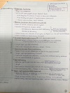
A patient has a blood transfusion (the corrrect blood type was transfused) and gets anaphylactic shock. What underlying immuno disorder might the patient have?
IgA deficiency
(these patients have antibodies against IgA in blood products)
What immuno disorder is due to a defect in the STAT 13 signaling pathway? How does it present?
Hyper-IgE syndrome (Job’s syndrome)
defect in STAT 13 signaling pathway—> defective Th17 cells—> no IL-17–> loss of attraction of neutrophils to inflammatory sites
- increased IgE (decreased IFN-gamma, poorly understood)
- presentation: newborn baby, deformed face/ 2 rows of teeth (retain baby teeth), cold skin abscesses, eczema, infections

What immuno disorder is due to a lack of IL-17? How does it present?
Hyper-IgE syndrome (Job’s syndrome)
defect in STAT 13 signaling pathway—> defective Th17 cells—> no IL-17–> loss of attraction of neutrophils to inflammatory sites
- increased IgE (decreased IFN-gamma, poorly understood)
- presentation: newborn baby, deformed face/ 2 rows of teeth (retain baby teeth), cold skin abscesses, eczema, infections
Baby with an extra row of teeth is having frequent infections. Also has dry, red skin. Diagnosis? Explain the cause and typical presentation.
Hyper-IgE syndrome (Job’s syndrome)
defect in STAT 13 signaling pathway—> defective Th17 cells—> no IL-17–> loss of attraction of neutrophils to inflammatory sites
- increased IgE (decreased IFN-gamma, poorly understood)
- presentation: newborn baby, deformed face/ 2 rows of teeth (retain baby teeth), cold skin abscesses, eczema, infections
Cause of hyper IgM syndrome?
Due to failed CD40 L (T-cell)—CD40 receptor (B-cell) interaction
(remember, this interaction enables T-cells to secrete cytokines to enable class switching. So if there’s a problem with this interaction, B-cells will only be able to make IgM, the 1st antibody that always gets made, no other—“MD before you AGE”)

Patient has recurrent infections. All immunoglobulin levels are low except IgM is high. Diagnosis? Cause?
Hyper IgM syndrome
Due to failed CD40 L (T-cell)—CD40 receptor (B-cell) interaction (*usually a T-cell problem)—> w/o this interaction, T-cells can’t secrete cytokines to enable class-switching, so B-cells can only make IgM (the 1st antibody that always gets made)
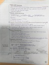
What immuno disorder is due to a defective WASP protein? How does it present?
Wiskott-Aldrich syndrome
X-linked (boys) disorder of WASP gene—> no WASP protein—> abnormal T-cell cytoskeleton—> T-cell dysfunction (they can’t react well to APCs)
-presents with triad: (1) eczema, (2) bleeding (dec platelets), and (3) recurrent infections

6 month old boy presents with recurrent infections, dry red skin, and increased bleeding time. Diagnosis? Cause?
Wiskott-Aldrich syndrome
X-linked (boys) disorder of WASP gene—> no WASP protein—> abnormal T-cell cytoskeleton—> T-cell dysfunction (they can’t react well to APCs)
-presents with triad: (1) eczema, (2) bleeding (dec platelets), and (3) recurrent infections

What is Wiskott-Aldrich syndrome? Cause and triad it typically presents with
Wiskott-Aldrich syndrome
X-linked (boys) disorder of WASP gene—> no WASP protein—> abnormal T-cell cytoskeleton—> T-cell dysfunction (they can’t react well to APCs)
-presents with triad: (1) eczema, (2) bleeding (dec platelets), and (3) recurrent infections

Explain positive and negative selection in the thymus. What are AIRE genes?
Positive selection: do you recognize MHC? If yes—> go on. If no—> apoptosis.
Negative selection: do you bind self-antigen? If yes—> apoptosis. If no—> go on.
*negative selection occurs on dendritic cells or medullary epithelial cells. AIRE genes= TF necessary for medullary epithelial cells to express/ show self-antigen to T-cells to see if they bind it (so they are an important part of carrying out negative selection).

What immuno disorder occurs due to a defect in AIRE genes (autoimmune regulatory genes)? What 2 problems occur in this disorder?
(AIRE genes= TF necessary for medullary epithelial cells to express/ show self-antigen to T-cells to see if they bind it—so they are an important part of carrying out negative selection)
Chronic Mucocutanous Candidiasis aka
Autoimmune Polyendocrine Syndrome
-defect in AIRE genes that help with negative selection in the thymus and response to Candida infections—> you get autoimmune T-cells that attack endocrine cells and cause endocrine dysfunction + recurrent Candida infections

Endocrine problems and recurrent Candida infections. Diagnosis? Cause?
Chronic Mucocutanous Candidiasis aka
Autoimmune Polyendocrine Syndrome
-defect in AIRE genes that help with negative selection in the thymus and response to Candida infections—> you get autoimmune T-cells that attack endocrine cells and cause endocrine dysfunction + recurrent Candida infections

What immuno disorder is due to a defect in the ATM gene on chromosome 11? How does it present?
Ataxia Telangiectasia
defective ATM gene on chromosome 11–> can’t repair non-homologous end-joining/ dsDNA breaks
- ataxia (cerebellar dysfunction)
- telangiectasia (dilated blood vessels on skin)
- inc sensitive to radiation/ inc skin CA risk
- recurrent sinopulmonary infections (VDJ recombination/ immune cells shuffling their DNA to create diversity involves DNA breaks…so if you can’t repair these breaks—> lack of immune cell diversity—> inability to fight off variety of infections—> recurrent infections)

Describe negative selection for immature B-cells in the bone marrow. (What 2 options can occur if the cell fails this step?)
do you bind self-antigen (on dendritic cells)?
if no—> go on
if yes—> (1) receptor editing—> then go on OR
(2) apoptosis (via mitochondrial pathway)

Increased Neisseria infections. What immuno problem?
C5-C9 deficiency—> can’t for MAC
What immuno problem results in hereditary angioedema?
C1 inhibitor deficiency


