Pathoma Ch. 1 (Cell Growth/ Injury/ Death) Flashcards
Hyperplasia can lead to —> dysplasia —> cancer. Give an example and an exception to this ‘rule.’
Example: endometrial hyperplasia—> endometrial CA
Exception: BPH does NOT increase prostate CA risk

What is myositis ossificans and what is it an example of?
Myositis ossificans- mesenchymal/ connective tissue—> bone during healing post-trauma
Example of Metaplasia
What’s primary vs secondary amyloidosis? (What type of amyloid protein is deposited, what protein it’s derived from, example of disease)
Primary amyloidosis- systemic deposition of AL amyloid, derived from Ig light chain, ex: Multiple Myeloma
Secondary amyloidosis- systemic deposition of AA amyloid, derived from serum-amyloid associated protein, ex: Familial Mediterranean Fever

Explain liquefactive necrosis.
Enzymes act on dead tissue—> liquified
(ex: BRAIN infarction, abscess, pancreatitis)
Hallmark of cell death?
Loss of nucleus
(1. Pyknosis*- nucleus shrinks, 2. *Karyorrhexis*- nucleus breaks up, 3. *Karyolysis- nucleus further breaks down into building blocks)

Explain coagulative necrosis.
Dead tissue that stays firm/ keeps its shape (in any tissue but the brain)
pale infarct= cut blood supply, wedge-shaped
red infarct= blood re-enters (ex: testicular infarction…the twisting/ torsion prevents blood from being drained through the collapsible veins, but the thick-walled artery remains open so blood can keep coming into the testicle)
What is keratomalacia and what is it an example of. Explain.
Karatomalacia- metaplasia of conjunctiva lining the eye
-you need vitamin A to maintain the specialized epithelium of the eye. Vit A deficiency—> metaplasia of these specialized cells lining the eye= karatomalacia

What’s the difference between slow-developing vs. acute ischemia? Give an example for each. What happens to the cells in both cases?
Slow-developing (cells have time to adapt)—> atrophy (ex: renal artery atherosclerosis)
Acute (cells don’t have time to adapt)—> cellular injury (ex: renal artery embolus)
(*causes of cellular injury: inflammation, nutritional deficiency or excess, hypoxia (low O2), trauma, genetic mutations)

Dystrophic vs metastatic calcification?
Dystrophic calcification- calcium binds up fat in the setting of normal serum calcium levels
Metastatic calcification- calcium binds up fat in the setting of high serum calcium levels
(ex: hyperparathyroidism…you have too much PTH—> too much calcium reabosorption into blood)

Where are free radicals seen (in normal physiology and in pathology)?
- ETC in ox phos (cyt C oxidase/ complex 4 transfers electrons to O2)
- ionizing radiation
- inflammation (NADPH oxidase in oxidative burst to kill bacteria/ foreign objects/ dead cells)
- metals
- drugs (Tylenol is a big one), chemicals (P450 system, CCl4 used in the cleaning industry)

What is re-perfusion injury?
When you re-perfuse (ex: open up a blood vessel with a stent so blood can flow through coronary arteries again following an MI), you are introducing blood flow= introducing oxygen back to the area.
Oxygen brings in free radicals. This combined with the surrounding area of necrosis/ inflammation (+ loss of antioxidants to deal with free rads) can cause further damage mediated by the free-radicals.
(Re-perfusion injury would present like this: patient had MI. You placed a stent or gave a fibrolytic drug, re-perfusing the site. Now patients cardiac troponin levels are rising, indicating more cell death/ necrosis after you re-perfused.)

Hallmark of reversible cell injury?
Cellular swelling
(loss of membrane microvilli, membrane blebbing, decreased protein synthesis)

What are the 3 pathways of apoptosis? Explain them all.
- Intrinsic/ mitochondrial pathway- cell injury—> knocks out Bcl2 (a regulator of apoptosis)—> cyt C leaks out of mitochondria—> cyt C activates caspases
- Extrinsic receptor-ligand pathway- FAS (or TNF) binds to FAS death receptor aka CD95 (or TNF receptor)—> the binding activates caspases (ex: this type of apoptosis happens in negative selection of tymocytes the thymus)
- Cytotoxic CD8+ T-cell pathway- CD8+ T-cels poke holes in the membrane of the target cell—> granzymes leak out—> activates caspases
*For all pathways:caspases= enzymes that mediate apoptosis. They do this by activation ofproteasesto break down the cytoskeleton of the cell andendonucleases to break down its DNA—> dying cell shrinks, becomes more pink/ eosinophilic, nucleus condenses, macrophages remove debris.

What is amyloid? What sheets is it made of? What is it’s staining?
Misfolded protein
made of beta-pleated sheets
congo-red staining
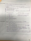
What is dialysis-associated amyloidosis?
Dialysis—> beta-2 microglobulin not filtered well—> this misfolded protein deposits in joints

Explain caseous necrosis.
Dead tissue that looks like cottage cheese
it is coagulation + liquefactive necrosis
(ex: TB or fungal infection)
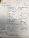
Explain fat necrosis.
Dead tissue that is chalky-white (due to calcium deposition)
fat dies—> FAs released—> fat can bind to calcium (saponification)
(ex: trauma to breasts (fat tissue) and pancreatitis damaging surrounding fat)
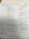
Decreased blood= decreased O2= decreased ATP. What channels/ biochemical processes does this disrupt? What electrolyte changes will occur in a cell that lacks ATP?
Disrupts Na+/K+ pump and Ca2+ pump—> Na+/ water/ Ca2+ build up in the cell—> cell swells (—> loss of microvilli, membrane blebbing, dec protein synthesis—the cell is injured)
Also disrupts oxidative phosphorylation (since you have low O2 and O2 is needed to run this aerobic process)—> switch to anaerobic glycolysis—> you get less ATP + lactic acid build up—> decreased pH (denatures proteins, precipitates DNA)

What amyloid deposits are seen in T2DM? Alzheimer’s disease?
T2DM—> amyloid deposits in pancreatic islet cells
Alzheimers—> A-beta amyloid deposits in the brain

Talk about Barrett esophagus. How is this an example of metaplasia? What specific type of cancer can it lead to and how?
Esophageal cells switch from squamous—> columnar to handle the increased stress (the acid refluxing up from GERD)
Can progress to esophageal cancer: metaplasia (Barrett esophagus)—> dysplasia—> CA (adenocarcinoma affecting the lower 1/3rd of the esophagus)

Are the PaO2 and SaO2 levels high, low, or normal in anemia? CO poisoning?
(PaO2= oxygen that gets into blood, SaO2= oxygen that saturates/ binds to Hb)
Anemia- PaO2 is normal, SaO2 is normal
(plenty of oxygen in the blood, plenty of oxygen bound to Hb—problem is there’s decreased Hb)
CO poisoning- PaO2 is normal, SaO2 is low
(plenty of oxygen in the blood, decreased oxygen bound to Hb bc CO is stealing it away)
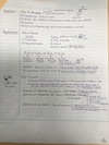
Give the definitions for: hypertrophy, hyperplasia, atrophy, metaplasia, dysplasia, aplasia, and hypoplasia.
Hypertrophy= increase size of cells
Hyperplasia= increase number of cells
Atrophy= decrease size and number of cells
Metaplasia= change in stress—> change in cell type
Dysplasia= disordered cell growth (pre-cancer)
Aplasia= no growth (—> no organ)
Hypoplasia= decreased growth (—> underdeveloped organ)
Hyperplasia and hypertrophy often occur together. What’s an example of this?
The uterus in pregnancy

How does medullary carcinoma of the thyroid relate to amyloidosis?
Malignancy of C-cells in the thyroid—> increased calcitonin, amyloid is in the background on histo
Necrosis vs. apoptosis? Definition, small or large groups of cells involved?, inflammation?
Necrosis- “cell murder,” large groups of cells involved, followed by inflammation
Apoptosis- “cell suicide,” small groups of cells involved, NOT followed by inflammation

Explain how CCl4 (carbon tetrachloride—found in the cleaning industry) can lead to fatty change of the liver.
CCl4 free radical damage—> cell swelling—> ribosome los (ribosomes, the protein-making factories, pop off)—> decreased protein synthesis—> this includes decreased apolipoprotein synthesis—> fat stays (can’t get re-packaged/ sent out of the liver)—> fatty change of the liver

Example of hypoplasia?
Streak ovary in Turner syndrome
(hypoplasia= decreased growth—> underdeveloped organ)

Caspases are the enzymes that drive apoptosis…but how? (They use 2 things). What changes do we see in a cell dying by apoptosis?
Caspases mediate apoptosis by:
- Activation of proteases to break down the cytoskeleton of the cell
- Activation of endonucleases to break down its DNA
The dying cell shrinks—> becomes more pink/ eosinophilic—> nucleus condenses—> macrophages remove debris

Name all the causes of hypoxia and hypoxemia.
Hypoxia (dec oxygen to tissues)-
- Hypoxemia
- Decreased cardiac output (or ischemia: atherosclerosis, Budd-Chiari syndrome, shock)
- Anemia
- CO or CN poisoning
Hypoxemia (dec oxygen in blood) w/ normal A-a gradient-
- High altitude
- Hypoventilation
Hypoxemia (dec oxygen in blood) w/ high A-a gradient (diffusion issue)-
- V/Q mismatch
- R—> L shunt (bypasses lungs for oxygenation)

What has to happen for hypertrophy to take place? What has to happen for hyperplasia to take place?
Hypertrophy (inc size of cells)- 1. Gene activation, 2. Protein synthesis, 3. Cell has to make more organelles
Hyperplasia (inc number of cells)- stem cells make new cells

What’s the difference between: FiO2, PAO2, PaO2, and SaO2?
FiO2- oxygen you breathe in
PAO2- oxygen that gets to alveoli
PaO2- oxygen that gets to blood
SaO2- oxygen that saturates Hb (percent of Hb bound to O2)
What deposits are seen in senile cardiac amyloidosis? Familial amyloid cardiomyopathy? How do these 2 conditions differ?
Senile cardiac amyloidosis—> serum transthyretin deposits (asymptomatic)
Familial amyloid cardiomyopathy—> misfolded serum transthyretin deposits (leads to restrictive cardiomyopathy- the heart is restricted from filling properly due to these infiltrates)
What are the 3 permanent cells? Do they do hypertrophy, hyperplasia, or both?
- Cardiac cells (ex: myocytes in response to HTN)
- Skeletal cells
- Nerves (neurons)
They do HYPERTROPHY (inc size of cells) only! (Cannot inc number of these cells)

What’s an exception to the ‘rule’ that metaplasia—> dysplasia—> cancer?
Metaplasia of the breast (fibrocystic change) does NOT increase risk for breast cancer
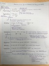
Explain fibrinoid necrosis.
Dead blood vessel wall—> leakage of proteins (fibrin) into vessel wall
(in vasculitis, etc.)
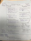
Hallmark of irreversible cell injury?
Membrane damage (leads to cell death)
(leakage of enzymes like Troponin in cardiac cells, increased calcium in cell, loss of ETC, cyt C leaks out of damaged mitochondrial membranes—> apoptosis)

Explain gangrenous necrosis.
Dead tissue that looks like a mummy
Dry gangrene- type of coagulative necrosis
Wet gangrene- type of liquefactive necrosis (get it if there’s an infection of dead tissue)
(this type of necrosis happens in the lower limbs + GI tract)

In atrophy, what is responsible for decrease in size? What is responsible for decrease in number of cells?
Dec size—> ubiquitin-proteosome degradation (degrades cytoskeleton. Other parts of the cell are degraded by autophagy, where vacuoles fuse w/ lysosomes)
Dec number—> apoptosis
