Pathology of Mouth and Esophagus Flashcards
What infectious organisms typically infect the mouth and the esophagus?
HSV 1 and 2 CMV Fungal: Candida, Aspergillus, Mucor
What Lesion is this? Describe what you see on this lesion.

Herpetic vesicle
What Lesion is this? Describe what you see on this lesion.

herpetic ulcer
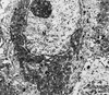
What Lesion is this? Describe what you see on this lesion.

Diagnosis of HSV infection. Multinucleate, intra-nuclear smudgy/steel gray inclusions
What type of cells does HSV infect?
epithelial cells
What Lesion is this? Describe what you see on this lesion.

Intra-nuclear and cytoplasmic inclusions “owl eye”
What type of cells does HSV infect?
Endothelial and mesenchymal cells
What Lesion is this? Describe what you see on this lesion.

Oral thrush
What is candida?

Budding yeast and pseudohyphae Most common –> C. albicans
What lesion or organism is this? Give a description.

Aspergillus: - hyphal forms only - septate hyphae with parallel walls - branching at acute angles of 45 degrees - also angioinvasive
What are the common pathogenic species of Aspergillus?
A. niger, A. fumigatus, A. flavus
What are the other species of infectious candida?
C. tropicalis, C. krusei, C. parapsilosis, C. guillermondii
What is this lesion? Describe what you see.

Mucormycosis: - hyphal forms only - bold, bulbous, non-septate hyphae - Right Angle branching - Also angioinvasive - Mucor, Rhizopus, Absidia
Key terms for HSV infections:
epithelial cells, multinucleation, nuclear inclusions, tzanck test
key terms for CMV infections:
endothelial and mesenchymal cells, nuclear (owl) and cytoplasmic inclusions
key terms for candida:
budding yeast, pseudohyphae
key terms for aspergillus:
hyphae, 45 degree branching
key terms for mucormycosis:
hyphae, 90 degree branching
what lesion is this?

Pyogenic granuloma - a type of oral cavity lesion
What Lesion is this? Describe what you see on this lesion.

Pyogenic granuloma: - Lobular capillary hemangioma with surface ulceration - Inflammation is secondary
What Lesion is this? Describe what you see on this lesion.

Hair leukoplakia: EBV associated lesion - benign
Which patients do you usually see hairy leukoplakia?
immunocompromised patients; HIV 80% organ transplant patients on radiation and chemotherapy
What Lesion is this? Describe what you see on this lesion.

hyperkeratosis, acanthosis and balloon cells
What Lesion is this? Describe what you see on this lesion.

Oral leukoplakia; 5-25% are pre-malignant; range from hyperplasia and hyperkeratosis to high grade dysplasia