Pathology Flashcards
What is pathology?
The study of the structural, molecular, and functional manifestation of disease, and the mechanisms that cause a disease
Vacutainer Tubes
Tool used by pathologists to test skin, cell, or body fluid for disease
In a clinical setting, pathologists are responsible for:
- Evaluation of surgical, cytologic, hematologic, and autopsy specimens 2. Genetic testing and tissue typing 3. Blood transfusions, apheresis, stem cell and donor services 4. Microbiology, immunology, coagulation, and biochemical testing
Mechanism of Disease
pathogenesis
Etiology mnemonic and what it stands for
VINDICATEP Vascular Inflammation Neoplasm Drug/Toxin Infection Congenital/Genetic Auto-immune/immune Trauma/Physical Endocrine/Nutrition/Metabolic Psychological
Disease
Molecular, cellular, tissue, organ, and organismic damage caused by etiology (VINDICATEP) and mediated by pathogenic mechanisms
Diagnosis
the name for the disease
Pathogenesis
the sequence of events that leads from etiology to manifestation of disease
Symptom
Disease manifestation of disease as perceived and reported by the patient
Sign
Manifestation of the disease that can be identified by the physical examination, laboratory tests, imaging studies, and other methods
Differential Diagnosis
A ranked list of most likely diagnoses based on the signs and symptoms of disease in a given patient
Sub-cellular responses to an injury
Occur in a reversibly injured cell 1. Increased intracellular volume 2. Mitochondria swelling and calcification 3. Disaggregated ribosomes 4. Cell membrane bleb 5. Aggregated cytoskeletal elements 6. Dilated, vesicular endoplasmic reticulum
Hypertrophy
Increased size of cells, which also results in increased organ or tissue size; Cellular response to injury
Hyperplasia
Cellular response to injury; Non-neoplastic increase in the number of cells in an organ or tissue
Atrophy
Cellular response to injury; Reduced size of cells or organs
Metaplasia
Cellular response to injury; Conversion of one differentiated cell type to another
Neoplasm
Cellular response to injury; Autonomous growth of cell proliferation
Bengin Neoplasm
Neoplasm that remains localized
Malignant Neoplasm
Neoplasm that spreads or is capable of spreading to distant sites (metastasize)
Hydropic Degeneration
Abnormal swelling because of increased water within organelles usually caused by toxin or injury
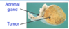
What cellular response to injury is this image portraying?

hypertrophy
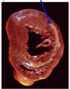
What cellular response to injury is this image portraying?

hypertrophy
Dysplasia
Cellular response to injury; Disorded growth and maturation of the cellular components of a tissue. May be a precursor to malignant neoplasia.
What cellular response to injury is the cellular tissue portraying?

hyperplasia