Orthopedics Flashcards
Draw and name the types of ulnar fracture
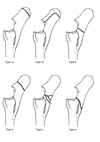
Draw and name the types of Salter-Harris fracture

What are the options for pastern arthrodesis (medical and surgical)? Include names/descriptions of implants also
Injection of 75% ethyl alcohol
Casting
Ideally: PIP arthrodesis LCP - 3 hole narrow LCP with 2 transarticular cortex screws
Dorsal 3-hole narrow DCP or LCP combined with 2 transarticular cortex screws
2 places, T-plate, Y-plate also possible (requires 5.5mm transarticular screws)
Describe the tarsal drilling technique for tarsal arthrodesis
GA, lateral/dorsal recumbency
From dorsomedial aspect of tarsus
Sterile prep and drape
3cm skin incision on dorsal medial aspect of TMT and DIT joints
Drill entry midway between line extending form groove between proximal MTII and MTIII and most dorsal asset of distal tarsus (plantar to saphenous vein).
Needles used to identify joint spaces with rads/fluoroscopy
Tracts drilled in pairs (TMT and DIT). 4.5mm drill bit
20mm directed to lateral palpable extremity of MTIV
20mm angled 30 degree to first tract in plantar direction
35mm tract angled 30 degrees to first in dorsal direction
Incision closed subcutaneous (continuous 2-0 absorbable) and skin (interrupted 2-0 absorbable)
Which is the most common digit to be a supernumerary digit
Medial aspect of forelimb in 80% of cases
List the causes of exostosis of the splint bones
Trauma:
Subperiosteal hemorrhage
Elevation of periosteum
Instability between MCIII and MCII
MCII fractures
Inflammation of intercarpal ligament:
Can result from circles on a hard surface
Or conformation abnormalities (bench knees)
Carpal varus
Describe the post-op care after splint bone removal
Post-op:
Pressure bandage for 2 weeks
Stall rest 1 month
2 months handwalking/small paddock turnout
Radiography to assess stability of proximal fragment
Drain may be required for 2-3 days
NSAIDs
ABs depending on drainage and incision
Full limb cast may be required for recovery and post-op if whole MTIV removed
What is the endurance limit of metallic implants
Maximum stress below which a material can endure an infinite number of stress cycles
What is shot peening
Done before electropolishing
Implant subjected to high-velocity impaction by metallic or ceramic particles
Produces roughened surface with increased residual compressive stress for enhanced fatigue life
List all the medical and surgical options for management of strain-induced tendinitis
Non-surgical therapies:Physical therapies:
Cold therapy:
Compression and coaptation:
Corrective shoeing
Controlled exercise:
Extracorporeal shock wave therapy:
Therapeutic ultrasound, laser and magnetic fields:
Counter-irritation: (not effective)
Pharmacologic management:Systemic medication:
Corticosteroids:
NSAIDs:
DMSO
Intralesional medication:
PSGAGs:
HA:
Component of tendon matrix
Beta-aminopropionitrile fumarate
Methylprednisolone: (avoid)
New advances: Tissue engineering approaches:
IGF-1:
Recombinant equine growth hormone:
TGF-B:
PRP:
TGF-B
VEGF
ACELL VET:
Bone marrow:
MSCs:
Surgical therapies:
Tendon splitting:
Desmotomy of the accessory ligament of the superficial digital flexor tendon:
Tenoscopy:
Bursoscopy:
Annular ligament desmotomy:
Fasciotomy and neurectomy of the deep branch of the lateral plantar nerve for the treatment of proximal suspensory ligament desmopathy:
Desmotomy or desmectomy of the accessory ligament of the deep digital flexor tendon
List the methods of diagnosis of strain-induced tendinitis
Clinical history
Palpation
Ultrasonography
Molecular markers:
PICP
COMP
Why do intrathecal tendon lesions heal more slowly than other tendon lesions
No paratenon
Reduced extrinsic repair
Synovial fluid slows repair
Describe the phases of tendon healing
Inflammatory reaction:
Increased blood flow
Edema
Neutrophils, macrophages, monocytes
Proteolytic enzymes
Also further damages tendon
Reparative phase:
After a few days, lasts several months
Angiogenesis
Fibroblastic cellular infiltration (extrinsic repair)
Limited intrinsic repair
Scar:
Higher ratio of Collage III to collage I (50% cf 10% in normal tendon)
Higher hydration
Higher GAGs
Reparative phase:
Type III to type I collagen as scar matures
Thicker collagen fibrils and cross-links increase
Mature scar less stiff than tendon but as there is more tissue, scar tissue actually more stiff
Result: strong but functionally inferior tendon, predisposing to reinjury
What are the most common intrathecal tendon lesions in the forelimb and the hindlimb
Forelimb: Bursting of lateral border of DDFT
Hindlimb: Manica flexor of SDFT
What is the prognosis for horses with a peroneus tertius rupture
78% midbody/insertion returned to previous level of work
21.7% euthanized
Premature return to exercise assocaited with re-injury
Monitor with U/S
If avulsion fracture: guarded
Age, open/closed injury, U/S size, location, duration of rehab had no influence on return on exercise
Racing at time of injury reduced prognosis
If additional structures damaged, 8 times less likely to return to soundness
Describe the procedure for semiteninosus tenectomy
GA, lateral recumbency
Landmarks:
Tibial insertion of muscle on caudomedial aspect of tibia just distal to medial femorotibial joint and caudal to saphenous vein overlying gastrocnemius muscle
8cm vertical incision made over palpable tendon and through subcutaneous and crural fascia until tendon exposed
Kelly/crile forceps passed under tendon to isolate from muscle and tendon transected
Resection of 3cm segment (prevents of delays recurrence)
Fascial layers closed with interrupted or continuous synthetic absorbable sutures
Skin closed with interrupted or continuous non-absorbable suture
Pull limb forward; if tendon of insertion of semitendinosus muscle onto calcaleal tuber taut:
3-4cm incision directly over tendon (caudal and distal to first incision)
Isolate and transect
How does fibrotic myopathy occur
Adhesions and fibrosis of semitendinosus (or semimembranosus, biceps femoris, gracilis) muscle
Secondary to IM injections, trauma (lacerations, slipping, kicks) or tearing insertion of semitendinosus while barrel racing, lameness
Can be caused temporarily by breach bar
Can occur as neonates
If involves both limbs, likely neuropathy is the cause
Ossifying myopathy: when bone forms in affected tissue
Describe the procedure of a lateral digital extensor penectomy and partial myectomy
Remove distal 2-10cm of LDE muscle and entire tendon
Standing or GA (can remove more muscle under GA)
Sites:
Junction of LDE tendon with log digital extensor tendon on lateral aspect of metatarsus
LDE 2cm proximal to lateral malleolus
Distal incision made directly over tendon just proximal to junction with long digital extensor tendon
Blunt dissection beneath tendon with curved kelly or Ochsner forceps
Proximal incision on lateral aspect of limb 6cm above lateral malleolus (skin, subQ and fascia directly over lateral digital muscle parallel with muscle fibers)
Blunt dissection to expose muscle belly and heavy curved instrument placed underneath it
Sever at distal incision and pull through by traction on proximal section with curved Ochsner forceps of Mayo scissors
Muscle severed at proximal aspect of incision, ensuring at least 2cm muscle removed
Close fascia proximally with simple interrupted or continuous USP 0 absorbably suture
Subcutaneous 2-0 absorbable simple continuous
Skin non-absorbable simple continuous
Distal incision with skin sutures only
Sterile dressing and whole limb bandage 10-14 days
Stall rest 2 weeks
1 week hand-walking
Normal exercise in 2-4 weeks
What are the treatments for cribbing (medical and surgical)
Non-surgical:
Pasture turnout
Remove objects that horse cribs on
Cribbing straps
Acupuncture
Aversion therapy
Surgical:
Forssell procedure
Modified Forssell procedure
Bilateral neurecomy of the ventral branch of the spinal accessory nerves
Surgery of choice is to combine modified Forssell procedure with bilateral neurectomy
Puncture of the sole of the foot by a nail can involve which structures
P3
Navicular bone
DIP jt
Navicular bursa
DDFT
DFTS
Sole
Digital cushion
Laminae
Heel bulbs
Palmar cartilages of P3
Collateral ligaments of DIP jt
Impar ligament
How should keratomas be managed
Remove keratoma up to origin:
Can resolve inflammatory process first or can remove immediately - depends on level of lameness
Altered horn and altered sensitive lamina must be removed
Surgery:
Tourniquet
Standing/ring block or GA
Remove as much horn as possible with horse standing until Dremel tool exchanged for scalpel and curettes
Aseptic prep
Altered lamina and entire keratoma removed in toto
Aseptic pressure bandage applied to plalangeal region
Bandage changes at 3-4 days intervals under aspectic conditions
Support to hoof wall
Medication plate can be applied as soon as granulation tissue
Fill hoof wall defect with artificial horn as soon as sensitive lamina healed
Shoe with large clips on either side of defect
Post-op:
Stall rest 4-6 weeks
Reshod
Light walking after 2-4 months if healing good
If re-infection, remove all affected tissues and start process again
List the methods of treating canker
Removal of abnormal tissue surgically using knife/blade
Cryotherapy
Surgery likely to be repeated
Clean with povidone-iodine with bandage changes every 2-3 days
Apply shoe with pad
Daily bandage changes with 20g iodoform iodine, 20g zinc oxide, 20g tannic acid, 40g metronidazole
OR Chloramphenicol + metronidazole
Systemic ABs if more than one hoof affected (doxycycline or oxytetracycline)
Biotin and zinc added to feed
List and draw the types of P3 fracture
What are the surgical options for treating laminitis
DDF tenotomy (mid carpal or pastern approach)
Hoof wall resection
List all the causes of ALD
Perinatal:Incomplete ossification:Mare:
Placentitis
Metabolic disease
Parasite infection
Colic
Foal:
Premature
Twins
Uneven loading of joints due to mild ALD
Osteochondral fractures may occur in severely dysmature (esp dorsal aspect small tarsal bones)
Laxity of periarticular structures:
If ALDs of several regions and rotational deformities
Due to laxity or soft tissue trauma
Laxity lead to abnormal loading and can incide ALD if incomplete ossification
Causes:
Hormonal imbalance
Intrauterine positioning
Aberrant intrauterine ossification:
Deformed long bone at birth
Caused by mechanical factors leading to deformation of precursor cartilage
Distal physeal region on MTIII involved; triangular epiphysis and varus deformity
Developmental:Unbalanced nutrition:
Excessive intake - too much grain from crib feeding
Unbalanced trace minerals:Caused by:
Zinc toxicity
Copper deficiency
Ca:P
Excessive exercise and trauma:
Microfractures/crushing of proliferative zone
Type V Salter-Harris
Epiphyseal fractures from kicks
Physeal trauma:
Salter-Harris type V or IV
Local retardation on medial or lateral aspect
Compensatory:
Proximal phalanx due to prolonged loading distal to deviation
List the methods of treatment for ALD
Stall rest
Controlled exercise
Splints and casts
Hoof manipulation
Radial pressure wave therapy
HCPTE
Transphyseal staple
Transphyseal bridge (screws and wire)
Transphyseal screw
At what age does the rapid growth stage end for: MCIII/MTIII and proximal phalanx; tibia and radius
MCIII/MTIII proximal phalanx: 2 months
Tibia: 4 months
Radius: 6 months
What are the landmarks for PE at the distal radial physis
3cm vertical incision between CDE and LDE tendons, starting from point 4-5cm proximal to distal physis of radius and continuing proximally
Which ALD, when it is mild, is protective against carpal fracture and effusion
Carpal valgus
List the causes of congenital flexural limb deformity
Intrauterine malpositioning:
Rare
If large foal
Diseases acquired by mare during pregnancy
Agents ingested during pregnancy:
Locoweed
Hybrid Sudan grass
Dominant gene mutation in sire
Equine gioter
Influenza
Neuromusclar disorders
Lathyrism:
Defects in cross-linking of elastin and collagen
Glycogen branching enzyme deficiency:
QH (transient flexural deformity)
What are the treatment options for digital hypertension deformities
Swimming
Farriery:
Shorten toe
Rasp palmar half of foot
Heel extensions
Bandaging :
Phalangeal region
Tenoplasty: Mini foals - NOT recommended
How does oxytetracycline work
3g in 250-500mL physiologic saline administered slowly IV. Administer 2-3 times in first week.
Induces dose-dependent inhibition of collagen gel contraction by equine myofibroblasts and indices a dose-depended decrease in MMP-1 mRNA expression by myofibroblasts. Basically oxytetracycline inhibits tractional structuring of collagen fibrils by equine myofibroblasts through an MMP-1-mediated mechanism.
What are the treatment options for treating acquired flexural limb deformity of the DIP jt
Nutrition:
Early weaning
Decrease mare ration
Decrease foal concentrate ration
Evaluate soil and water for trace mineral composition
Physiotherapy and exercise:
Controlled exercise
Analgesia:
Farriery:
Protect toe
Toe extension if small deformity
Rasp heels if in contact with ground
Cast:
10-14 days maximum
Surgery:
Desmotomy of ALDDFT
DDFT tenotomy
Which vessel can be damaged and is usually ligated in a medial approach to performing desmotomy of the ALSDFT
Cephalic
List the type of sesamoid fracture
Apical
Midbody]Basal
Abaxial
Sagittal
Comminuted
Regarding all types of sesamoid fractures, what percentage returned to racing after surgical and conservative management
Surgery: 64%
Conservative: 37%
What are the 4 types of P2 fracture
Dorsal or palmar/plantar intra-articular osteochondral chip fractures
Palmar/plantar eminence fractures
Axial fractures
Comminuted fractures
What are the treatment options for lunation/subluxation of the PIP jt
Arthrodesis
But some cases are due to excessive tension on DDFT so transection of medial head of DDFT can help
What the tenoscopy landmarks for transection of the PAL
The arthroscope is inserted just distal to the PAL halfway between the digital neurovascular bundle and the ergot. The lateral or medial entrance portal is positioned lateral or medial to the respective edge of the SDFT.
The instrument portal is made 5 to 10 mm proximal to the PAL in the DFTS out pouching almost lateral to the SDFT.
Where should the distal screw be placed in a lateral condylar fracture
Centrally in the lateral condylar fossa
What is the prognosis for return to racing for displaced lateral condylar fractures
50%
Overall, what percentage of condylar fractures return to racing
70-80%
Where should plates be positioned for diaphyseal fractures of MCIII
Dorsolaterally and dorsomedially
Define a saucer fracture
A dorsal cortical fracture that curves proximal and courses back to the dorsal cortex
List the ultrasonographic features that indicate tendon injury
Enlargement
Hypoechogenicity
Reduced striated pattern
Changes in shape, margin, position
Irregular striated pattern indicates fibrosis
Heterogenous pattern indicates chronic tendinopathy
What intralesional medications can be used in tendon injury
PSGAGs:
Inhibit collagenases and metalloproteinases
Inhibit macrophage activation
Intralesion or M
76% return to work vs 46% controls
Improved echogenicity of U/S
HA:
Component of tendon matrix
Contains:
D-glucuronic acid
N-acetyl-D-glucosamine
Peritendinous, intralesional, intrathecal, systemically
No difference in reinjury
Less tendon enlargement cf controls
Peritendinous injection may reduce lameness
Decreases adhesions when administered intrathecally
Decreases inflammation and hemorrhage
Beta-aminopropionitrile fumarate
Methylprednisolone:
Dystrophic mineralization and tissue necrosis - avoid
Ultrasonographic guidance standing or GA
2.5cm 22Ga needle for not treatments
NOT for first 3 days after injury as can increase hemorrhage
Large volumes can be damaging
New advances: Tissue engineering approaches:IGF-1:
Stimulates extracellular tendon matric synthesis
Mitogen
Decreases initial swelling
Recombinant equine growth hormone:
IM
Decreased yield point and ultimate tensile strength
TGF-B:
Fewer reinjuries at site but more on contralateral limbs
More tendon enlargement
PRP:
Plasma with at least twice the platelet concentration of normal plasma
Contains:
PDGF
TGF-B
VEGF
Stimulates cell proliferation and matrix synthesis
ACELL VET:
Intralesional therapy using acelllular tissue from porcine urinary bladder submucosa
Bone marrow:
Intralesional
MSCs:
Differentiate into tenocytes to regenerate tendon matrix
Functionally superior repear
Reinjury rate 26% (in 3 years) - improved from conventional treatment
What is the purpose of tendon splitting
Decompresses core lesion by evacuating serum or hemorrhage and facilitate vascular ingrowth, may reduce propagation of lesion
Faster resolution of lesion, quicker revascularization and increased collagen deposition cf controls
What is the prognosis for return to function following busoscopy for DDFT tears
28%
Describe the procedure for a neurectomy and fasciotomy for hindlimb proximal suspensory desmitis
GA, dorsal recumbency
4-6cm incision adjacent to lateral border of SDFT, originating proximally from level of chestnut
Plantar metatarsal fascia incised and incision extended deep to SDFT by blunt dissection, facilitated by retraction of SDFT
Deep branch of lateral plantar nerve located and transected using scalpel and 3cm section removed
Fasciotomy performed adjacent to lateral splint bone
Describe the pathophysiology of P2 plantar eminence fractures
Uniaxial or biaxial
Result of hyperextension of PIP jt, with tension on palmar/plantar attachments of SDFT, middle scutum and distal sesamoidean ligaments
Occasionally, soft tissues can be disrupted without bone damage
List the treatment options for a subchondral cystic lesion of P2
IA HA - temporary pain relief
Surgical curettage:
Transosseous drilling
Small drill bit under fluoroscopic control (inject saline into joint to confirm correct placement of drill)
Enlarge to 5.5mm drill bit
Allows access of curette to evacuate cyst
Lavage joint
Cyst and drill hole filled with tricalcium phosphate granules (or fibrous gel with PTH1-34)
Arthrodesis (but multiple lesions have a poor response)
What is the ethology of proximodorsal osteochondral fractures of P1
Common in racehorses and non-racehorses
Caused by hyperextension of MCP jt with impact of proximal and dorsal aspect of proximal phalanx onto dorsal region of MCIII
Does not seem to be genuine OC
List the consequences of not removing proximodorsal osteochondral fractures of P1
Erosion of opposing metacarpal condyle (lameness)
Synovitis
Cartilage degeneration
Villonodular synovitis
What are the two types of palmar/plantar osteochondral fractures of P1
Type I fractures:
Avulsed from axial, proximal, plantar or palmar rim of proximal phalanx and are mostly articular
Insertion of short sesamoidean ligament still attached to avulsed fragment
Minimal lameness, usually at trot
Type II fractures:
Larger, abaxially located, partly articular osteochondral fragments
Extend distad 2-3cm and contain minimal articular cartilage
No persistent lameness
May constitute delayed form of ossification
How should a closed fetlock lunation be treated?
Cast 6 weeks
Apply under GA
Discuss the treatment of chronic proliferative (villonodular) synovitis of the fetlock and how size of the lesion influences treatment
If <4mm:
IA atropine and steroids
If >4mm:Surgery:Arthroscopy:Small masses:
Synovectomy instruments, guarded scalpels, biopst suction punch rongeur, radiofrequency probe
Large masses:
Large biopsy punch rongeurs, CO2 or diode laser
Athrotomy
Describe the treatment of sagittal fractures of the proximal sesamoid bone
3.5mm cortex screws in lag fashion in lateral-to-medial orientation
MCP arthrodesis occasionally necessary:
If injury to intereseamoidean ligaments or P1 fracture also
Post-op:
Cast for 2-3 weeks
Then heavy bandage
Retire from racing as usually concurrent condylar fracture
What is the prognosis for return to function for basilar sesamoid fractures?
Poor
Inverse relationship between dorsopalmar fragment length and likelihood of return to racing
Basal osteochondral fragments that do not extend to palmar surface: 59% return to racing
57% return to racing if fragment involves <25% of base
40% return to racing if fragment involves >25% of base
What is the aetiology of palmar osteochondral disease (POD) of the fetlock joint?
Trauma:
Accumulated stress and sclerosis developing during racing, particularly hyperextension
Degenerate metacarpal condyle has acellular and necrotic bone, with zone of new bone formation deep to this: remodelling and fracture changes
Early lesions have little cartilage damage, however, eventual fracture and displacement of subchondral bone results in complete bone and cartilage loss
