Orthopaedic for the MRCS part A Flashcards
Define ankle fractures
A fracture around the tibia-talar joint of any malleolus(lateral,medial,or posterior) with or without disruption to the syndesmosis
What is the location of the ankle fracture?
A fracture involving the
(1) Lateral malleolus and/or
(2) Medial malleolus and/or
(3) Posterior malleolus
What is the incidence of ankle fractures?
Affect men and women equally
1st/Generally-ankle fractures are common and account for approx.10% of all fractures seen in trauma setting
2nd/Sex:(1)Men-have a higher rate as young adults due to sports and contact injuries
(2)Women-have a higher rate old or menopausal causing fragility type fracture
What is the general incidence of ankle fractures?
(1) ankle fractures are common and account for approx.10% of all fractures seen in trauma setting
(2) affect men and women equally
What is the sex incidence of ankle fractures?
(1) Men-have a higher rate as young adults due to sports and contact injuries
(2) Women-have a higher rate old or menopausal causing fragility type fracture
What is the incidence of ankle fractures in men?
have a higher rate as young adults due to sports and contact injuries
What is the age incidence of ankle fractures?
(1) Men-young adults
(2) Women-old or post menopausal
What is the incidence of ankle fractures in women?
have a higher rate old or post-menopausal causing fragility type fracture
Why ankle fractures occur in men?
Due to sports and contact injuries
What is most common period ankle fractures occur in women?
Old or post menopausal
What ankle fractures cause during post menopausal period in women?
Fragility type fractures
What are the types of ankle fractures?
(1) Tillaux-fracture occurs during the unique closure pattern of the distal tibial physis
(2) Pilon-occurs at the bottom of the tibia(shinbone)which is the tibial plafond,i.e.,tibial articular surface.
- involves the weight-bearing surface of the ankle joint -is a separate injury
Define Tillaux ankle fracture
fracture occurs during the unique closure pattern of the distal tibial physis
Define Pilon ankle fracture
- occurs at the bottom of the tibia(shinbone)which is the tibial plafond,i.e.,tibial articular surface
- involves the weight-bearing surface of the ankle joint -is a saparate injury
Discuss osseos anatomy in relation to ankle fractures

Discuss ligamentous anatomy in relation to ankle fractures

What is the other name of medial side of the ankle in relation to the ligamentous anatomy as explanation of ankle fracture?
Deltoid ligament
What is the division of medial side of the ankle in relation to the ligamentous anatomy as explanation of ankle fracture?
Divided into
(1) Superficial portion
(2) Deep portion
What is the function of medial side of the ankle in relation to the ligamentous anatomy as explanation of ankle fracture?
Is the primary restraint to valgus tilting of the talus
What are the components of lateral side of the ankle in relation to the ligamentous anatomy as explanation of ankle fracture?
The lateral side of the ankle consists from anterior to posterior of:-
(1) Anterior talofibular ligament(ATFL)
(2) Calcaneofibular ligament(CFL)
(3) Posterior talofibular ligament(PTFL)
What is the other name of lateral side of the ankle in relation to the ligamentous anatomy as explanation of ankle fracture?
Lateral ligament complex
What is the function of lateral side of the ankle in relation to the ligamentous anatomy as explanation of ankle fracture?
All the 3 ligaments on the lateral side of the ankle(ATFL,CFL and PTFL)
(1) resist valgus stress to the ankle
(2) are a restrain to the anterior translation of the talus within the Morris joint
Discuss syndesmosis

Define syndesmosis
Is a ligament complex between the distal tibia and fibula,holding the two bones together and consists of a very strong fibrous structure
What is the location of the syndesmosis?
Between the distal tibia and fibula
What is the function of the syndesmosis?
(1) Holds the tibia and fibula together
(2) Stability of the ankle-It is fundamental to the integrity of the ankle joint,and its disruption leads to instability
What does syndesmosis of ankle joint consist of?
Mnemonic;A TIP
It consists of(from anterior to posterior)the:-
(1) Anterior-inferior tibiofibular ligament(AITFL)
(2) Transverse tibiofibular ligament(TTFL)
(3) Interosseous membrane
(4) Posterior inferior tibiofibular ligament(PITFL)
Discuss clinical picture of ankle fractures

What is the usual presentation of a traumatic ankle fracture?

Discuss Ottawa rules

Discuss imaging of ankle fracture

Discuss XRs for ankle fractures

What are the indications of AP-lateral and mortise views in ankle fractures?

Define mortise view in imaging of ankle joint
20 degrees internal rotation
What are the indications(i.e.,how do we know) of imaging in syndesmosis injury?

What kind of imaging is done for syndesmosis injury in ankle fractures?
XRs(AP,lateral and mortise view(20 degrees internal rotation))
What should be the position of the ankle joint on imaging and why?
Dorsiflexed

What are the indications of stress radiographs in ankle fractures?

What is the indication of CT in syndesmosis injury?
used for surgical planning

When is appropriate time of doing plain radiographs in ankle fractures?

Discuss classification of ankle fractures

Discuss anatomical classification of ankle fracture?

Discuss Lauge Hansen classification of ankle fractures

What are the parts of Lauge-Hansen classification of ankle fractures?

What are the types of Lauge-Hansen classification of ankle fractures?

What are the indications of Lauge-Hansen classification?

What is the classical feature of the Lauge-Hansen classification system?
used widely in orthopaedic practice as it is much more detailed than Denise-Weber classification
Discuss Denis-Weber classification of ankle fractures

What is the frequency of use of Denise-Weber classification in ankle fractures?
Commonly used
What are the indications of Denis-Weber classification of ankle fractures?

What are the types of Denis-Weber classification of ankle fractures?

plain radiography demonstrating Denise-Weber classification system

Compare with a picture between Lauge-Hansen and Denis-Weber classification of ankle fractures

Discuss management of ankle fractures

What is the initial management of ankle fractures?

Discuss general principles of initial management of ankle fractures

How do you manage high energy ankle injuries?
Management should follow ATLS principles to identify more significant injuries first
How do you manage open ankle injuries?
Management should be in line with BOAST 4 principles
How do you manage ankle deformities and dislocation?
(1) Reduce obvious deformity with appropriate analgesia or conscious sedation
(2) Radiographs of clearly deformed or dislocated joints are not necessary
(3) Removing the pressure on the surrounding soft tissues from the underlying bony deformity is the priority
Enumerate indications of conservative management of ankle fractures

How do you define stability of ankle fracture and what is the treatment of each definition?
+often involves (1)stress radiographs (2)a trial of mobilisation (3)repeat radiographs
+defining unstability is a subject of much ongoing research

How do you define stability of ankle fractures?
+often involves (1)stress radiographs (2)a trial of mobilisation (3)repeat radiographs
+defining unstability is a subject of much ongoing research
What is the definition and treatment of Weber A ankle fracture ?

What is the definition and treatment of Weber B ankle fracture ?

What is the definition and treatment of Weber C ankle fracture ?

Summerise management of different Weber classifications

Discuss some examples for defining stability of ankle fracture to underpin the treatment decision

Discuss operative fixation of ankle fractures

Discuss operative fixation of ankle fractures

Discuss internal fixation of ankle fractures?

What is the method of internal fixation of ankle fractures?

What is the prerequisite of internal fixation of ankle fractures?

Why open reduction and internal fixation(ORIF) is often required for ankle fractures?
(1)To achieve stable anatomical reduction of the talus within the the ankle mortise. (2)The type of operative procedure peformed depends on the specific type of ankle fracture sustiand
Enumerate indications of open reduction and internal fixation(ORIF)

Discuss external fixation of ankle fractures

What is the method of external fixation?
External fixation,or with a hind foot nail
What is the prerequisite for external fixation?
Where soft tissue or bone quality is poor
Discuss post operative management of ankle fractures

What is the duration and reason for casts post operatively in ankle fractures?
Duration 6 weeks
Reason (a)6 weeks is an appropriate time period to keep cast on in a conservatively managed patient
(b) ankle fractures generally take 6 weeks to(1)unite enough
(2) prevent secondary displacement
What are the factors that weight bearing post operatively depends on?

What is the time taken for a patient with ankle fractures to return to activities and what does it require?

What is the differential diagnosis of the ankle fracture?

Define ankle sprain?
Ligamentous injury
What is the incidence of ankle sprain?
Much more common
What is the aetiology of ankle sprain?
Inversion injury on a plantarflexed ankle
Discuss classification of ankle sprains
(1) High ankle sprains Injuries to the syndesmosis
(2) Low ankle sprains Injuries to the: (1)Anterior inferior talofibular ligament(AITFL) (2)Calcaneofibular ligamnet(CFL)-the commonest (3)Posterior inferior talofibular ligament(PITFL)
Define high ankle sprains
Injuries to the syndesmosis
Define low ankle sprains
Injuries to the: (1)Anterior inferior talofibular ligament(AITFL) (2)Calcaneofibular ligament(CFL)-the commonest (3)Posterior inferior talofibular ligament(PITFL)
What is the most common ligament to be injured in low ankle sprains?
Calcaneofibular ligament(CFL)
Discuss the clinical picture of ankle sprains
(1)Significant ankle pain and swelling. (2)No weight bearing (3)Finger tenderness distal to the malleoli over the affected ligament
Discuss investigations of ankle sprain
Plain film radiograph +the image of choice +to rule out any bony injury
Discuss management of ankle sprains
Conservative(Mnemonic;ICE/A)
(1) Early Immobilisation
(2) Cold compression and ice
(3) Elevation
(4) Analgesia
Enumerate complications of ankle fractures

Discuss Maisonneuve fracture
Definition A combination of: (1)High proximal tibia fracture(high Weber C) (2)Unstable ankle injury
Significance (1)It is a high fibula fracture which is above the syndesmsis(high Weber C).Therefore,it may be an ankle fracture. (2)Indicates unsable ankle injury with likely injury to the iterosseous membrane.Consequently,it can be associated with ankle instability
Imaging Plain radiograph shows evidence of syndesmotic widening Management Surgical fixation-to reduce and stabilise the syndesmosis
Define Maisonneuve fracture
A combination of: (1)High proximal tibia fracture(high Weber C) (2)Unstable ankle injury
What is the significance of Maisonneuve fracture?
(1)It is a high fibula fracture which is above the syndesmsis(high Weber C).Therefore,it may be an ankle fracture. (2)Indicates unsable ankle injury with likely injury to the iterosseous membrane.Consequently,it can be associated with ankle instability
What does a plain radiograph show in Maisonneuve fracture?
Plain radiograph shows evidence of syndesmotic widening
What is the management of Maisonneuve fracture?
Surgical fixation-to reduce and stabilise the syndesmosis
Define avascular necrosis

Enumerate causes of avascular necrosis

Explain the clinical picture of avascular necrosis of the bone

Discuss in brief imaging of avascular necrosis of the bones

Discuss treatment of avascular necrosis of bone

What are the other names of Perthes disease?
(1)Legg-Calve-Perthes disease. (2)Avascular necrosis of the femoral head
Define Perthes disease
Idiopathic avascular necrosis of the femoral epiphysis of the femoral head causing a self limiting disease of the femoral head comprising of: (1)Necrosis. (2)Collapse
(3)Repair and remodelling
Discuss incidence of Perthes disease?
(1) In general-approximately 1:10000 (2)Sex-males 4 times greater than females(male to female ratio 4:1)
(3) Age-those who are small for their ge and between the overall age 2-12
- rare < 4 years
- common in average 4-8 years(in some other resources 5-7 years) with a limp. -the younger the age of onset,the better the prognosis

What is the aetiology of Perthes disease?
A transient disruption in the blood supply to the femoral head
What is the pathogenesis of Perthes disease?

What is the clinical picture of Perthes disease?
(1)Limping with hip pain(may be referred to the knee)
(2)Bilateral in 20%
(3)Decreased abduction and internal rotation
Mnemonic;PIRAB=Perthes….Internal rotation….Abduction

What are the symptoms of Perthes disease?

What is the chance of Perthes disease being bilateral?
10-20%
Describe hip pain of a patient with Perthes disease
(1)Onset:starts and worsens over few weeks to months (2)On activity, especially,on internal and external rotation (3)Intermittent + no history of trauma
Describe knee pain in a patient with Perthes disease?
(1)Chronic (2)With normal knee examination (3)Lasts for several hours
What are the investigations(diagnosis) of Perthes disease?
(1) Plain XRs
(2) Technitium-99 bone scan-shows earliest avascular change
(3) MRI-Indications:a)if normal XRs
b) symptoms persist

Discuss catteral staging of Perthes disease?

What is catteral stage 1 in Perthes disease?
Clinical and histological features only
What is catteral stage 2 in Perthes disease?
(1)Sclerosis with or without cystic changes (2)Preservation of the articular surface
What is catteral stage 3 in Perthes disease?
Loss of structural integrity of the femoral head
What is catteral stage 4 in Perthes disease?
Loss of acetabular integrity
Discuss the role of plain XRs in diagnosing Perthes disease?

What is the disadvantage of plain XRs in a patient with Perthes disease?
Early disease can be missed on XRs
What are the early changes that could be seen on plain XRs of a patient?

What are the changes that could be seen in more advanced cases of Perthes disease?
Fragmentation of the femoral head
What is the role of MRI in diagnosing Perthes disease?

What are the indications of MRI in diagnosing Perthes disease?
(1)If normal XRs and (2)Symptoms persist
What are the findings that could be seen on MRI of a patient with Perthes disease?

What is the role of technitium 99 bone scan in diagnosing Perthes disease?
It is an alternative option
What is the management of Perthes disease?
(1) Remove pressure from joint to allow normal development
(2) Physiotherapy
(3) Usually self limiting if diagnosed and treated promptly

What is the indication of treatment of perthes disease?

What is the main objective of management of Perthes disease?
To keep the femoral head within the acetabulum by cast,braces or surgery
What should be done in managing a patient with perthes disease < 6 years?
Observation and symptomatic treatment
What should be done in managing a patient with Perthes disease between 6-8 years?
Brace or surgical management with moderate results
What should be done in managing a patient with perthes disease > 8 years?
Surgical containment:(femoral/pelvic)osteotomy
What should be done in managing a patient with perthes disease and has severe deformities?
Operate
What is the prognosis of Perthes disease?
Early diagnosis improves outcome

Define ankylosing spondylitis?
A type of artheritis in which there is a long term or chronic inflammation of the joints of the spine or the axial skeleton
What are the feature of ankylosing spondylitis?

What are the general features of ankylosing spondylitis?

What are the clinical features of ankylosing spondylitis?

What the other name for the early cases of ankylosing spondylitis?
Uncomplicated cases
What are the clinical features of early cases of the ankylosing spondylitis?

What the other name for the advanced cases in ankylosing spondylitis?
Complicated cases
What are the clinical features of advanced cases of the ankylosing spondylitis?

What are the sites affected by ankylosing spondylitis?

What are the joints affected by ankylosing spondylitis?

Define the typical joints affected by ankylosing spondylitis?
Where the spine joins the pelvis
What are the other joints affected by ankylosing spondylitis?
e.g.,shoulder
What condition affects the joints in association with ankylosing spondylitis?
Psoriatc artheritis
What the sites,other than the joints,that are affected by ankylosing spondylitis?
Eyes(acute unilateral anterior uveitis) and bowel(IBD,especially ulcerative colitis in which there is a strong association with HLA B27 in patient with ankylosing spondylitis)problems may also occur
What condition affects the eyes in patients with ankylosing spondylitis?
acute unilateral anterior uveitis
What condition affects the bowel in patients with ankylosing spondylitis?
inflammatory bowel disease(IBD),especially ulcerative colitis in which there is strong association with HLA B27 in patients with ankylosing spondylitis
What type of inflammatory bowel disease affects patients with ankylosing spondylitis?
Ulcerative colitis in which there is a strong association with HLA B27 in patients with ankylosing spondylitis
Comment on the back pain in ankylosing spondylitis
(1)The characteristic symptoms of ankylosing spondylitis (2)Often comes and goes(i.e.,on and off)
What is the characteristic symptom of ankylosing spondylitis?
Back pain
What is the character of back pain in ankylosing spondylitis?
Often comes an goes(i.e.,on and off)
Comment on the joint stiffness in ankylosing spondylitis
Worsens over time
What are the typical spinal features of ankylosing spondylitis?
Typical spinal features which may be seen in a young patient and are suggestive of ankylosing spondylitis: (1)loss of lumbar lordosis (2)restrictions of spinal movement
(3)progressive spinal deformities (4)progressive kyphosis of the cervico-thoracic spine
Comment on the typical spinal features in ankylosing spondylitis
(1)seen in young patients (2)suggestive of ankylosing spondylitis
What is the age incidence of the typical spinal features in ankylosing spondylitis?
young patients
What the presence of the typical spinal features of ankylosing spondylitis actually means?
Suggestive of ankylosing spondylitis
What are the investigations of ankylosing spondylitis?

Name one specific physical test for ankylosing spondylitis
Schober test
What are the blood tests for investigating ankylosing spondylitis?

Comment on the ESR in ankylosing spondylitis
Raised
What is the association of ankylosing spondylitis?
HLA B27 in up to 20%
Comment on HLA B27 association in ankylosing spondylitis

What is the incidence of HLA B27 association in ankylosing spondylitis
20%
What is the other disease associated with HLA DR27 only if the patient is affected by ankylosing spondylitis?
Ulcerative colitis
What do the radiographs show in patients with ankylosing spondylitis?
the classical bamboo spine appearance

What is the one specific radiographic sign suggestive of ankylosing spondylitis?
the classical bamboo spine appearance

What is the treatment of ankylosing spondylitis?

What is the treament of the early cases in ankylosing spondylitis?

What the symptomatic treatment of ankylosing spondylitis?
NSAIDs
Comment on the use of NSAIDs in ankylosing spondylitis?
Should be carefully used in patients with IBD who may be taking steroids
What is the treatment of advanced cases of ankylosing spondylitis?

What is the indication of spinal decompression in patients with ankylosing spondylitis?
For complicated cases with progressive neurological deficit
Draw a diagram to illustrate the difference between early and advanced case in ankylosing spondylitis

Define spondylolysis
Congenital or acquired deficiency of the pars interarticularis of the neural arch of a particular vertebral body,usually affects L4/L5
What are the vertebrae affected by spondylolysis?
L4/L5
What is the incidence of spondylolysis?
Up to 5% of the population
What are the symptoms of spondylolysis?
(1) Asymptomatic
(2) Spondylolysis is the commonest cause of spondylolisthesis in children
What is the treatment of spondylolysis?
Asymptomatic cases do not require treatment
Define spondylolisthesis
This occurs when one vertebra is displaced relative to its immediate inferior vertebral body leading to an abnormal forward slip of one vertebral body on another
What is the incidence of sponylolisthesis?
a young atheletic female with a background of spondylolysis and presents with a sudden pain
What is the aetiology of spondylolisthesis?
(1) Stress fracture
(2) Spondylolysis-is a risk factor for a young atheletic female with a background of spondylolysis and presents with a sudden pain
(3) Trauma
What are the investigations(diagnosis) of spondylolisthesis?
Plain films-traumatic cases show the classic ‘Scotty Dog’ appearance on plain films
What are the factors on which treatment of spondylolisthesis depend on?
(1) Extent of deformity
(2) Associated neurological symptoms
What is the treatment of spondylolisthesis?
(1) Active observation-Minor cases may be actively monitored
(2) Surgery with spinal decompression and stabilisation-Individuals with radicular symptoms or signs require spinal decompression and stabilisation
What is characteristic feature of observation of a patient with spondylolisthesis?
Active observation or monitoring
What is the indication of active observation of a patient with spondylisthesis?
Minor cases should be actively monitored
What type of sugery is performed for a patient with spondylolisthesis?
Surgical decompression and stibilisation
What is the indication of surgery in spondylolisthesis?
Radicular symptoms or signs
What other names for sheuermann’s disease?
(1)Juvenile kyphosis (2)Juvenile discogenic disease. (3)Vertebral epiphysitis
Define Scheuermann’s disease
Epiphysitis of the vertebral joints
What is the main pathological process in Scheuermann’s disease?
Epiphysitis of the vertebral joints
What is the incidence of Scheuermann’s disease?
Predominately affects adolescents
What is the clinical picture of Scheuermann’s disease?
(1) Back pain
(2) Stiffness
(3) Progressive kyphosis(at least 3 vertebrae must be involved)
What is the imaging in Scheuermann’s disease and what does it show?
XRs changes include
(1) Epiphyseal plate
(2) Anterior wedging
What is the management(treatment) of Scheuermann’s disease?
(1) Minor cases-managed with physiotherapy and analgesia
(2) More severe cases-require bracing or surgical stabilisation
Define scoliosis
Lateral curvature of the spine in the coronal plane
Discuss the types of scoliosis?
(I)Structural
+Feature:1)affects more than 1 vertebral body
2)not corrected by alterations in posture
+Types:1)Idiopathic-the most common type
2)Congenital
3)Neuromuscular
+Management:Severe or progressive structural disease is managed surgically with bilateral rod stabilisation of the spine
(II)Non structural(postural)
+Incidence:commonest in adolescent females who develop minor postural changes only
+Feature:typically disappear on manoeuvres such as bending forwards
Discuss structural scoliosis
+Feature:1)affects more than 1 vertebral body
2)not corrected by alterations in posture
+Types:1)Idiopathic-the most common type
2)Congenital
3)Neuromuscular
+Management:Severe or progressive structural disease is managed surgically with bilateral rod stabilisation of the spine
What are the features of structural scoliosis?
1) affects more than 1 vertebral body
2) not corrected by alterations in posture
What are the types of structural scoliosis?
1) Idiopathic-the most common type
2) Congenital
3) Neuromuscular
What is the most common type of structural scoliosis?
Idiopathic
What is the management of structural scoliosis?
Severe or progressive structural disease is managed surgically with bilateral rod stabilisation of the spine
What is the other name for non structural scoliosis?
Postural
Discuss non structural(postural)scoliosis
+Incidence:commonest in adolescent females who develop minor postural changes only
+Feature:typically disappear on manoeuvres such as bending forwards
What is the incidence of non structural(postural)scoliosis ?
commonest in adolescent females who develop minor postural changes only
What is the feature of non structural(postural)scoliosis?
typically disappear on manoeuvres such as bending forwards
Define spina bifida
No fusion of the vertebral arches during embryonic development
What are the types of spina bifida?
(1) Myelomeningocele
(2) Spina bifida occulta
(3) Meningocele
Discuss myelomeningocele
(1) the most severe type of spina bifida
(2) associated with neurological defects that may persist in spite of anatomical closure of the spina bifida defect
Discuss spina bifida occulta
+Incidence:up to 10% of population
+C/P:(1)the skin and tissues(but not the bone) develop over the distal cord
(2)the site is identified by a birth mark or hair batch
What is the incidence of spina bifida occulta?
up to 10% of population
What are the clinical features of spina bifida occulta?
(1) the skin and tissues(but not the bone) develop over the distal cord
(2) the site is identified by a birth mark or hair batch
What is the treatment of spina bifida?
The incidence of spina bifida is reduced by the use of folic acid during pregnancy
Discuss dorsal column lesion
+Feature:loss of vibration and proprioception
+e.g:Tabes dorsalis,SACD
Discuss spinothalamic tract lesion
Loss of pain,sensation and temperature
Discuss osteomyelitis
Aetiology
(1) Staph aureus in IVDU
(2) Fungal infections in immunocompromised
Features
(1) Normally progressive
(2) Normally cervical region affected
(3) Thoracic region affected in TB
What is the aetiology of osteomyelitis?
(1) Staph aureus in IVDU
(2) Fungal infections in immunocompromised
What are the features of osteomyelitis?
(1) Normally progressive
(2) Normally cervical region affected
(3) Thoracic region affected in TB
What are the features of infarction of spinal cord?
Dorsal column signs(loss of proprioception and fine discrimination)
What are the features of cord compression?
(1) UMN signs
(2) Haematoma
(3) Fracture
(4) Malignancy
What is the cause of central cord lesion?
Usually seen in older patients with cervical spondylolysis
What are the features of central cord lesion?
(1)Flaccid paralysis of the upper limbs
(2)Preserved motor and sensory fibres to lower limb(these are located prepherally)
What is the aetiology of anterior cord syndrome?
(1) Common after compression fractures
(2) Often damage to anterior spinal artery,so neurological damage is a combination of direct trauma with ischaemic damage
What are the features of anterior cord syndrome?
(1) Corticospinal-loss of power
(2) Spinothalamic-pain and temperature
What is the aetiology of posterior cord syndrome?
(1) Posterior column affected
(2) Proprioception is affected-ataxia
Define Brown sequard syndrome?
Hemisection of the spinal cord
What is the aetiology of Brown sequard syndrome?
(1)Stab wound (2)Gun shot. (3)Lateral vertebral fractures
What are the features of Brown sequard syndrome?
The following manifestations are because of the spinothalamic tract decussation below the level of the cord transection (1)Ipsilateral paralysis(pyramidal tract lesion)
(2)Ipsilateral loss of proprioception and fine discrimination sense(dorsal columns) (3)Contralateral loss of pain and temperature sensation(spinothalamic tract)
What is the explanation of manifestations of the Brown sequard syndrome?
spinothalamic tract decussation below the level of the cord transection
Define cauda equina syndrome?
a surgical emergency causing compression of the cauda equina below the connus medullaris
Discuss anatomy of cauda equina

Define cauda equina?
A bundle of spinal nerves that arise from the distal end of the spinal cord
What is the location of the cauda equina?
Inferior to the spinal cord below connus medullaris
What is the course of the cauda equina?

What is the distribution of the cauda equina?

What forms the cauda equina?
Lower motor neurons containing
(1)Motor and sensory impulses to the lower limbs (2)Motor innervation to the anal sphincter (3)Parasympathetic innervation for the bladder
Where does the spinal nerves of the cauda equina run?
The spinal nerves run in the subarachnoid space
Where does the cauda equina end?
They taper to an end +known as the conus medullaris +approximately at the L1 +nerve roots L1-S5 leave at this region
Where does the cauda equina exit?
(1)They pass down the spinal canal as the cauda equina (2)They exit at their respective foramina and their appropriate vertebral level
What is the incidence of the cauda equina syndrome?

What is the general incidence of the cauda equina syndrome?
Approximately 4 in every 10000 patients presenting with lower back pain are ultimately diagnosed with the cauda equina syndrome
What is the age incidence of auda equina syndrome?
Peak age onset=40-50 years of age
What is the peak age of onset of cauda equina syndrome?
40-50 years of age
What are the aetiology and pathophysiology of the cauda equina syndrome?

What is the most common cause of cauda equina?
Disc herniation(or intervertebral disc proplapse)
What is the most common disc herniates in relation to cauda equina syndrome?
most commonly occurs between L5/S1 and L4/L5 level
What is the most common trauma causing cauda equina syndrome?
vertebral fracture and subluxation
What are the types of neoplasms causing cauda equina syndrome?
(1)Primary cord tumours (2)Metastatic(i.e.,extrinsic)cord tumours
What are the most common metastatic neoplasms or cancers causing cauda equina syndrome?
The most common cancers that spread to spinal vertebrae (1)Thyroid (2)Breast (3)Lungs 🫁 (4)Renal (5)Prostate
Examples of infection causing cauda equina syndrome
Mnemonic;PAD
(1)Potts disease (2)Abscess formation (3)Discitis
Example of a chronic inflammation causing cauda equina syndrome?
Ankylosing spondylitis
Example of an iatrogenic cause of cauda equina syndrome
Haematoma secondary to spinal anaesthesia
What is the next step to be taken if no obvious cause of cauda equia is evident?
If no obvious cause of cauda equina is evident,a thorough history and examination may reveal the aetiology and pathophysiology,such as (mnemonic;LAW)
(1)Living in an area of endemic tuberculosis (2)A sign of metastatic disease (3)Weight loss
Discuss the classification of cauda equina

What are the manifestations of the cauda equina syndrome with retention(CESR)?
(1)Back pain with (2)Unilateral or bilateral sciatica (3)Lower limb motor weakness (4)Sensory disturbance in the saddle region (5)Loss of anal tone, and (6)Loss of urinary control
What are the manifestations of the incomplete cauda equina syndrome(CESI)?
As cauda equina with retention, however only altered urinary sensation (e.g. loss of desire to void, diminished sensation, poor stream, and need to strain); painful retention may precede painless retention in some cases.
Incomplete cauda equina has a greater potential for nuerological recovery
What are the manifestations of the suspected cauda equina syndrome(CESS)?
(1)Cases of severe back and leg pains with (2)Variable neurological symptoms and signs, and (3)A suggestion of sphincter disturbance
What is the clinical picture of cauda eqina?

Comment on the bladder dysfunction or loss of control on bladder caused by cauda equina syndrome

Comment on the bowel dysfunction or incontinence caused by cauda equina syndrome
should be investigated during the history taking
Define the saddle area anaesthesia caused by cauda equina syndrome?
Perianal or lower limb anaesthesia(the lower sacral dermatomes,termed saddle anaesthesia)
A diagram ilustrating the distribution of the saddle area anaesthesia

Comment on the loss of anal tone and urinary retention caused by cauda equina syndrome
As part of the examination for suspeced CES,regardless of symptoms,patients will require
(1)PR to check for loss of anal tone (2)Post-void bladder scan to check for urinary retention
Comment on lower limb weakness caused by cauda equina syndrome
usually associated with
(1)hyporeflexia (2)paralysis with or without sensory loss
What should be done in the full peripheral neurological examination of a patient with cauda equina syndrome?

Comment on radiculopathy as a differential diagnosis for cauda equina syndrome
presents with radiating back pain,however there will be no faecal,urinary,or sexual dysfunction in these patients
Comment on cord compression as a differential diagnosis for cauda equina syndrome
a surgical emergency with a similar pathophysiology to CES, however is characterised by upper motor neurone signs
Comment on muscloskeletal pain as a differential diagnosis for cauda equina syndrome
relating to strain of paraspinal muscles, with severe pain that may lead to limited movement, but no other focal neurological signs
What are the investigations of cauda equina syndrome?

Comment on the emergency lumbar-sacral spine MRI for investigation of patients with cauda equina syndrome

A diagram illustrating an MRI for cauda equina syndrome
What is the indication of further imaging in patients with cauda equina syndrome?
may be required dependent on the underlying cause
What is the treatment of cauda equina syndrome?

What is the indication of urgent surgical decompression in patients with cauda equina syndrome?
Any confirmed case must be sent for surgical decompression wihin 36 hours of first presentation of the symptoms
What is the maximum duration that should be taken for undergoing surgical decompression for a patient with cauda equina syndrome?
this intervention should take place as soon as possible, including out of hours (24-36 hrs)
What is the reason for undergoing an early surgerical decompression ,within 24 hrs, for patients with cauda equina syndrome?
Indeed, a retrospective study examined the case for early surgery and found that patients who were in theatre within 24 hours from onset of autonomic dysfunction had reduced bladder problems at long-term follow up.
What should be done before undergoing urgent surgical decompression for patients with cauda equina syndrome?
(1)An early neurosurgical review for urgent decompression must be initiated, especially for those with incomplete CES as the prognosis is potentially more favourable. (2)The neurosurgical team will discuss plans for surgical decompression, risks and benefits with the patient.
What is the purpose of urgent surgical decompression for patinets with cauda equina syndrome?
All acute CES patients will usually be recommended for surgical decompression, aiming to prevent permanent sphincter and lower limb dysfunction
What is the indication of radiotherapy and/or chemotharapy for patients with cauda equina syndrome?
In certain rarer situations, such as malignancy, radiotherapy and/or chemotherapy may be used (especially if the patient is not suitable for surgery) after consultation with specialist teams.
What should be done before initiating radiotherapy and/or chemotherapy for patients with cauda equina syndrome?
consultation with specialist teams.
Discuss prognosis of cauda equina syndrome
The prognosis of cauda equina syndrome is variable depending on both aetiology and the time taken from symptom onset to surgery.
Most cases will be progressive in nature and will cause complete compression on the cauda equina if left untreated. This is important for the management, as incomplete cauda equina syndrome has a greater potential for neurological recovery. Additionally, speed of symptom onset is important, as acute rather than subacute onset has a better prognosis when promptly treated.
What is the location of the lumbar disc herniation?
The commonest site for sliped disc

What is the herniating structure in lumbar disc herniation?
Nucleus polposus
What is the clinical picture of the lumbar disc herniation?

What is the nature of the back pain caused by lumbar disc herniation?
(1)sudden (2)radiating to one of the lower limbs
What is the cause of lumbar lordosis in lumbar disc herniation?
may occur due to spasm and contraction of prevertebral muscles
What is the effect of lumbar disc herniation on the spinal movements?
No effect
What is the sensory effect of lumbar disc prolapse on the lower back and limbs?
Numbness on the lower back and limbs
What is the effect of lumbar disc prolapse on the bladder?
Inability to pass urine
What is the investigation of lumbar disc herniation?
MRI
Why MRI is used to investigate lumbar disc herniation?
Diagnostic
What is the treatment of lumbar disc herniation?
Depending upon severity of disease
(1)Conservative (2)Surgery
On what factor does the treament of lumbar disc herniation depend?
Severity of the disease
Discuss dermatomes
1st/C2-C4
(1) C2-occiput and top part of the neck
(2) C3-lower part of the neck to the clavicle
(3) C4-the area just below the clavicle
2nd/C5-T1(situated in the arms)
(1) C5-lateral arm at and above the elbow
(2) C6-forearm and the radial(thumb)side of the hand
(3) C7-middle finger
(4) C8-medial aspect of the hand
(5) T1-medial side of the forearm
3rd/T2-T12(the thoracic covers the axillary and chest regions)
(1) T3-T12-chest and back to the hip girdle
(2) T4-the nipples are situated in the middle of T4
(3) T10-umbilicus
(4) T12-ends just above the hip girdle
4th/L1-L5
(1) L1-the cutaneous dermatome representing the hip and groin area
(2) L2-L3-front part of the thighs
(3) L4-L5-medial and lateral aspects of the lower leg
5th/S1-S5
(1) S1-heel and middle back of leg
(2) S2-back of thighs
(3) S3-medial side of buttocks
(4) S4-S5-perineal region
(5) S5(the lowest dermatome)-skin immediately at and adjacent to the anus
What C2 dermatome covers?
occiput and top part of the neck
What C3 dermatome covers?
lower part of the neck to the clavicle
What C4 dermatome covers?
The area just below the clavicle
What is the location of dermatome C5-T1?
Situated in the arms
What dermatome C5 covers?
lateral arm at and above the elbow
what dermatome C6 covers?
forearm and radial(thumb)side of the hand
what dermatome C7 covers?
middle finger
What dermatome C8 covers?
medial aspect of the hand
What dermatome T1 covers?
medial side of the forearm
What dermatome T2-T12 covers?
the thoracic myotomes cover the axillary and chest region
What dermatome T3-T12 covers?
chest and back to the hip girdle
What dermatome T4 covers?
The nipples are situated in the middle of T4
What dermatome T10 covers?
Umbilicus
What dermatome T12 covers?
Ends just above the hip girdle
What dermatome L1-L5 called?
Cutaneous dermatome
What dermatome L1-L5 covers?
Hip girdle and groin area
What dermatome L2-L3 covers?
Front of thighs
What dermatome L4-L5 covers?
Medial and lateral aspects of the lower leg
What dermatome S1 covers?
Heel and middle back of leg
What dermatome S2 covers?
Back of thighs
What S3 covers?
Medial side of buttocks
What dermatome S4-S5 covers?
Perineal region
What myotome S5 called?
The lowest dermatome
What myotome S5 covers?
Skin immediately at and adjacent to anus
Discuss myotomes

What myotome C5 indicated?
Elbow flexors/biceps
What myotome C6 indicate?
Wrist extensors
What myotome C7 indicates?
Elbow extensors/triceps
What myotome C8 indicates?
Long finger flexors
What myotome T1 indicates?
Small finger abductors
What myotome L1 and L2 indicates?
Hip flexors(psoas)
What myotome L3 indicates?
Knee extensors(quadriceps)
What myotome L4 and L5 indicates?
Ankle dorsiflexors(tibialis anterior)
What myotome L5 indicates?
Toe extensors(hallucis longus)
What myotome S1 indicates?
Ankle plantar flexors(gastrocnemius)
What the Scottie dog sign refers to?

What is the other name of Colles’ fracture?
Dinner fork deformity
What is the cause of Colles’ fracture?
Fall onto an extended outstretched hand
What is the incidence of distal radius fracture?
(1) Common
(2) Elderly females with osteoporosis
What is the usual cause for distal radius fracture?
Fall onto an extended outstretched hand
What is the location of the Colles’ fracture?
Mnemonic;ED 1
(1) Extra-articular
(2) Distal radius fracture
(3) 1 inch proximal to the radio-carpal joint(wrist joint)
What is the feature of Colles’ fracture?
(1) Dorsal angulation and displacement of the fracture fragment
(2) The distal end of the ulna is sometimes involved
What are the factors favouring instability of the distal radius(i.e.,wrist joint)?
(1) Dorsal tilt of more than 20 degrees
(2) Comminuted fracture
(3) Injury to ulnar styloid
(4) Intra-articular disruption
Discuss the management of Colles’ fracture
I)Conservative
(1)Reduction of the fracture under either a haematoma block or Biers block
(2)Immobilisation in a cast
(3)In the elderly with osteoporosis
II)Surgical fixation for unstable injuries
What is the conservative management of Colles’ fracture?
(1) Reduction of the fracture under either a haematoma block or Biers block
(2) Immobilisation in a cast
(3) In the elderly with osteoporosis
What is the other name for Smith fracture?
Reverse Colles’ fracture
What is the location of Smith’s fracture?
(1) Extra-articular
(2) Distal radius fracture
(3) 1 inch proximal to the radio-carpal joint(wrist joint)
What is the cause of Smith’s fracture?
Falling backwards onto the palm of an outstretched hand or falling with wrists flexed
What is the feature of Smith’s fracture?
Volar angulation and displacement of distal radius fragment(Garden spade deformity)
What is the deformity produced by Smith’s fracture?
Garden spade deformity
Define Garden spade deformity?
- Volar angulation and displacement of distal radius fragment
- Produced by Smith’s fracture
What is the other name for Barton fracture?
Colles’/Smith fracture
What is the cause of Barton fracture?
Fall onto extended and pronated wrist
What is the location of Barton fracture?
(1) Intra-articular
(2) Distal radius fracture
What is the defining feature of Barton fracture?
(1) Dorsal or Volar angulation and displacement of fracture segment
(2) Radio-carpal(wrist joint)dislocation
(3) Involvement of the joint is the defining feature
What are the classical features of Colles’ fracture?
(1) Transverse radial fracture
(2) 1 inch proximal to the radio-carpal joint(wrist joint)
(3) Dorsal displacement and angulation
What is the cause of Bennett’s fracture?
Impact on flexed metacarpal caused by fist fights
Define Bennett’s fracture
Intra-articular fracture of the first carpometacarpal joint
Define Rolando fracture?
Comminuted intra-articular fracture of the first carpometacarpal joint
Compare using a picture between Bennett’s and Rolando fracture

Define Monteggia’s fracture
Dislocation of the proximal radioulnar joint in association with ulnar fracture
What is the cause of Monteggia’s fracture?
Fall onto an outstretched hand with forced pronation
What is the management of Monteggia’s fracture?
Needs prompt diagnosis to avoid disability
What are the features of Galeazzi fracture?
(1) Radial shaft fracture
(2) Distal radioulnar joint dislocation
(3) Direct blow
What are the features of Monteggia’s fracture?
(1) Ulna fracture
(2) Proximal radioulnar joint dislocation
Compare between Monteggia’s. and Galeazzi fractures

Define Holstein Lewis fracture
Fracture of the distal 1/3rd of humerus resulting in entrapment of the radial nerve
What is the management of Holstein Lewis fracture ?
I)Conservative
(1)Reduction
(2)Functional brace
II)Open surgery for vascular injury
What is the conservative management of Holstein Lewis fracture?
(1) Reduction
(2) Functional brace
What is the indication of surgical treatment of Holstein Lewis fracture?
Vascular injury requires open surgery
Define Pott’s fracture
Bimalleolar ankle fracture
What is the cause of Pott’s fracture?
Forced foot eversion
What is the complications of Holstein Lewis fracture?
Radial nerve injury(with temporary concussion of the nerve,90% of injuries recover within 3-4 months)
What causes bony injury or fractures?
(1) Trauma(excessive forces applied to bone)
(2) Stress related(repetitive low velocity injury)
(3) Pathological(abnormal bone which fractures during normal use of following minimal tauma)
Define trauma in relation to fracture management
Excessive forces applied to the bone
Define stress fracture in relation to fracture management
Repetitive low velocity injury
Define pathological fracture in relation to fracture management
Abnormal bone which fractures during normal use of following minimal trauma
What are the points to be evaluated or assessed in any fracture?
Mnemonic;STAD
(1) Site of injury
(2) Type of injury
(3) Associated injuries
(4) Distal neurovascular deficits
What are the points evaluated or assessed in the XRs of any fracture?
Mnemonic;CARP
(1) Changes in length of the bone
(2) Angulation of the distal bone
(3) Rotational effects
(4) Presence of material such as glass
Define types of fractures in general

Define oblique fracture
Fracture lies obliquely to long axis of bone
Define comminuted fracture
>2 fragments
Define segmental fracture
> 1 fracture along a bone
Define transverse fracture
Perpendicular to long axis of bone
Define spiral fracture
Severe oblique fracture with rotation along long axis of bone
Discuss Gustilo and Anderson classification system for open vs closed fractures

What Gustilo and Anderson classification system is used for in orthopaedic?
(1) To distinguish between open from closed injuries
(2) Mainly to classify open fractures
What is grade 1 Gustilo and Anderson classification system for open vs closed fractures?
Low energy wounds<1cm
What low energy wound<1cm represents in Gustilo and Anderson classification system for open vs closed fractures?
Grade 1
What grade 2 represents in Gustilo and Anderson classification system for open vs closed fractures ?
(1) Greater than 1cm wound with
(2) moderate soft tissue damage
What grade 3 represents in Gustilo and Anderson classification system for open vs closed fractures?
(1) High energy wound >10cm with
(2) Extensive soft tissue damage
What do wounds greater than 1 cm with moderate soft tissue damage represents in Gustilo and Anderson classification system for open vs closed fractures?
Grade 2
Give examples for grade 2 Gustilo and Anderson classification system
Mnemonic;FAMSS
(1) Flaps
(2) Avulsion
(3) Minimum to moderate crushing component
(4) Simple transverse fractures
(5) Short oblique fractures with minimum comminution
Give examples for grade 1 Gustilo and Anderson classification system for open vs closed fractures
(1) Quite clean wounds most likely from inside to outside
(2) Minimum muscle contusion
(3) Simple transverse fracture
(4) Short oblique fracture
What grade 3A represents in Gustilo and Anderson classification system for open vs closed fractures?
(1) Grade 3(High energy wound >10 cm with extensive soft tissue damage)
(2) Adequate soft tissue or bone coverage
Give examples for grade 3A in Gustilo and Anderson classification system for open vs closed fractures
(1) Segmental fractures
(2) Gunshot injuries
What grade 3B represents in Gustilo and Anderson classification system for open vs closed fractures?
(1) Grade 3(High energy wound >10 cm with extensive soft tissue damage)
(2) Inadequate soft tissue or bone coverage
Give examples for grade 3B in Gustilo and Anderson classification system for open vs closed fractures
(1) Periosteal stripping and bone exposure
(2) Massive contamination
What does grade 3B requires in Gustilo and Anderson classification system for open vs closed ?
Soft tissue coverage
What grade 3C represents in Gustilo and Anderson classification system for open vs closed fractures?
Vascular injury requires repair
What does grade 3C requires in Gustilo and Anderson classification system for open vs closed fractures?
Vascular injury requires repair
Mention some key points in management of fractures in general

What is the management of open fractures?
When do we usually start antibiotics in open fractures and who is giving them?
Started immediately
Usually given by ambulance staff in the UK
When and how do we immobilise an open fracture?
I)Pre-hospital:initial splinting by ambulance staff
II)On arrival to the hospital
+1st/imaging
+2nd/correction of deformities under sedation in the ED
+3rd/immobilisation in splint or plaster
What are the indications of CT trauma series for open fractures?
(1) Polytrauma patients
(2) High energy trauma
What are the steps of wound dressing in any open fractures?
I)Prior to formal debridement
1st/Allow photography
2nd/Remove gross contamination from the wound
3rd/Dress the wound with a saline soaked gauze
4th/Cover the wound with an occlusive film
II)For debridement
+Prerequisite:(1)should be done in theatre
(2)should be performed using fasciotomy lines for wound extension
+Timing:1st/immediately for-(a)highly contaminated wounds(agricultural,aquatic,sewage)
(b)vascular compromise(compartment syndrome or arterial disruption producing ischaemia)
2nd/Within 12hrs of injury for-other solitary high energy open fractures
3rd/Within 24hrs of injury for-all other low energy open fractures
+Method:(1)In most cases the wound is left open
(2)The wound is irrigated by 6 litres of normal saline
(3)Initially the fracture should be stabilised with an external fixator
How the wounds of open fractures are dressed prior to debridement?
1st/Allow photography
2nd/Remove gross contamination from the wound
3rd/Dress the wound with a saline soaked gauze
4th/Cover the wound with an occlusive film
How the wound after debridement of any open fracture is dressed ?
+Prerequisite:(1)should be done in theatre
(2)should be performed using fasciotomy lines for wound extension
+Timing:1st/immediately for-(a)highly contaminated wounds(agricultural,aquatic,sewage)
(b)vascular compromise(compartment syndrome or arterial disruption producing ischaemia)
2nd/Within 12hrs of injury for-other solitary high energy open fractures
3rd/Within 24hrs of injury for-all other low energy open fractures
What is the indication of definitive internal stabilisation for open fractures?
Only be carried out when it can be immediately followed with definitive soft tissue cover
Discuss the incidence of neck of femur fracture?
(1)the most common reason for admission to an orthopaedic trauma ward in the UK
(2)over 65000 fractures annually in the UK
(3)bimodal age distribution
+young patients-high energy injury(e.g.,RTA,horse riding)associated with
-vertical fracture orientation
-femoral shaft fractures
+elderly-predominantly females with osteoporotic fracture,i.e.,fragility fracture,due to low energy injury like fall from
standing height
What is bimodal age distribution in neck of femur fracture?
+young patients-high energy injury(e.g.,RTA,horse riding)
+elderly-predominantly females with osteoporotic fracture,i.e.,fragility fracture,due to low energy injury like fall from
standing height
What is the angle of neck of femur?
Normal neck-shaft angle is 130+/-7 degrees,and 10+/-7 degrees of neck anteversion
What is the blood supply to the femoral head and neck?
+Retinacular branches from the medial and lateral femoral circumflex arteries(branches of profunda femoris)
+These anastomose and pierce the joint capsule at the base of the neck ,mainly posteriorly
+There is a small vascular contribution from the artery of the ligament teres
Why should we understand the blood supply to the neck of femur?
Understanding the blood supply is fundamental to the decision making process in treating neck of femur(NOF)fractures
Define hip/neck of femur fracture
A fracture of the proximal femur(proximal to 5cm below the lesser trochanter)
What is the mode of injury in hip/neck of femur fractures?
+young patients-high energy injury(e.g.,RTA,horse riding)associated with
-vertical fracture orientation
-femoral shaft fractures
+elderly-predominantly females with osteoporotic fracture,i.e.,fragility fracture,due to low energy injury like fall from
standing height
What are the types of hip/neck of femur(NOF)fracture?
(a)Intracapsular fractures-femoral neck and head blood supply disruption is common with intracapsular NOF fractures and rare
with extracapsular fractures
(b)Extracapsular trochanteric fractures
I)pertrochantric
II)subtrochantric(within 5cm distal to the lesser trochanter)
III)Reverse oblique fractures
IV)Isolated trochanteric avulsion fractures
Result from sudden violent force avulsing the insertion of:(1)gluteus medius from greater trochanter
(2)iliopsoas from lesser trochanter
What is the classification systems of hip/neck of femur fracture?
(1)Named(there has been a move away from named classification systems towards descriptive classification systems)
+Elderly intracapsular-Garden classification
+Young intracapsular-Pauvels(or Pauwels)classification
+Extracapsular intertrochantric(or pertrochantric)-Evans
+Extracapsular subtrochantric-Russell Taylor
(2)Descriptive
Draw classification of hip/neck of femur fracture

Discuss with pictures the named classification systems of hip/neck of femur(NOF)fractures

Discuss intracapsular hip/neck of femur fracture
Location
Involve the femoral neck between the edge of the femoral head and insertion of the capsule of the hip joint
Incidence
Around 1/2 of all hip fractures are intracapsular
Complications
(1)Disrupt the blood supply to the femoral head,leading to avascular necrosis
(2)Femoral neck and head blood supply disruption is common with intracapsular NOF fractures and rare with extracapsular
fractures
What is the location of the intracapsular hip/neck of femur(NOF)fracture
Involve the femoral neck between the edge of the femoral head and insertion of the capsule of the hip joint
What is the incidence of hip/neck of femur(NOF)fracture?
Around 1/2 of all hip/neck of femur(NOF)fractures are intracapsular
What are the complications of hip/neck of femur(NOF) fracture?
(1)Disrupt the blood supply to the femoral head,leading to avascular necrosis
(2)Femoral neck and head blood supply disruption is common with intracapsular NOF fractures and rare with extracapsular
fractures
Discuss extracapsular hip/neck of femur(NOF)trochantric fracture
Location
Distal to the insertion of the capsule,involving or between the trochanters
Types
I)Intertrochantric or pertrochantric
II)Subtrochantric(within 5 cm distal to the lesser trochanter)
III)Reverse oblique fractures
IV)Isolated trochanteric avulsion fractures
Result from sudden violent force avulsing the insertion of:(1)gluteus medius from greater trochanter
(2)iliopsoas from lesser trochanter
Complication
femoral neck and head blood supply disruption is common with intracapsular NOF fractures and rare with extracapsular fractures
What is the location of the extracapsular trochantric fractures?
Distal to the insertion of the capsule,involving or between the trochanters
What are the types of extracapsular trochanteric fractures?
I)Intertrochantric or pertrochantric
II)Subtrochantric(within 5 cm distal to the lesser trochanter)
III)Reverse oblique fractures
IV)Isolated trochanteric avulsion fractures
Result from sudden violent force avulsing the insertion of:(1)gluteus medius from greater trochanter
(2)iliopsoas from lesser trochanter
What is the location of extracapsular subtrochantric fracture?
within 5 cm distal to the lesser trochanter
What is the cause of isolated trochanteric avulsion fractures?
Result from sudden violent force avulsing the insertion of:
(1) gluteus medius from greater trochanter
(2) iliopsoas from lesser trochanter
What is the clinical picture of hip/neck of femur fracture?
(1)Pain-in the outer upper thigh or in the groin
-pain may be particularly aggravated by flexion and rotation of the leg
-where there is a preceding stress injury or bone pathology(e.g.,metastasis)there may be a preceding history of aching
in the groin or thigh
(2)Inability to bear weight
(3)Inability to straight leg raise
(4)The affected leg may be shortened,abducted and externally rotated(due to the unopposed pull of muscles that act across hip).
(5)With undisplaced fractures,signs are more subtle
(6)There may be no history of injury,especially in an elderly patient with confusion or dementia
What are the features of pain caused by hip/neck of femur(NOF)fracture?
(1)in the outer upper thigh or in the groin
(2)pain may be particularly aggravated by flexion and rotation of the leg
(3)where there is a preceding stress injury or bone pathology(e.g.,metastasis)there may be a preceding history of aching
in the groin or thigh
Discuss imaging in hip/neck of femur(NOF)fracture
I)Plain films-(1)AP and cross table lateral plain:are sufficient to diagnose the majority of NOF fractures
(2) Full length femur views:to plan surgery if (a)the fracture extends below the level of lesser trochanter
(b)pathological fracture
II)MRI-if plain films are inconclusive and hip/neck of femur(NOF)fractures are highly suspected
III)CT-done if MRI is not available within 24 hrs or contraindicated(e.g.,pacemaker)
-the majority of fractures can be seen with CT so it is becoming the 1st line in many hospitals
Discuss the use of plain films to diagnose hip/neck of femur(NOF)fractures
(1) AP and cross table lateral plain:are sufficient to diagnose the majority of NOF fractures
(2) Full length femur views:to plan surgery if (a)the fracture extends below the level of lesser trochanter
(b) pathological fracture
What is the indication of full length femur views in the diagnosis of hip/neck of femur(NOF)fracture?
to plan surgery if (a)the fracture extends below the level of lesser trochanter
(b)pathological fracture
What is the feature of AP and cross table lateral plain films in diagnosis of hip/neck of femur(NOF)fracture?
are sufficient to diagnose the majority of NOF fractures
What are the indications of MRI in the diagnosis of hip/neck of femur(NOF) fractures?
if plain films are inconclusive and hip/neck of femur(NOF)fractures are highly suspected
What are the indications of CT in diagnosis of hip/neck of femur(NOF) fractures?
(1) done if MRI is not available within 24 hrs or contraindicated(e.g.,pacemaker)
(2) the majority of fractures can be seen with CT so it is becoming the 1st line in many hospitals
What is the feature of CT in diagnosis of hip/neck of femur(NOF) fractures?
the majority of fractures can be seen with CT so it is becoming the 1st line in many hospitals
What is the management of hip/neck of femur(NOF)fractures in general?
Method-Treated operatively except if the patient is unlikely to survive anaesthesia
Timing-Best practice tarif(BPT)dictates surgery within 36hrs as delay of more than 48hrs is associated with increased
morbidity and mortality
Discuss management of intracapsular and extracapsular hip/neck of femur(NOF)fracture

Discuss management of intracapsular hip/neck of femur(NOF)fractures

Discuss management of extracapsular hip/neck of femur(NOF)fractures

What is the aim of management of intracapsular hip/neck of femur(NOF) fractures in young patients?
Aim to preserve bone in young patients(internal fixation)or consider total hip replacement(THR)in displaced fractures with high risk of avascular necrosis(AVN)
What is the other name for the tibial collateral ligament?
Medial collateral ligament
Discuss anatomy of the tibial(medial)collateral ligament
Shape
(1) Broad
(2) Flat
Attachment
(1) Upper end-attaches to the medial epicondyle of the femur
(2) Some fibres-project onto the adductor magnus muscle
(3) The deepest fibres-are fused with the medial meniscus
Direction of fibres
The ligament passes downwards,forwards to the medial side of the tibia
What is the shape of the tibial(medial)collateral ligament?
(1) Broad
(2) Flat
What is the attachment of the medial collateral ligament?
(1)Upper end-attaches to the medial epicondyle of the femur
(2)Some fibres-project onto the adductor magnus muscle
(3)The deepest fibres-are fused with the medial meniscus
What is the direction of fibres of the tibial(medial)collateral ligament?
The ligament passes downwards,forwards to the medial side of the tibia
Discuss anatomy of the fibular collateral ligament
Shape
(1) Round
(2) Cord like
Attachement
(1) Stands clear of the thin,lateral part of the fibrous capsule
(2) It is enclosed within the fascia lata
(3) It splits the tendon of biceps femoris
(4) On the lateral side of the joint the fibres-are short and weak
- bridge the interval between the femoral and tibial condyles
(5) The popliteus tendon intervenes between the lateral meniscus and the capsule
Direction of fibres
(1) Downwards and backwards
(2) In font of its highest point-It passes from the lateral epicondyle of the femur to the head of the fibula
What is the shape of the fibular(lateral)collateral ligament?
(1) Round
(2) Cord like
What is the attachment of the fibular(lateral)collateral ligament?
(1) Stands clear of the thin,lateral part of the fibrous capsule
(2) It is enclosed within the fascia lata
(3) It splits the tendon of biceps femoris
(4) On the lateral side of the joint the fibres-are short and weak
- bridge the interval between the femoral and tibial condyles
(5) The popliteus tendon intervenes between the lateral meniscus and the capsule
What is the direction of fibres of the fibular(lateral)collateral ligament?
(1) Downwards and backwards
(2) In font of its highest point-It passes from the lateral epicondyle of the femur to the head of the fibula
What is the other name for the fibular collateral ligament?
Lateral collateral ligament
What are the functions of both tibial(medial)and fibular(lateral)collateral ligaments?
(1) Prevent disruption of the joint at the sides
(2) They are most tightly stretched in extension
(3) Their direction of fibres prevents rotation of the tibia laterally or the femur medially.Rotation is demonstrated in flexed knee.
What is the incidence of collateral ligaments injury?
(1) Common
(2) The medial is most commonly affected
(3) Associated injuries to both the tibial plateau or minisci are not uncommon
What is the cause of collateral ligaments injury?
Significant force to strike the side of the leg such as
(1) Sporting tackle
(2) Motor vehicle accident
What are the grading and treatment of collateral ligaments injury?

Discuss knee injury

Discuss schatzker classification system for tibial plateau fractures

What is the cause of ruptured anterior cruciate ligament(ACL)?
Sport injury
What is the mechanism of ruptured anterior cruciate ligament(ACL)?
High twisting force applied to a bent knee(POP sound)
What is the clinical picture of ruptured anterior cruciate ligament (ACL)?
(1) Loud crack/POP sound
(2) Joint pain
(3) Rapid joint swelling(haemarthrosis)
(4) Poor healing
What are the investigations(diagnosis)of anterior cruciate ligament(ACL)?
(1) Anterior drawer test
(2) Lachman test
What is the management of ruptured anterior cruciate ligament(ACL)
Intense physiotherapy or surgery
What is the mechanism of ruptured posterior cruciate ligament(PCL)?
Hyperextension injuries
What is the clinical picture of ruptured posterior cruciate ligament(PCL)?
(1) Tibia lies back on the femur
(2) Paradoxical anterior drawer test
What are the investigations(diagnosis) of ruptured posterior cruciate ligament(PCL)?
Posterior drawer test
What is the mechanism of ruptured medial collateral ligament?
Leg forced into valgus via force outside the leg
What is the clinical picture of ruptured medial collateral ligament?
Knee unstable when put into valgus position
What is the mechanism of menisceal tear?
Rotational sporting injuries
What is the clinical picture of menisceal tear?
(1) Delayed knee swelling
(2) Joint locking(patient may develop skills to unlock the knee
(3) Recurrent episodes of pain and effusions are common,often following minor trauma
What are the investigations(diagnosis)of menisceal tear?
McMurray’s test
What is the sex incidence of chondromalacia patellae?
Teenage girls
What is the mechanism chondromalacia patellae?
Following an injury to knee e.g.,dislocation patella
What is the clinical picture of chondromalacia patellae?
(1) Typical of pain ongoing downstairs or at rest
(2) Tenderness,quadriceps wasting
What is the mechanism of dislocation of the patella?
Most commonly occurs as a traumatic primary event,either through
(1) Direct trauma or
(2) severe contraction of quadriceps with knee stretched in valgus and external rotation
What are the risk factors of dislocation of patella?
(1) Genu valgum
(2) Tibial torsion
(3) High riding patella
What is the clinical picture of dislocation of patella?
(1) Osteochondral fracture in 5%
(2) 20% recurrence rate
What is the incidence of tibial plateau fracture?
Occurs in the elderly or following significant trauma in the young
What is the percentage of recurrence rate in dislocation of patella?
20%
What are the mechanisms of fractured patella?
(1) Direct blow to patella causing undisplaced fragments
(2) Avulsion fracture
What is the mechanism of tibial plateau fracture?
Knee forced into valgus or varus,but the knee fractures before the ligaments rupture
What is the clinical picture of tibial plateau fracture?
(1) Valgus injury affects lateral plateau causing depressed fracture
(2) Varus injury affects medial plateau
(3) Classified according to Schatzker classification
What is the classification of tibial plateau fracture?

Define open fractures
Disruption of bony cortex associated with a breach in overlying skin
What is the method of wound debridement in open fractures?
(1) In most cases the wound is left open
(2) The wound is irrigated by 6 litres of normal saline
(3) Initially the fracture should be stabilised with an external fixator
What system is used in type 3C Gustilo and Anderson classification system for open vs closed fractures?and what is used for?
The mangled extremity scoring system(MESS)to predict the need for primary amputation
What are the basics of osteomalacia?

What are the types of osteomalacia?

What are the features of osteomalacia?
I)Rickets:(1)knock-knee
(2) bow leg
(3) features of hypocalcaemia
II)Osteomalacia:(1)bone pain
(2) fractures
(3) muscle tenderness
(4) proximal myopathy
What are the investigations(diagnosis)of osteomalacia?
(1) Serum calcium-low or normal(high in hypophosphatasia)
(2) Serum phosphate-low or normal
- low or normal Ca++x Pi >30 if albumin normal(high in renal osteodystrophy)
(3) 25(OH)vitamin D:low
(4) Alkaline phosphatase:elevated except in hypophosphatasia
(5) Urinary calcium-normal or low(high in hypophosphatasia
(6) Bone biopsy-tetracycline labels abnormal
What is the most important biochemical test for osteomalacia which differentiates it from other disease?

What is the level of serum calcium in osteomalacia?
Low or normal(high in hypophosphatasia)
What is the level of serum phosphate in osteomalacia?
(1)Low or normal
(2)Low or normal Ca++ x Pi > 30 if albumin normal(high in renal osteodystrophy)
What is the level of 25(OH)vitamin D in osteomalacia?
Low
What is the level alkaline phosphatase in osteomalacia?
Elevated except in hypophosphatasia
What is the level of urinary calcium in osteomalacia?
Low or normal(high in hypophosphatasia)
What is the state of bone biopsy in osteomalacia?
Tetracycline labels abnormal
What is the imaging used to diagnose osteomalacia?and what are findings?
XRs
1st/Children:cupped,ragged metaphyseal surfaces
2nd/Adults:appendicular predominance-(1)translucent bands(Looser’s zones or pseudofractures)
(2) complete fractures
Enumerate causes of pseudofractures
(1) Paget’s disease of bone(rare cause)
(2) Hyperparathyroidism
(3) Renal osteodystrophy
(4) Osteogenesis imperfecta
(5) Fibrous dysplasia
(6) Hypophosphatasia
What is the other name of looser zones?
Cortical infarctions
What is the other name for pseudofractures?
Milkman lines or Loozer’s zones
Describe pseudofractures
Transverse lucencies
I)wide
II)with sclerotic borders:(1)travesing partway through a bone
(2)perpendicular to the involved cortex
(3)associated with osteomalacia
Define osteomalacia
Bone mass variable,meneralisation decreased
What is the age of onset of osteomalacia?
Any age
What are the clinical picture of osteomalacia?
+Symptoms-Generalised bone pain
+Signs-(1)Tenderness at fracture site
(2)Generalised tenderness
What are the symptoms of osteomalacia?
Generalised bone pain
What are the signs of osteomalacia?
(1) Tenderness at fracture site
(2) Generalised tenderness
What the XRs shows in children with osteomalacia?
cupped,ragged metaphyseal surfaces
What the XRs shows in adults with osteomalacia?
appendicular predominance-(1)translucent bands(Looser’s zones or pseudofractures)
(2) complete fractures
What is the treatment of osteomalacia?
Calcium with vitamin D tablets
Define rickets
the childhood form of osteomalacia
What is the aetiology of rickets?
(1)Vit.D deficiency(dietary or metabolic) causing failure of the osteoid to ossify (2)Intestinal malabsorption (3)Renal disease. (4)Liver disease
What are the clinical picture of rickets?

What is the age affected by rickets?
(1)Symptoms start about the age of one (2)The child is small for age. (3)History of failure to thrive
What are the bone deformities associated with rickets?

What is the effect of rickets on the femur and tibia?
Bowing of the femur and tibia
Define the large head in rickts
Bossing of the skull due persistence of suture lines and fontanelles
What is the reason for large head in patients with rickets?
Because of bossing of the skull due persistence of suture lines and fontanelles
Define rickettary rosary
Deformity of the chest wall with tickening of the costochondral junction
Define Harrison’s sulcus in rickets
Transverse sulcus in the chest caused by the pull of the diaphragm
What is the effect of rickets on the epiphysis?
Enlarged epiphysis
What is the effect of rickets on growth?
Stunted growth
What is the effect of rickets on the teeth?
Delayed dentition
What are findings that could be found in an X-rays film of a patent with rickets?
These findings are most readily apparent in the wrist: widening and cupping of the long bones
Define osteoporosis
(1) A bony atrophy in which the volume of bone tissue per unit volume of anatomical bone is reduced
(2) Bone mass decreased,mineralisation normal
What are the types of osteoporosis?
1st classification
(1) Generalised-most common
(2) Localised-following pressure or disuse
2nd classification
(1)Type I-incidence:most common
site:Affects cancellous bone of femoral neck and vertebral body
(2)TypeII-incidence:age related=70years
site-both cancellous and cortical bone mass are deficient
What is the pathogenesis of osteoporosis?

What is the hallmark of osteoporosis?
Reduction of the amount of osteoid matrix which is normally mineralised
What is the clinical picture of osteoporosis?
(1) Commoner in-old age-Generally elderly
- postmenopausal women
(2) Asymptomatic
(3) Increased risk of pathological fracture(hip,wedge fractures of vertebrae,Colles’ fracture)
(4) Pain referrable to fracture site
(5) Tenderness at the fracture site
What are the causes of osteoporosis?
Mnemonic;CA/IDEA
(1) Calcium deficiency
(2) Alcoholism
(3) Idiopathic inactivity
(4) Disuse
(5) Endocrine abnormality
(6) Age
What are the investigations(diagnosis) of osteoporosis?
(1) Serum calcium-normal
(2) Serum phosphate-normal Ca+++ x Pi >30
(3) Alkaline phosphatase-normal
(4) Urinary calcium-high or normal
(5) Bone biopsy-Tetracycline labels normal
What are the imaging to diagnose osteoporosis?
(1) Plain films-only visible when calcium content approximately halved
- axial predominance
- show osteopenia,i.e.,loss of bone density,and cortical thickening,when 30-40% of bone mass has been lost
(2) DEXA scan-standard for evaluation
- shows more subtle changes
What is the level of serum calcium in osteoporosis?
Normal
What is the level of serum phosphate in osteoporosis?
Normal Ca+++ x Pi >30
What is the level of alkaline phosphatase in osteoporosis?
Normal
What is the level of urinary calcium in osteoporosis?
High or normal
What is the result of bone biopsy in osteoporosis?
Tetracycline labels normal
What is the DDx of osteoporosis?
Osteomalacia-it is distinct from osteomalacia in which there is abundant osteoid which is poorly calcified
What is the difference between osteoporosis and osteomalacia?
Osteoporosis is distinct from osteomalacia in which there is abundant osteoid which is poorly calcified
What is the complication of osteoporosis?
Pathological fracture(hip,wedge fractures of vertebrae,Colles’ fracture)
What is the the treatment of osteoporosis?
(1) Bisphosphonate
(2) Non bisphosphinate
(3) Vitamin D and calcium supplements
Discuss bisphosphonate

Discuss the combination of Alendronate,residronate and etidronate for treatment of osteoporosis

Discuss Alendronate for treatment osteoporosis

Discuss raloxifene for treatment of osteoporosis?

Discuss strontium ranelate for treatment of osteoporosis

Discuss secondary prevention of osteoporosis

Compare osteomalacia and osteoporosis

Define paget’s disease of bone?

What is the incidence of Paget’s disease of bone?

What is the age incidence of Paget’s disease?

What is the commonest age for Paget’s disease?
50 years of age
What is the sex incidence of Paget’s disease?

What is the aetiology of Paget’s disease?
Unknown
What is the pathogenesis of Paget’s disease?

What is the clinical picture of Paget’s disease?

What are the sites of Paget’s disease?

What are the symptoms of Paget’s disease?

What is the investigations of Paget’s disease?
(1)Elevated alkaline phosphatase(ALP) (2)Normal calcium,phosphate,and PTH
What are the complications of Paget’s disease?

What are the compressive symptoms of Paget’s disease?
(1) Blindeness
(2) Deafness
(3) Cranial nerve entrapment
(4) nerve compression
What is the treatment of Paget’s disease of bone?

Discuss paediatric fractures

Define complete fracture in paediatric
Both sides of cortex are breached
Define toddlers fracture in paediatric
Oblique tibial fracture in infants
Define plastic deformity or bowing fractures in paediatric
Stress on bone resulting in deformity without cortical disruption
What is the pathogenesis of plastic deformity or bowing fractures?
The increased flexibility of paediatric bones makes them more likely to bend rather than break
What is the other name for plastic deformity?
Bowing fracture
Define green stick fracture in paediatric?
Unilateral cortical breach only
What is the pathogenesis of green stick fractures?
Occur when the bone bends and partially breaks but does not extend through the width of the bone,giving it a tented appearance
What is the other name for buckle fracture?
Torus fracture
Define buckle or torus fracture
Incomplete cortical disruption resulting in periosteal haematoma only
What is the pathogenesis of buckle or torus fracture?
The bones of paediatric patients are more porous than mature bone,placing them at greater risk for compression fractures
What is the pathogenesis of avulsion type fracture?
The tendons and ligaments in paediatric patients are proportionally much stronger than the bones, leading to an increased incidence of avulsion
XRs illustrating different types of paediatric fractures

What is the other name for toddler fracture in paediatric?
Childhood accidental spiral tibial fracture
What is the cause of toddler fracture?
Twisting injury while
(1) tripping
(2) stumbling
(3) falling
What are the general features of toddler fracture?
(1) Minimally or undisplaced spiral fracture
(2) Usually of the tibia
(3) Typically in toddlers
(4) It is difficult to diagnose provided that symptoms and imaging findings are subtle
What is the clinical picture of toddler fracture?
Limping child refusing to walk
What is the treatment of toddler fracture?
Plaster
Discuss paediatric growth plate fractures/epiphyseal fractures

What is Salter Harris classification used for?
Fractures involving growth plate and epiphysis in children are classified using Salter Harris classification system
What is type 1 salter Harris means for growth plate/epiphyseal fractures?
Slip-Transverse fracture through the growth plate/physis only
What is type 2 salter Harris means for growth plate/epiphyseal fractures?
Above-Fracture through the growth plate/physis to the metaphysis(commonest type)
What is the commonest type of Salter Harris classification for growth plate/epiphyseal fractures?
Type 2
What is type 3 Salter Harris means for growth plate/epiphyseal fractures?
Lower-Fracture through the growth plate/physis and epiphysis with metaphysis spared
What is type 5 Salter Harris means for growth plate/epiphyseal fractures?
What is type 4 Salter Harris means for growth plate/epiphyseal fractures?
Through everything-Fracture involving the growth plate/physis,metaphysis and epiphysis
What is the incidence of proximal humerus fractures?
(1) Very common injury
(2) Often in the elderly(3rd most common fragility fracture in the elderly)
(3) Usually through the surgical neck
What is the cause of proximal humerus fractures?
I)Indirect violence,i.e.,fall on the shoulder,often in the elderly
II)Low energy fall in elderly females
III)High energy trauma in young males
What are the types of proximal humerus fractures?
I]Anatomical neck fractures:
(a)rare
(b)if displaced >1cm carry a risk of avascular necrosis of the humeral head
II]Surgical neck fractures:
greenstick fracture through surgical humeral neck is the commonest in children
III]Undisplaced
IV]Displaced
What is the management of proximal humerus fractures?
I]Undisplaced or minimally displaced humerus fractures(vast majority)are treated conservatively by
(a)immobilisation in a polysling with a collar and cuff(for gravitational traction)and
(b)progressive mobilisation followed by physiotherapy.
(c)Pendular exercise can commence at 14 days and active abduction from 4-6 weeks
II]Significantly displaced avulsion of the tuberosities or anatomical neck of humerus fractures-
(1)internal fixation (2)repair of rotator cuff
III]Surgical neck fractures-
(a)impacted:collar and cuff for 3-6weeks followed by physiotherapy
(b)significantly displaced:(1)open reduction and fixation or
(2)use of intramedullary device
IV]Operative management indications
(a)Irreducible fracture dislocation
(b)Large displacement
(c)Young patient
(d)Head splitting(intra-articular fracture)
V]PROFHER trial
(a)has suggested no benefit to operative intervention on patient outcome
(b)it must be applied cautiously as majority of patients were elderly with extraarticular fractures
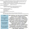
How do you manage Undisplaced proximal humerus fractures?
Undisplaced or minimally displaced humerus fractures(vast majority)are treated conservatively by
(a) immobilisation in a polysling with a collar and cuff(for gravitational traction)and
(b) progressive mobilisation followed by physiotherapy.
(c) Pendular exercise can commence at 14 days and active abduction from 4-6 weeks
How do you manage anatomical humeral neck fractures?
Significantly displaced avulsion of the tuberosities or anatomical neck of humerus fractures-
(1) internal fixation
(2) repair of rotator cuff
How do you manage significantly displaced avulsion of proximal humerus tuberosities?
(1) internal fixation
(2) repair of the rotator cuff
How do you manage surgical neck of humerus fractures?
(a) impacted:collar and cuff for 3-6weeks followed by physiotherapy
(b) significantly displaced:(1)open reduction and fixation or
(2) use of intramedullary device
How do you manage impacted surgical neck of humerus fractures?
collar and cuff for 3-6 weeks followed by physiotherapy
How do you manage significantly displaced surgical neck of humerus fractures?
(1) open reduction and fixation or
(2) use of intramedullary device
What are the complications of proximal humerus fractures?
(1) Axillary nerve damage(common)
(2) Axillary vessel damage
(3) Shoulder stiffness
(4) Fracture dislocation of the humeral head
For the above complications detailed neurological assessment is essential for all upper limb injuries
What is the incidence of supracondylar fractures?
(1) One of the most common types of fractures
(2) Peak incidence in children between ages 5-8 years
What are the types and causes of supracondylar humerus fractures?
1st/Extension type fracture(95-98%)
+Mechanism-Typically occurs from a fall on outstretched hand with elbow in extension or hyperextension
+Complication-Anterior interosseous nerve injury:causing neuropraxia
2nd/Flexion type fracture(2-5%)
+Mechanism-Typically occurs from a fall onto the flexed elbow
+Complications-Ulnar nerve injury:(a)the most common nerve injury from flexion type
(b)iatrogenic from medial pinning
What is the incidence of extension type supracondylar fracture?
95-98%
What is the mechanism of extension type supracondylar humerus fractures?
Typically occurs from a fall on outstretched hand with elbow in extension or hyperextension
What are the complications of the extension type supracondylar humeral fractures?
Anterior interosseous nerve injury:causing neuropraxia
What is the most common nerve affected by supracondylar fracture of the humerus?
Anterior interosseous nerve causing neuropraxia
What is the most common symptom caused by anterior interosseous nerve in supracondylar humerus fractures?
Neuropraxia
What is the incidence of flexion type supracondylar humerus fractures?
2-5%
What is the mechanism of flexion type supracondylar humerus fractures?
Typically occurs from a fall onto the flexed elbow
What are the complications of flexion type supracondylar humerus fractures?
Ulnar nerve injury:(a)the most common nerve injury from flexion type
(b)iatrogenic from medial pinning
What is the most common nerve injury from flexion type supracondylar humerus fractures?
Ulnar nerve injury
What are the most common nerve injuries,in order, from supracondylar humerus fractures?
1stly/Anterior interosseos nerve(AIN) injury-from extension type supracondylar humerus fractures causing neuropraxia
2ndly/Radial nerve injury-the second most common neuropraxia after anterior interosseous nerve(AIN)palsy
What are the investigations(diagnosis)of supracondylar humerus fractures?

Why should we examine AP views in supracondylar humerus fractures?
For Baumann angle
What should be done after doing AP view for a patient with supracondylar humerus fractures?
(1) Examine for Baumann angle
(2) We need to compare with contralateral arm
Why should we examine lateral views in supracondylar humerus fractures?
To see if the anterior humeral line intersects the middle 1/3rd of the capitellar ossification centre
What is the use of posterior fat pad displacement in supracondylar humerus fractures?
Posterior fat pad displacement is always pathological and can indicate a nondisplaced fracture
Discuss Gartland classification for supracondylar humerus fractures?

Picture illustrating Gartland classification of supracondylar humerus fractures

What are the complications of supracondylar humerus fractures?
1st/Neurovascular structures at risk
I]Anterior interosseous nerve(AIN)injury-most common for extension type supracondylar humerus fractures
-causing neuropraxia
II]Ulnar nerve injury-the most common nerve injury from flexion type supracondylar humerus fractures
-iatrogenic from medial pinning
III]Angulation:(1)Posteromedial angulation-associated with radial nerve injury(the 2nd most common neuropraxia after
AIN injury)
(2)Posterlateral angulation-associated with(a)brachial artery injury
(b)median nerve injury
2nd/Vascular injuries(1%)
What is the the 2nd most common neuropraxia in supracondylar humerus fractures?
Radial nerve injury neuropraxia after AIN injury or palsy neuropraxia
What are the complications of posteromedial angulation in supracondylar humerus fractures?
Radial nerve injury(the 2nd most common neuropraxia after AIN palsy)
What are the complications of posterolateral angulation in supracondylar humerus fractures?
(1) Brachial artery injury
(2) Median nerve injury
What is the management of supracondylar humerus fractures?

What is type I Gartland classification of supracondylar humerus fractures?
Nondisplaced(beware of subtle medial comminution)
What is the management of type I supracondylar humerus fractures?
Treated closed in a long arm cast for 2-3 weeks
What is the duration of the cast in type I Gartland classification of supracondylar humerus fractures?
2-3weeks
What is type II Gartland classification of supracondylar humerus fractures?
Displaced,posterior cortex and periosteal hinge intact
What is the management of type II Gartland classification of supracondylar humerus fractures?

What are the criteria for conservative management in type II Gartland classification of supracondylar humerus fractures?

What is type III Gartland classification of supracondylar humerus fractures?
Completely displaced
What is the management of type III Gartland classification of supracondylar humerus fractures?

What is the other name for non accidental injury?
(1) Battered child
(2) Child abuse
Discuss non accidental injury

What is the incidence of non accidental injury?

What is the incidence of repeated child abuse if not diagnosed?
30-50% there is a chance to repeat the child abuse if not diagnosed
What is mortality rate of child abuse(non accidental injury) if not diagnosed?
5-10% chance of death from subsequent abuse
What is incidence of fractures in non accidental injury(child abuse)?
(1) Children younger than 1 year = 50% chance of fractures
(2) Children younger than 3 years =30% chance of fractures
What is incidence of fractures in non accidental injury(child abuse) in children younger than 1 year?
50%
What is incidence of fractures in non accidental injury(child abuse) in children younger than 3 year?
30%
What is the most cause of femur fractures in non ambulatory children?
Non accidental injuries(child abuse)
When to suspect non accidental injury(NAI)?

What is the other name for osteopetrosis?
Marble bone disease
Define osteopetrosis
Hard and more dense bones
What is the genetic predisposition of osteopetrosis?
Autosomal recessive condition
What is the incidence of osteopetrosis?
Commonest in young adults
What is the pathogenesis of osteopetrosis?
Osteoclast dysfunction results in too much bone density
What does imaging show in osteopetrosis?
Radiology reveals a lack of differentiation between the cortex and the medulla described as marble bone
Picture illustrating osteopetrosis

What are the causes of pathological fracture?

What are the metastatic tumours causing pathological fracture?

What are the bone diseases causing pathological fracture?

What are the local benign conditions causing pathological fracture?

What are the primary malignant tumours causing pathological fracture?

Define osteogenesis imperfecta
Inherited condition causing increased bone fragility
What are the common sites affected by osteogenesis imperfecta?

Discuss pathogenesis of osteogenesis imperfecta

What are the subtypes of osteogenesis imperfecta?
Type I osteogenesis imperfecta - people with type I
O1 have less collagen than normal. This makes their
bones fragile, but they don’t have bone deformities.
The first break usually happens when a child starts
walking. Fractures typically decrease after puberty.
Type I osteogenesis imperfecta-
babies with type
Il Ol usually are born with many fractures, are very
small, and have severe breathing problems. As a
result, most will not survive.
Type Ill osteogenesis imperfecta
- people with
type Ill O1 usually will be shorter than their peers,
and may have severe bone deformities, breathing
problems (which can be life-threatening), brittle
teeth, a curved spine, ribcage deformities, and other
problems.
Type IV osteogenesis imperfecta - people with
type IV O1 can have mild to serious bone
deformities, short stature, frequent fractures (which
may lessen after puberty), and a curved spine.

Discuss type I of osteogenesis imperfecta
Type I osteogenesis imperfecta - people with type I
O1 have less collagen than normal. This makes their
bones fragile, but they don’t have bone deformities.
The first break usually happens when a child starts
walking. Fractures typically decrease after puberty.

Define type I of osteogenesis imperfecta?
The collagen is
(1) normal quality
(2) insufficient quantity
What is the specific characteristic of type I osteogenesis imperfecta?
Mildest form
What is the incidence of type I osteogenesis imperfecta?
60% of all cases
What is the mechanism of type I osteogenesis imperfecta?

What is the clinical picture of type I osteogenesis imperfecta?
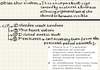
What is the cause of blue thin sclera in type I osteogenesis imperfecta?
It is an important sign caused by sceleral thinness allowing pigmented coat of the choroid to become visible
Discuss fractures in type I osteogenesis imperfecta
Can occur any time from the perinatal period onwards:-
(1) In adults:There is(a)a 7x greater incidence of overall fracture rate than normal,with
(b) Reduced vertebral bone mineral content in adults
(2) In children:Fractures may be numerous but rarely lead to deformity
Discuss type II osteogenesis imperfecta?
Type I osteogenesis imperfecta-
babies with type
Il Ol usually are born with many fractures, are very
small, and have severe breathing problems. As a
result, most will not survive.

What is the mechanism of type II osteogenesis imperfecta?

What is the clinical picture of type II osteogenesis imperfecta?

Discuss multiple fractures in type II osteogenesis imperfecta
Frequently occurring in utero
What is the cause of short limbs in type II osteogenesis imperfecta?
Due to faulty conversion of normal mineralised cartilage to defective bone matrix
What is the clinical picture of type II osteogenesis imperfecta?

Discuss type III osteogenesis imperfecta

What is the characteristic feature of type III osteogenesis imperfecta?
Lethal form
What is the characteristic feature of type III osteogenesis imperfecta?
Severely progressive deforming subtype
Discuss fractures in type III osteogenesis imperfecta
Type Ill osteogenesis imperfecta
- people with
type Ill O1 usually will be shorter than their peers,
and may have severe bone deformities, breathing
problems (which can be life-threatening), brittle
teeth, a curved spine, ribcage deformities, and other
problems.

Discuss the progressive deformity occurs in type III osteogenesis imperfecta

What is the period of occurrence of progressive deformity in type III osteogenesis imperfecta?
Occurs during early years and increases with age
What are the sites where progressive deformity occurs?
(1) Skull
(2) Chest
(3) Spine
(4) Long bones
(5) Pelvis
What is the appearance of the face in type III osteogenesis imperfecta?
Triangular with
(1) Large vault
(2) Prominent eyes
(3) Small jaw
Discuss the inability to walk in type III osteogenesis imperfecta
Patients rarely walk,even after multiple surgical procedures
What is the reason for extreme short stature of patients with type III osteogenesis imperfecta?
Due to repeated childhood fractures
Discuss dentinogenesis imperfecta(DI) associated with type III osteogenesis imperfecta

Define dentinogenesis imperfecta(DI) associated with type III osteogenesis imperfecta
Impaired dentition
What is the aetiology of dentinogenesis imperfecta associated with type III osteoporosis imperfecta?
Secondary to type I collagen defect
What is colour of sclera in type III osteogenesis imperfecta according to the age?
(1) blue in infancy
(2) normal colour in childhood
Descuss type IV osteogenesis imperfecta
Type IV osteogenesis imperfecta - people with
type IV O1 can have mild to serious bone
deformities, short stature, frequent fractures (which
may lessen after puberty), and a curved spine.

Define type IV osteogenesis imperfecta
Sufficient collagen quantity but poor quality
What is the characteristic feature of type IV osteogenesis imperfecta?
Moderately severe form
What is the clinical feature of type IV osteogenesis imperfecta?

What is the period in which type IV osteogenesis imperfecta will apparent?
This may be apparent at birth with
(1) Fractures
(2) Recurrent fractures on walking
(3) Bowing of leg
What are the features of the sclera in type IV osteogenesis imperfecta?
Normal white colour in childhood with
(1) reduced stature
(2) variable disability
What are the complications of type IV osteogenesis imperfecta?

What is the differential diagnosis of type IV osteogenesis imperfecta?

How to differentiate between type IV and type I osteogenesis imperfecta?
Type IV is differentiated from type I by having white sclera
How to differentiate between type IV and type III osteogenesis imperfecta?
Type IV is differentiated from type I by having autosomal dominant inheritance
What are the findings of radiology in osteogenesis imperfecta?

Discuss Ehler’s Danlos syndrome
Ehlers Danlos:
- Multiple sub types
- Abnormality of types 1 and 3 collagen
- Patients have features of hypermobility.
- Individuals are prone to joint dislocations and pelvic organ prolapse. -In addition to many other diseases related to connective tissue defects.
What is the other name of Osgood Schlatter disease?
Runner’s knee
What is the genetic predisposition of Osgood Schlatter disease/runner’s knee?
Autosomal recessive condition
What is the cause of Osgood Schlatter disease/Runner’s knee?
Involves traction apophysis of tibial tubercle-due to repeated microtrauma to tibial apophysis
What is the incidence of Osgood Schlatter disease/Runner’s knee?
(1) Age=10-15 years
(2) Sex=M>F
What is the age incidence of Osgood Schlatter disease/Runner’s knee?
10-15 years of age
What is the clinical picture of Osgood Schlatter disease/Runner’s knee?
Symptoms
I)No history of trauma
II)Knee pain after activity
Signs
I)Tenderness-very specific point of tenderness over the tibial tubercle
II)Lump-(1)tender
(2)palpable
(3)over proximal tibia
what are the symptoms of Osgood Schlatter disease?
I)No history of trauma
II)Knee pain after activity
What are the signs of Osgood Schlatter disease?
I)Tenderness-very specific point of tenderness over the tibial tubercle
II)Lump-(1)tender
(2)palpable
(3)over proximal tibia
What are the investigations of Osgood Schlatter disease?
XRs- shows fragmentation of apophysis
What is the treatment of Osgood Schlatter disease?
(1) Rest
(2) Plaster cast for 6-8 weeks
What is the location of pes anserinus bursa?
(1) At the medial aspect of the knee
(2) At the level of the joint space
(3) Deep to pes anserinus tendons(SGS-Sartorius,Gracilis,Semiteninosus)
Define pes anserinus bursitis
Symptomatic inflammation of the pes anserinus bursa
What are the clinical picture of pes anserinus bursitis
Pain
(1) Site-along the proximal medial tibia
(2) Associated with-swelling
(3) Exacerbated by-particular activities such as ascending and descending stairs
What is the treatment of pes anserinus bursitis?
Physiotherapy-most cases resolve with physiotherapy
What is the other name for gout and pseudogout?
Crystal induced arthropathies
Define gout
What is the incidence of gout and pseudogout?
Common
What is the cause of gout?
Monosodium urate monohydrate crystals
What is the cause of pseudogout?
Pseudogout is a form of microcrystal synovitis caused by the deposition of calcium pyrophosphate dihydrate in the synovium
What are the risk factors for pseudogout?

What are the symptoms and signs for gout and pseudogout?
I)Podagra (initial) joint manifestation in 50% of gout cases and eventually
involved in 90%; also observed in patients with pseudogout and other conditions II)Gout is a form of inflammatory artheritis charaterised by recurrent attacks of a red,tender,hot and swollen joint.The joint at the base of the big toe(i.e.,1st metatarsophalangeal joint) is most commonly affected but many other joints including those of hands my be affected III)Arthritis in other sites
- In gout:(1)the instep,
(2)ankle,
(3)wrist,
(4)finger joints, and
(5)knee;
- In gout,the joint is
(1) swollen
(2) hot
(3) tender
(4) red
(3)shows white chalky patches in the skin through which crystals can often be expressed
-In pseudogout, large joints,eg.,the: (1)knee,
(2)wrist,
(3)elbow, or
(4)ankle
(5)shoulder
IV)Monoarticular involvement
(1)most commonly, though polyarticular acute flares are not rare, and
(2)many different joints may be involved simultaneously or in rapid succession
V)In gout, attacks that begin abruptly and typically reach maximum intensity
within < 8-12 hours;
VI)In pseudogout, attacks resembling those of acute gout or a more insidious onset
that occurs over several days
VII)Without treatment, symptom patterns that change over time; attacks can
(1)become more polyarticular,
(2) involve more proximal and upper-extremity joints,
(3)occur more often, and
(4)last longer
VIII)In some cases, eventual development of chronic polyarticular arthritis that can
resemble rheumatoid arthritis
What is the most common site for gout?
Podagra
(1) initially in 50% of cases
(2) Eventually in 90% of cases
Define Podagra
Podagra, which in Greek translates to ‘foot trap’, is gout which affects the joint located between the foot and the big toe, known as the metatarsophalangeal joint.
What is the incidence of gout in Podagra?
Initially 50%
Eventually 90%
What other conditions affecting Podagra other than gout?
Pseudogout gout and other conditions
Which joints are affected by arthritis in gout?
(1) instep,
(2) ankle,
(3) wrist,
(4) finger joints, and
(5) knee
Which joints are affected by pseudogout?
large joints,eg.,the
(1) knee,
(2) ankle,
(3) wrist, or
(4) elbow
(5) shoulder
Does gout and pseudogout have monoarticular or polyarticular involvement?
Monoarticular involvement most commonly, though polyarticular acute flares are not rare, and many different joints may be involved simultaneously or in rapid succession
What is the time expected for polyarticular involvement of gout and pseudogout to occur?
different joints may be involved simultaneously or in rapid succession
What is the time expected for attacks of gout and pseudogout to occur?
(1)In gout, attacks that begin abruptly and typically reach maximum intensity
within 8-12 hours;
(2)in pseudogout, attacks resembling those of acute gout or a more insidious onset
that occurs over several days
What is the time expected for attacks of gout to occur?
In gout, attacks that begin abruptly and typically reach maximum intensity within 8-12 hours
What is the time expected for attacks of pseudogout to occur?
in pseudogout, attacks resembling those of acute gout or a more insidious onset that occurs over several days
What happens to patients with gout or pseudogout if left without treatment?
Without treatment, symptom patterns that change over time; attacks can (1)become more polyarticular,
(2) involve more proximal and upper-extremity joints,
(3) occur more often, and
(4) last longer
What is the complication of gout and pseudogout?
Eventual development of chronic polyarticular arthritis that can resemble rheumatoid arthritis
Chronic renal failure in gout
What are the investigations(diagnosis) of gout?
(1) Elevated uric acid
(2) negative birefringent crystals on synovial fluid examination
What are the investigations(diagnosis) of pseudogout?
(1)Joint aspiration:weakly positively birefringent rhomboid shaped crystal (2)XRs:Chondrocalcinosis
What does the joint aspiration show in pseudogout?
weakly positively birefringent rhomboid shaped crystal
Compare between gout and pseudogout
(1) WBCs 3000-50000 in both gout and psedogout
(2) good response to colchicine in gout and weak in pseudogout
(3) red compensator when parallel is yellow in gout and blue in pseudogout

What is the management of gout?
1st/Acute gout
(1) NSAID
(2) Colchicine
2nd/Chronic gout or for prophylaxis
Allopurinol
What is the management of pseudogout?
(1) Aspiration of joint fluid to exclude septic arthritis
(2) NSAID
(3) Steroids-intra-articular or intramuscular or oral as for gout
(4)Weak response to colchicine
What is the incidence of scaphoid fractures?
(1) The commonest carpal fracture
(2) Incidence of scaphoid fractures in the UK ranges from 12.4-29/100000
What is the incidence of scaphoid fractures in the UK?
Incidence of scaphoid fractures in the UK ranges from 12.4-29/100000
What is the reason that scaphoid fractures risk blood supply causing avascular necrosis?
Because surface of scaphoid is covered by articular cartilage with small area available for blood vessels
What is the anatomical importance of scaphoid bone?
Forms floor of the anatomical snuffbox
What is the cause or risk factor for scaphoid fractures?
Fall onto outstretched hand
(1) tubercle
(2) waist
(3) proximal 1/3rd
What is the imaging that should be done for a suspected scaphoid fractures?
1st/A series of 4 radiographs should be done
(1)PA view
(2)Pronated oblique view
(3)Ziter view-a PA view with the wrist in ulnar deviation and beam angulated
at 20 degrees
(4)Lateral view
2nd/Repeat imaging should be done at 10 days
3rd/MRI-should be done in case of diagnostic uncertainty where the 4 scaphoid
view radiographs can not exclude scaphoid fractures if negative
Define Ziter view
A PA view with
(1) the wrist in ulnar deviation and
(2) beam angulated at 20 degrees
What is the sensitivity of scaphoid radiographs in the 1st week?
80%
What is the period required to do a repeat imaging for a suspected scaphoid fracture?
At 10 days
What should be done in case of diagnostic uncertainty in scaphoid fractures?
MRI
What should be done in case of diagnostic uncertainty in scaphoid fractures?
4 Scaphoid view radiographs can not exclude scaphoid fractures if negative
When the MRI should be done in scaphoid fractures?
In cases of diagnostic uncertainty where 4 scaphoid view radiographs can not exclude scaphoid fractures if negative
What is the classification of scaphoid fractures?
(1) Scaphoid tubercle
(2) Distal pole
(3) Waist
(4) Proximal pole
What is the management of scaphoid fractures?

What are the complications of scaphoid fractures?

Image shows classification of scaphoid fractures,scaphoid blood supply and scaphoid fixation

What is the commonest organisms causing septic arthritis?
(1) Staph.aureus overall
(2) Neisseria gonorrhoea-in sexually active young adults
(3) Staph.aureus-in paediatric patients
What is the commonest organism causing septic arthritis in the sexually active young adults?
Neisseria gonorrhoea
What is the most common organism causing septic arthritis in paediatric patients?
Staph.aureus
What are the investigations(diagnosis)of septic arthritis?
I)Plain XRs
II)Aspiration:Synovial fluids should be obtained before starting treatment
III)Kocher criteria
Define Kocher criteria

What is the expected ESR according to Kocher criteria for septic arthritis?
> 40 mm/hr
What is the expected WBC count according to Kocher criteria for septic arthritis?
> 12000 mm3
What is the prerequisite for Kocher criteria to be diagnostic in septic arthritis?
When 4/4 criteria are met,there is a 99% chance that the child has septic arthritis
What is the chance of having septic arthritis in a child with 4/4 Kocher criteria?
90%
What is the treatment of septic arthritis?
I)Needle aspiration should be used to decompress the joint
II)Arthroscopic lavage
III)Repeated procedures are necessary in some cases
V)Antibiotics:(1)urgent washout and antibiotics otherwise high risk of joint
destruction.
(2)Antibiotics should be given for 6-12 weeks
(3)IV antibiotics cover gram+ve cocci.BNF currently recommends
flucloxacillin or clindamycin if penicillin allergic
V)Surgical drainage of the affected joint,this should be as soon as possible since
permenant damage to the joint may occur
What is the purpose of needle aspiration in septic arthritis?
to decompress the joint
Discuss the use of antibiotics in the treatment of septic arthritis?
(1)Urgent washout and antibiotics otherwise high risk of joint destruction (2)Antibiotics should be given for 6-12 weeks
(3)IV antibiotics cover gram+ve cocci.
BNF currently recommends flucloxacillin or clindamycin if penicillin allergic
What is the duration of antibiotics in treatment of septic arthritis?
6-12 weeks
What is the reason for using antibiotics in septic arthritis?
urgent washout and antibiotics otherwise high risk of joint destruction.
What is the organism covered by IV antibiotics in septic arthritis?
IV antibiotics cover gram+ve cocci.BNF currently recommends flucloxacillin or clindamycin if penicillin allergic
What are the antibiotics recommended to treat septic arthritis?
IV antibiotics cover gram+ve cocci.BNF currently recommends flucloxacillin or clindamycin if penicillin allergic
What is the purpose of surgical treatment in septic arthritis?
Surgical drainage of the affected joint,this should be as soon as possible since permenant damage to the joint may occur
What are the complications of Perthes disease?
(1)Flattening and fragmentation of epiphysis:due to osteonecrosis of proximal femoral epiphysis
(2)AVN….Deformity….Subsequent revascularisation(2-4 years cycles)
What does a plain XRs film show in Perthes disease?
XRs normal in early stage

Define Gage’s sign in Perthes disease

Discuss the other staging of Perthes disease?(NOT THE CATTERALL STAGING)

What are the causes of stress fractures?
The following may result in small hairline stress fractures
(1) Repetitive activity
(2) Loading of normal bone
What is the clinical picture of stress fractures?
(1) Painful
(2) Stress fractures are seldom displaced
(3) Surrounding soft tissue injury is unusual
(4) Stress fractures may present late following the injury,in which case callus formation may be identified on radiographs
What is the management of stress fractures?
(1)Stress fractures may not require formal immobilisation
(2)Injuries associated with severe pain and presenting at an earlier stage may benefit from immobilisation tailored to the site
of injury
What are the causes of pathological fractures among children?
(1) Osteogenesis imperfecta
(2) Osteopetrosis
(3) Osgood-Schlatter disease/Runner’s knee
(4) Pott’s fracture
(5) Bone cyst
(6) Bone tumour
(7) Non accidental injury(NAI) or child abuse

What are the causes of growth plate fractures/epiphyseal fractures?
I)Non accidental injury(NAI) or child abuse
II)Pathological fractures
(1)Osteogenesis imperfecta
(2)Osteopetrosis
(3)Osgood-Schlatter disease/Runner’s knee
(4)Pott’s fracture
(5)Bone cyst
(6)Bone tumour
What is the other name for transient tenosynovitis?
Irritable hip
What is the incidence of Transient tenosynovitis(irritable hip)?
A common childhood condition affecting children between 3-8 years of age
What is the clinical picture of transient synovitis(irritable hip)?
(1) History of URTI or other similar viral infections
(2) Fever
(3) Pain-usually in the knee or thigh
(4) Hip pain
(5) Restricted movement in one of the hip joints
What are the complications of transient tenosynovitis(irritable hip)?
(1) Effusion
(2) Synovitis
What are the investigations(diagnosis)of transient tenosynovitis(irritable hip)?
Blood tests and radiology are normal
What is the treatment of transient tenosynovitis/irritable hip?
(1) Self resolving
(2) Analgesics
(3) Rest
Define developmental dysplasia of the hip
Congenital abnormality of 2 types:
(1) Mild dysplasia acetabulum
(2) Irreducible dislocation
What are the types of developmental dysplasia of the hip(DDH)?
(1) Mild dysplasia acetabulum
(2) Irreducible dislocation
What is the incidence of developmental dysplasia of the hip (DDH)?
More common in
(1) Extended breach babies
(2) Females
What is the cause or risk factors of developmental dysplasia of the hip (DDH)?
Mnemonic;MBC OFF
(1) Multiple pregnancies
(2) Breach position
(3) Certain ethnic groups-native American
(4) Oligohydromnios
(5) Female sex
(6) First born child with prematurity
(1) Usually diagnosed in infancy by screening tests
(2) Left hip more affected but may be bilateral
(3) Leg length inequality when disease is unilateral
(4) Slight external rotation
(5) As disease progresses child may limp and then early onset arthritis
(6) Trendelenberg test positive
What are the clinical tests to confirm developmental dysplasia of the hip(DDH)?
1st/Barlow’s test(BAD)
- *Mnemonic;Barlow….Adduction….Dislocation(BAD)**
(1) Adduct the hip
(2) Apply light pressure on the knee directing the force posteriorly trying to dislocate the joint
(3) If the hip is dislocatable,the test is considered positive
2nd/Ortolani test(ABO)
Mnemonic;Abduction….Ortolani(ABO)
+Purpose:The Ortolani manoeuvre is then used,to confirm that the positive findings(positive Barlow’s test,i.e.,that the hip
actually dislocated)
+Manoeuvre:(1)It is performed by gently abducting the infant’s leg by the examiner’s thumb
(2)Apply anterior pressure on the greater trochanter using the examiner’s index and forefinger
(3)A positive sign is a distinctive ‘clunk’ which can be heard and felt as the femoral head relocates anteriorly into
the acetabulum
Discuss Barlow’s test
Mnemonic;Barlow….Adduction….Dislocation(BAD)
(1) Adduct the hip
(2) Apply light pressure on the knee directing the force posteriorly trying to dislocate the joint
(3) If the hip is dislocatable,the test is considered positive
Discuss Ortolani test
Mnemonic;Abduction….Ortolani(ABO)
+Purpose:The Ortolani manoeuvre is then used,to confirm that the positive findings(positive Barlow’s test,i.e.,that the hip
actually dislocated)
+Manoeuvre:(1)It is performed by gently abducting the infant’s leg by the examiner’s thumb
(2)Apply anterior pressure on the greater trochanter using the examiner’s index and forefinger
(3)A positive sign is a distinctive ‘clunk’ which can be heard and felt as the femoral head relocates anteriorly into
the acetabulum
Picture illustrating Barlow’s and Ortonali test

What are the investigations(diagnosis) of developmental dysplasia of the hip(DDH)?

What is the initial findings on plain XRs of developmental dysplasia of the hip(DDH)?
(1)Initially no obvious change on plain films but small femoral head
may be present
(2)On plain films shentons line should form a smooth arc
What are the findings in ultrasound of developmental dysplasia of the hip(DDH)?
(1)The most effective
(2)USS gives best resolution until 3 months of age
(3)In recent years,hip ultrasonography(US)has appeared as an effective tool for the
early diagnosis of developmental dysplasia of the hip(DDH)in the newborns
(4)US Is an effective and noninvasive method without radiation
(5)Due to the high cost of US,there are still some controversial issues to use US as
a screening method
What is the most effective method of diagnosing developmental dysplasia of the hip(DDH)?
US
What is the characteristic feature of US in diagnosing developmental dysplasia of the hip(DDH)?
(1) USS gives best resolution until 3 months of age
(2) The most effective method
(3) Effective tool for early diagnosis of developmental dysplasia of the hip
What is the treatment of developmental dysplasia of the hip(DDH)?

What is the treatment of developmental dysplasia of the hip in the age 0-6m?
(1) Pelvic harness
(2) Surgery is needed in case of dislocated and irreducible cases
What is the treatment of developmental dysplasia of the hip in the age 6m-18m?
(1)Close reduction+/-adductor tenotomy
(2)If fail then arthrogram+open reduction with hip spica cast 600 abduction and
900 flexion
What is the treatment of developmental dysplasia of the hip in the age 18m-3yrs?
OR+/-femoral varus de rotation osteotomy
What is the treatment of developmental dysplasia of the hip in the age 3yrs-8yrs?
OR+/-Femoral varus de rotation osteotomy+pelvic osteotomy
What is the treatment of developmental dysplasia of the hip in the age > 8yrs?
THR when system justify surgical intervention
What is the general treatment of developmental dysplasia of the hip(DDH)?
(1)Splints and harnesses or traction
(2)In later years osteotomy and hip realignment procedures may be needed
(3)In arthritis a joint replacement may be needed.However,this is best deferred if
possible as it will almost certainly require
Discuss follow up of developmental dysplasia of the hip(DDH)?
At least until walking normally;WHO recommend up to 5 years
What is the other name of slipped capital femoral epiphysis(SCFE)?
(1) Slipped upper femoral epiphysis(SUFE)
(2) Displaced upper femoral epiphysis
What is the incidence of slipped capital femoral epiphysis(SCFE)?
Older obese male adolescents
Define slipped capital femoral epiphysis(SCFE)?
Displaced upper femoral epiphysis(head)from neck
Discuss clinical picture of slipped capital femoral epiphysis(SCFE)?
(1) Obese(remember,only this clue can guide you to the answer)
(2) Bilateral in 20%
(3) Associated with decreased GH and sex hormones
(4) Pain at the thigh and knee
(5) Knee pain at 2 months prior to hip slipping
(6) Mild shortening of limb
(7) Increased adduction and external rotation
(8) Decreased or limitation of abduction and internal rotation
(9) Chance of AVN increased
What does XRs show in slipped capital femoral epiphysis(SCFE)?
XRs

Picture illustrating slipped capital femoral epiphysis(SCFE)

Discuss treatment of slipped capital femoral epiphysis(SCFE)

What is the aim of bed rest and non weight bearing in the treatment of slipped capital femoral epiphysis(SCFE)?
To avoid avascular necrosis
What is the treatment of minor to moderate cases of slipped capital femoral epiphysis(SCFE)?
Cannulated hip screw
What is the treatment of severe slippage of slipped capital femoral epiphysis(SCFE)?
Percutaneous pinning of the hip
What is the treatment if delayed cases of slipped capital femoral epiphysis(SCFE)?
Femoral neck osteotomy
What is the use of the southwick angle in slipped capital femoral epiphysis(SCFE)?
Gives an indication of the articular surface and disease severity
What is the other name for talipes equinovarus?
Club foot
What are the types of talipes equinovarus(club foot)?
(1) Equinus of the hindfoot
(2) Adduction and varus of the midfoot
(3) High arch
What is the incidence of talipes equinovarus(club foot)?
(1) Most cases in developing countries
(2) Incidence in the UK is 1/1000 live births
(3) More common in males
What is the sex incidence of talipes equinovarus(club foot)?
More common in males
What is the cause of talipes equinovarus(club foot)?
(1) Strong familial link
(2) Associated with other developmental disorders such as Down syndrome
What are the key anatomical deformities of talipes equinovarus(club foot)?
(1) Bilateral in 50%
(2) Adducted and inverted calcaneus
(3) Wedge shaped distal calcaneal articular surface
(4) Severe tibio-talar plantar flexion
(5) Medial talar neck inclination
(6) Displacement of the navicular bone(medially)
(7) Wedge shaped head of talus
(8) Displacement of the cuboid(medially)
What is the treatment of Talipes equinovarus(club foot)?
1st/Conservative(The Ponesti method)
*Advantage:gives comparable results to surgery
*It consists of:(1)Serial casting-to mold the foot into correct shape
(2)Achilles tenotomy-following casting around 90% requires
Achilles tenotomy
(3)Walking braces-done following Achilles tenotomy to maintain
the correction
2nd/Surgical correction
*Indication:reserved for those cases that fail to respond to conservative treatment
*Involves:(1)Multiple tenotomies
(2)Lengthening procedures
(3)Ilizarov frame-in patients who fail to respond surgically and gives
good results
What is the conservative management of talipes equinovarus(club foot)?
The Ponesti method
*Advantage:gives comparable results to surgery
*It consists of:(1)Serial casting-to mold the foot into correct shape
(2)Achilles tenotomy-following casting around 90% requires
Achilles tenotomy
(3)Walking braces-done following Achilles tenotomy to maintain
the correction
What is the advantage of the Ponesti method for treatment of talipes equinovarus(club foot)?
gives comparable results to surgery
What does Ponseti method,for the treatment of talipes equinovarus(club foot),consists of?
Mnemonic;SAW
(1) Serial casting-to mold the foot into correct shape
(2) Achilles tenotomy-following casting around 90% requires Achilles tenotomy (3)Walking braces-done following Achilles tenotomy to maintain the correction
What is the surgical treatment of talipes equinovarus(club foot)?
*Indication:reserved for those cases that fail to respond to conservative treatment
*Involves:(1)Multiple tenotomies
(2)Lengthening procedures
(3)Ilizarov frame-in patients who fail to respond surgically and gives
good results
What is the indication of surgical correction for the treatment of talipes equinovarus(club foot)?
reserved for those cases that fail to respond to conservative treatment
What does surgical correction involves in the treatment of talipes equinovarus(club foot)?
(1) Multiple tenotomies
(2) Lengthening procedures
(3) Ilizarov frame-in patients who fail to respond surgically and gives good results
What is the indication of Ilizarov frame reconstruction for treatment of talipes equinovarus(club foot)?
in patients who fail to respond surgically and gives good results
Picture illustrating types talipes equinovarus(club foot)

What are the causes of painful shoulder?

What is most common site for shoulder fracture?
Proximal humerus accounting for around 5% of all fractures.
What is the incidence of proximal humerus fracture?
5 %
What are the causes of proximal humerus fracture?
The majority of proximal humeral fractures are low energy injuries occurring in elderly patients falling onto an outstretched hand from standing. These injuries occur primarily in the context of an osteoporosis.
They also less commonly occur in younger patients usually the result of a high energy traumatic injury, therefore there are often associated soft tissue or neurovascular injuries.
What are the causes of proximal humerus fracture in the elderly?
The majority of proximal humeral fractures are low energy injuries occurring in elderly patients falling onto an outstretched hand from standing. These injuries occur primarily in the context of an osteoporosis.
What are the causes of proximal humerus fracture in the young patients?
They also less commonly occur in younger patients usually the result of a high energy traumatic injury, therefore there are often associated soft tissue or neurovascular injuries.
What are the risk factors of proximal humerus fracture?
The risk factors for low energy proximal humerus fractures are comparable to other osteoporotic fractures, including female gender, early menopause, prolonged steroid use, recurrent falls, and frailty.
What are the clinial picture of proximal humerus fracture?
1st/Symptoms (1)pain around the upper arm and shoulder, with (2)restriction of arm movement and (3)an inability to abduct their arm.
2nd/On examination, there is likely to be significant swelling and bruising of the shoulder, which can spread to the chest and down the arm.
Due to the close anatomical relationship with the axillary nerve and the circumflex vessels, is important to check the neurovascular status of the arm; damage to the axillary nerve can result in loss of sensation in the lateral shoulder (“Regimental Badge Area”) and loss of power of the deltoid muscle.
What are the symptoms of proximal humerus fracture?
(1)pain around the upper arm and shoulder, with (2)restriction of arm movement and (3)an inability to abduct their arm.
What are the signs of proximal humerus fracture?
On examination, there is likely to be significant swelling and bruising of the shoulder, which can spread to the chest and down the arm.
Due to the close anatomical relationship with the axillary nerve and the circumflex vessels, is important to check the neurovascular status of the arm; damage to the axillary nerve can result in loss of sensation in the lateral shoulder (“Regimental Badge Area”) and loss of power of the deltoid muscle.
What are the indications of conservative management of proximal humerus fracture?
(1) Minimally displaced fractures
(2) No neurovascular compromise
What are the steps of conservative management of proximal humerus fracture?
The patient requires immobilisation initially with early mobilisation including (1)pendular exercises at 14 days or 2-4 weeks post injury dependent on fracture pattern. (2)correctly applied polysling that allows their arm to hang; the effect of gravity on the arm will aid the reduction of the fragments of most humeral fractures. (3)Active abduction from 4-6 weeks
What are the indications of operative management of proximal humerus fracture?
(a) Irreducible fracture dislocation
(b) Large displacement
(c) Young patient
(d) Head splitting(intra-articular fracture) (e)Open fracture. (f)Neurovascularly compromised fracture
What are the factors that determine the type of surgery in proximal humerus fracture?
(1) Complexity of the fracture
(2) Patient factors
What does PROFHER trial suggest in the treatment of proximal humerus fractures?
(a) has suggested no benefit to operative intervention on patient outcome
(b) it must be applied cautiously as majority of patients were elderly with extraarticular fractures
What is the indication of ORIF and intermedullary nail ,in general,as operative options for the management of proximal humerus fractures?
multiple segment injuries
Discuss ORIF as an operative option for the management of proximal humerus fractures?
(1) Most commonly used
(2) Plate and screw fixation
(3) Can reconstruct complex fractures
What is the indication of ORIF as an operative option for the management of proximal humerus fractures?
often preferred in a head splitting fracture
Discuss indications of intramedullary nail as an operative option for the management of proximal humerus fractures?
(1) Suitable for extra-articular configuration,
(2) Predominately if the fracture involves surgical neck+/- greater tuberosity(GT)fractures (3)If the fracture is combined with a humeral shaft fracture
Discuss hemiarthroplasty as an operative option for the management of proximal humerus fractures?
Used for unreconstructable fractures in the older patient who has good glenoid quality
can be performed in a small number patients who experience complex injuries, or injuries that include splitting of the humeral head and are likely to have significant complications if the fracture is treated using ORIF.
Discuss total shoulder arthroplasty as an operative option for the management of proximal humerus fractures?
Unconstructable fractures where high functioning shoulder is required(hemiarthroplasty will cause glenoid erosion)
Discuss reverse shoulder arthroplasty as an operative option for the management of proximal humerus fractures?
Total shoulder arthroplasty that provides better functional outcome than conventional total shoulder replacement
Reverse shoulder arthroplasty (RSA) is an option for low demand patients, or patients who require revision after a failed previous procedure. RSA involves a total shoulder arthroplasty in which the ball and socket portions of the shoulder joint are reversed. Usually conservative management will be attempted before arthroplasty.
What is osteology of proximal humerus?
*Consists of
I)Articular head
II)Greater tuberosity
III)Metaphysis
IV)Diaphysis
V)Anatomical neck(previous physis):between the articular head and the tuberosities
VI)Surgical neck:between the tuberosities and the metaphysis
*Attachments
I)Greater tuberosity-attaches the following muscles
(1)Supraspinatus
(2)Infraspinatus
(3)Teres minor
II)Lesser tuberosity-attaches the subscapularis
What is the vascular supply of humeral head?
Anterior and posterior humeral circumflex arteries
What are the blood tests for proximal humerus fracture?
1st/for any trauma case, urgent bloods, including a coagulation and Group and Save, should be sent.
2nd/Where a pathological cause is suspected, further work-up bloods, such as a 1)serum calcium 2)myeloma screen, may be warranted.
What is the imaging of proximal humerus fractures?
*Aims
(1) Delineate the fracture pattern
(2) Confirm/exclude the presence of an associated dislocation
*Types of radiographs
I)Plain XRs film radiographs
+Feature:the required initial imaging modality for suspected shoulder fracture +Indication:to visulaise and classify aproximal humeral fracture +Options:1)True anteroposterior(AP)
2)Axillary lateral view and/or 3)Lateral scapular Y view
II)CT
1)better define intra-articular involvement
2)aid preoperative planning 3)if the position of any of the humeral segments is unclear.
III)MRI-is not useful for fracture imaging
Discuss classification of the proximal humerus fractures
The Neer classification system*
+Frequency:most commonly used
+Uses:used to characterise proximal humeral fractures based on the relationship between 4 main segments of the proximal humerus
+Aims:(1)Describe fractures as 2,3,or 4 depending on the number main fragments (2)Comments on the degree of displacement (3)Description of the fracture-more useful than classification
(4)Humeral alignment
(5)Fracture displacement
(6)Greater tuberosity position-rotator cuff will pull the greater tuberosity(GT)supero-posteriorly,which can cause impingement
problems with malunion
+Details:1st/Fragments
(1)Greater tuberosity
(2)Lesser tuberosity
(3)Articular surface or segment (anatomical neck)
(4)Humeral shaft (surgical neck)
2nd/Displacement
(1) > 1 cm or angulation
(2) > 45 degrees
*These segments are considered separate if there is displacement >1cm between segments, or if there is at least 45 degrees of angulation; it categorises injuries into either minimal displacement or two to four part injuries, dependent on the number of separate segments present.
What are the aims of proximal humerus fractures classification?
(1)Describe fractures as 2,3,or 4 depending on the number main fragments (2)Comments on the degree of displacement (3)Description of the fracture-more useful than classification
(4)Humeral alignment
(5)Fracture displacement
(6)Greater tuberosity position-rotator cuff will pull the greater tuberosity(GT)supero-posteriorly,which can cause impingement
problems with malunion
What is the type of classification of proximal humerus fractures?
The Neer classification system*
+Frequency:most commonly used
+Uses:used to characterise proximal humeral fractures based on the relationship between 4 main segments of the proximal humerus
+Aims:(1)Describe fractures as 2,3,or 4 depending on the number main fragments (2)Comments on the degree of displacement (3)Description of the fracture-more useful than classification
(4)Humeral alignment
(5)Fracture displacement
(6)Greater tuberosity position-rotator cuff will pull the greater tuberosity(GT)supero-posteriorly,which can cause impingement
problems with malunion
+Details:1st/Fragments
(1)Greater tuberosity
(2)Lesser tuberosity
(3)Articular surface or segment (anatomical neck)
(4)Humeral shaft (surgical neck)
2nd/Displacement
(1) > 1 cm or angulation
(2) > 45 degrees
*These segments are considered separate if there is displacement >1cm between segments, or if there is at least 45 degrees of angulation; it categorises injuries into either minimal displacement or two to four part injuries, dependent on the number of separate segments present.
What is the frequency of Neer classification?
Most commonly used
What are the details of Neer classification for proximal humerus fractures?
The Neer classification system*
+Frequency:most commonly used
+Uses:used to characterise proximal humeral fractures based on the relationship between 4 main segments of the proximal humerus
+Aims:(1)Describe fractures as 2,3,or 4 depending on the number main fragments (2)Comments on the degree of displacement (3)Description of the fracture-more useful than classification
(4)Humeral alignment
(5)Fracture displacement
(6)Greater tuberosity position-rotator cuff will pull the greater tuberosity(GT)supero-posteriorly,which can cause impingement
problems with malunion
+Details:1st/Fragments
(1)Greater tuberosity
(2)Lesser tuberosity
(3)Articular surface or segment (anatomical neck)
(4)Humeral shaft (surgical neck)
2nd/Displacement
(1) > 1 cm or angulation
(2) > 45 degrees
*These segments are considered separate if there is displacement >1cm between segments, or if there is at least 45 degrees of angulation; it categorises injuries into either minimal displacement or two to four part injuries, dependent on the number of separate segments present.
What are the complications of proximal humerus fracture
(1)reduced range of motion is the most common complication of a proximal humeral fracture , and extensive physiotherapy will be required to regain full function and reduce pain. Rehabilitation time for a proximal humeral fracture is around 1 year and is very dependent on how soon the patient was allowed to mobilise their shoulder.
(2)avascular necrosis of the humeral head following an injury disrupting the blood supply (from the anterior and posterior humeral circumflex arteries). In such cases, a hemiarthroplasty or reverse shoulder arthroplasty may be required.
Damage to the neurovascular supply is thankfully rare, however it remains essential to check the axillary nerve in these injuries.
What is the incidence of scapula fractures?
Uncommon
What is the cause of scapula fracture?
High energy trauma
What are the sites most commonly involved in scapula fractures?
(1) Scapula body(50%)
(2) Spine(50%)
(3) Glenoid fossa
(4) Glenoid neck
What should be excluded in scapula fractures?
Associated life threatening injury
What is imaging done for scapula fractures?
I)Plain radiographs
(1)True anteroposterior(AP)
(2)Axillary lateral and/or scapula Y view
II)CT scanning
(1)Define intra-articular involvement,displacement
(2)For three dimensional reconstruction
Discuss classification of scapula fractures?
Based on the location of the fracture
(1)Coracoid
(2)Acromion
(3)Glenoid neck-Floating shoulder:Beware of ipsilateral glenoid neck and clavicle
where limb is effectively dissociated from
axial skeleton
(4)Glenoid fossa
(5)Scapula body
What is the treatment of scapula fractures?
I]Conservative-sling immobilisation for 2 weeks followed by early rehabilitation
II]Surgical-(1)Floating shoulder:for fixation
(2)Intra-articular and displaced/angulated glenoid fractures
What are the types of dislocation around the shoulder joint?
(1) Glenohumeral dislocation
(2) Acromioclavicular joint disruption
(3) Sternoclavicular dislocation
What is the incidence of glenohumeral dislocation?
Commonly seen in the A&E with an incidence of up to 1.7% in the general population.
What is the recurrence rate of glenohumeral dislocation?
As high as 80% in teenagers
What is the cause of glenohumeral dislocation?
Usually a traumatic cause(multi-directional instability in frequent dislocation requires discussion with orthopaedics)

Discuss assessment of glenohumeral dislocation
(1) Careful history,examination,and documentation of neurovascular status of the limb,in particular the axillary nerve(regimental badge sensation)
(2) This should be reassessed post manipulation
(3) Early radiographs to confirm direction of dislocation
What is the initial management of glenohumeral dislocation?
(1) Emergent closed reduction under entanox and analgesics,but often requires conscious sedation
(2) Arm should then immobilised in a polysling
(3) XRs to confirm relocation
What is the imaging that should be done to diagnose glenohumeral dislocation?
(1) True anteroposterior(AP)
(2) Axillary
(3) Lateral and/or scapula Y view-reduced humeral head should lie between acromion and coracoid on lateral/scapula view
Discuss types of glenohumeral dislocation

What is the incidence of anterior glenohumeral dislocation?
Most common dislocation(> 90%)
What is the cause of anterior glenohumeral dislocation?
Usually traumatic anterior force on arm when shoulder is abducted,externally rotated

What is the examination of glenohumeral dislocation?
(1) Sulcus sign-Loss of shoulder contour
(2) Humeral head can be felt anteriorly
What is the reduction technique for the anterior glenohumeral dislocation?
(1) Hippocratic
(2) Milch
(3) Stimson
(4) Kocher-not advised due to complication of fracture
What is the incidence of posterior glenohumeral dislocation?
50% missed in A&E
What is the cause of posterior glenohumeral dislocation?
50% traumatic,but classically post seizure or electrocution

What is the examination of posterior glenohumeral dislocation?
(1) Shoulder locked in internal rotation
(2) XRs may show lightbulb appearance
What is the reduction technique of posterior glenohumeral dislocation?
Gentle lateral traction to adducted arm
What is the incidence of inferior glenohumeral dislocation?
Rare
What is the cause of inferior glenohumeral dislocation?
Associated with
(1) pectoral and rotator cuff tears
(2) glenoid fracture
What is the examination of inferior glenohumeral dislocation?
As for primary injury
What is reduction technique of inferior glenohumeral dislocation?
Management of primary injury
What is incidence of superior glenohumeral dislocation?
Rare
What is cause of superior glenohumeral dislocation?
Associated with acromion/clavicle fracture
What is the examination of superior glenohumeral dislocation?
As for primary injury
What is reduction technique of superior glenohumeral dislocation?
Management of primary injury
What are the associated injuries with glenohumeral dislocation ?

Define Bankart lesion
+Avulsion of the anterior glenoid labrum with an anterior shoulder dislocation
(reverse Bankart if poster labrum in posterior dislocation)
+Associated with glenohumeral dislocation
Discuss Hill Sachs defect
*Definition
Chondral impaction on posteriorsuperior humeral head from contact with glenoid labrum
(Reverse Hill Sachs in posterior dislocation)
*Treatment
Can be large enough to lock shoulder,requiring open reduction
What are the causes of rotator cuff tear?
(1) Chronic subacromial impingement-in older patients
(2) Avulsion injury-in younger patients
What is the clinical picture of rotator cuff tear?

What are the types of rotator cuff disease?
(1) Subacromial impingement
(2) Rotator cuff tears
(3) Rotator cuff arthropathy(a rtheritis)
Discuss anatomy of rotator cuff

Discuss action of rotator cuff muscles
The rotator cuff is composed of four muscles:
- Supraspinatus – abduction
- Infraspinatus – external rotation
- Teres minor – external rotation
- Subscapularis – internal rotation
What is the incidence of rotator cuff tear?
Acute full thickness tear
(1) In general-2.5 per 10000,approximately around 20% f the general population
(2) Age-40-70 years of age
Enumerate risk factors of rotator cuff tears?
1st/main risk factors for rotator cuff tears are(mnemonic;ROTA) (1)Age, (2)Trauma, (3)Overuse, and (4)Repetitive overhead shoulder motions (e.g. athletes, certain occupations).
2nd/Other risk factors include(mnemonic;OSD) (1)O***_besity (2)Smoking (3)D_***iabetes mellitus.
Discuss classification of rotator cuff tear
Rotator cuff tears are classified as either
1st/According to duration:(1)acute tears(lasting <3 months) or (2)chronic tears(lasting >3 months) .
2nd/According to thickness of the tear
(1)partial thickness tears or . (2)full thickness tears;Full thickness tears can be further classified into +small (<1cm), +medium (1-3cm), +large (3-5cm), or +massive (>5cm or involves multiple tendons)
Discuss pathophysiology of rotator cuff tear
1st/Acute tears +commonly occur within tendons with pre-existing degeneration +typically occurring alone following minimal force. However, +acute tears can occur in young individuals subjected to a larger force; these will therefore often occur alongside other injuries in the young.
2nd/Chronic tears +occur in individuals with degenerative microtears to the tendon +most commonly from overuse +seen in greater incidence with increasing age
Discuss clinical picture of the rotator cuff tear
1st/Symptoms (1)history of trauma with no fracture (2)pain over the lateral aspect of shoulder localised to the acromion (3)inability to initiate abduction or abduct the arm above 90 degrees but the patient can do minimal abduction (4)Tears are more common in the dominant arm.
2nd/On examination. (1)tenderness over the greater tuberosity and subacromial bursa regions. (2)Supraspinatus and infraspinatus atrophy can be seen in massive rotator cuff tears. (3)Specific Tests
There are specific tests that can be performed to help assess for the presence of a rotator cuff tear and elucidate which tendon(s) are affected:
a) Jobe’s test (the “empty can test”, tests supraspinatus) – place the shoulder in 90° abduction and 30° of forward flexion and internally rotate fully (as if you’re ‘emptying a can’), gently push downwards on the arm. +A positive test is present if there is weakness on resistance
b) Gerber’s lift-off test (tests subscapularis) – internally rotate the arm so the dorsal surface of hand rests on lower back, then ask the patient to lift hand away from back against examiner resistance +A positive test is weakness in actively lifting the hand away from back (compare to the contralateral side)
c) Internal rotation lag test(test subscapularis)
d) External rotation lag test(tests for infraspinatus)
e) Posterior cuff test (tests infraspinatus and teres minor) – the arm positioned at patient’s side, with the elbow flexed to 90°, then the patient is instructed to externally rotate their arm against resistance +A positive test is present if there is weakness on resistance
What are th specific tests used to diagnose rotator cuff tear
(1) Jobe’s test- positive for supraspinatus muscle
(2) Internal rotation lag sign-positive for subscabularis muscle
(3) Gerber’s lift-off test-positive for subscabularis muscle
(4) External rotation lag test-positive for infraspinatus muscle
Absense of these clinical signs make rotator cuff tear less likely
What are the investigations of rotator cuff tear?
(1) Urgent plain film radiograph +Patients presenting with clinical features of a rotator cuff tear should have an to exclude a fracture. +Whilst most plain film radiographs will be unremarkable, in chronic tears, there may be evidence of reduced acromiohumeral distance or sclerosis and cyst formation the rotator cuff insertion on the greater tuberosity.
(2) Ultrasonograhy. . +Once fracture has been excluded, rotator cuff tears can be assessed through further imaging. +can establish the presence and size of tear.
(3)MRI imaging +can also be used to detect the size, characteristics, and location of the tear.
What is the differential diagnosis of rotator cuff tear?
(1)shoulder fracture. (2)persistent glenohumeral subluxation (3)brachial plexus injury. (4)radiculopathy.
Discuss treatment of rotator cuff tear management?
*Prerequisite
When considering repair of a cuff tear the following should be considered when making a surgical plan
(1)age of patient
(2)functional status and activity of the patient
(3)nature of the tear(degenerative vs. acute traumatic)
(4)size and retraction of the tear (5)type of the tear
*Indications 1st/conservative (1)patients who are not limited by pain or loss of function (2)patients with significant co-morbidities (3)patients who are unsuitable for surgery (4)patients who are presenting within 2 weeks since injury (5)mild tears or tears in the elderly 2nd/surgical (1)patients who are presenting within 2 weeks since injury and remained symptomatic despite conservative management (2)Moderate tears
(3)Large or massive or retracted or complex tears
*Details
(1) Mild tears or tears in the elderly-managed conservatively
(2) Moderate tears-repaired arthroscopically allowing for earlier recovery
(3) Large or massive or retracted or complex tears-open repair(occasionally with a tendon transfer).Subacromial decompression is performed at the same time to reduce symptoms and recurrence
What are the prerequisite of rotator cuff tear management?
When considering repair of a cuff tear the following should be considered when making a surgical plan
(1) age of patient
(2) functional status and activity of the patient
(3) nature of the tear(degenerative vs. acute traumatic)
(4) size and retraction of the tear (5)type of the tear
What are the indications of conservative and surgical management for rotator cuff tear?
1st/conservative (1)patients who are not limited by pain or loss of function (2)patients with significant co-morbidities (3)patients who are unsuitable for surgery (4)patients who are presenting within 2 weeks since injury (5)mild tears or tears in the elderly 2nd/surgical (1)patients who are presenting within 2 weeks since injury and remained symptomatic despite conservative management (2)Moderate tears
(3)Large or massive or retracted or complex tears
What are the indications of conservative management for rotator cuff tear?
(1)patients who are not limited by pain or loss of function (2)patients with significant co-morbidities (3)patients who are unsuitable for surgery (4)patients who are presenting within 2 weeks since injury (5)mild tears or tears in the elderly
What are the indications of surgical management for rotator cuff tear?
(1) patients who are presenting within 2 weeks since injury and remained symptomatic despite conservative management (2)Moderate tears
(3) Large or massive or retracted or complex tears
What are the details of rotator cuff tear management?
(1) Mild tears or tears in the elderly-managed conservatively
(2) Moderate tears-repaired arthroscopically allowing for earlier recovery
(3) Large or massive or retracted or complex tears-open repair(occasionally with a tendon transfer).Subacromial decompression is performed at the same time to reduce symptoms and recurrence
What are the complications of rotator cuff tear?
(1)adhesive capsulitis The main complication from the condition is leading to stiffness of the glenohumeral joint.
(2)enlargement of tears Ocurrs within 5 years in 40% of those with age-related tears. Of those whose tears enlarge, 80% will become symptomatic.
Discuss prognosis of rotator cuff tear
(1)Good prognosis
Prognosis following surgical repair overall tends to be very good
(2)Bad prognosis in those with
(1)large or massive tears. (2)age >65yrs (3)poor compliance with rehabilitation programs (4)current smokers often have worse outcomes.
What is the location of the subacromial space?
(1) Below the subacromial arch
(2) Above the humeral head and greater tuberosity of the humerus
What are the structures that form the subacromial arch?
Consists of(lateral to medial)the:
(1)acromion. (2)coracoacromial ligament(anterior to the acromioclavicular joint) (3)coracoid process
What are the contents of the subacromial space?
Within the subacromal space run the (1)rotator cuff tendons (2)long head of biceps tendon (3)coraco-acromial ligament (4)all surrounded by the subacromial bursa which helps to reduce friction between these structures.
What are the other names for subacromial impingement syndrome(SAIS)?
1) PAINFUL ARC
2) SUBACROMION TENDINITIS
3) CHRONIC SUPRASPINATUS TENDINITIS
Define subacromial impingement syndrome(SAIS)?
inflammation and irritation of the rotator cuff tendons as they pass through the subacromial space, resulting in pain, weakness, and reduced range of motion within the shoulder.
What are the pathologies that constitute subacromial impingement syndrome(SAIS)
SAIS encompasses a range of pathology including
(1)rotator cuff tendinosis (2)subacromial bursitis (3)calcific tendinitis
All these conditions result in an attrition between the coracoacromial arch and the supraspinatus tendon or subacromial bursa.
What is the incidence of the subacromial impingement syndrome(SAIS)?
It occurs most commonly in patients under 25 years, typically in active individuals or in manual professions, and accounts for around 60% of all shoulder pain presentations, making it the most common pathology of the shoulder.
Discuss cause of subacromial impingement
The most common cause of shoulder pain which results from
(1) Impingement of the superior cuff on the undersurface of the acromion
(2) Inflammatory bursitis
What is the clinical picture of subacromial impingement?
I)Symptoms Pain-(1)insidious
(2)in the anterior superior shoulder (3)progressive (4)exacerbated by overhead activities abduction in the affected shoulder (5)relieved by rest (6)associated with weakness and stiffness secondary to the pain. (7)chronic pain in mid abduction(60-120)but no pain in early or late abduction
II)Two common examination signs can be elicited in cases of SAIS (specifically for subacromial impingement):
- Neers Impingement test – The arm is placed by the patient’s side, fully internally rotated and then passively flexed, and is positive if there is pain in the anterolateral aspect of the shoulder.
- Hawkins test – The shoulder and elbow are flexed forward to 90 degrees, with the examiner then stablising the humerus and passively internally rotates the arm, and the test is positive if pain is in the anterolateral aspect of the shoulder. III)Bigliani classification-associated with certain types of acromial morphology
Define rotator cuff arthropathy
Shoulder arthritis in the setting of rotator cuff dysfunction
What is the cause of rotator cuff arthropathy?
(1)Superior migration due to the loss of rotator cuff function and integrity
(2)Unopposed deltoid pulls the humeral head superiorly
(3)Associated with massive chronic cuff tears
What is the imaging of rotator cuff arthropathy?
1st/Plain radiographs
I]AP view of shoulder
(1)superior migration of the humerus with a cuff tear
(2)features of arthritis with arthropathy
(3)other causes of pain,e.g.,calcific tendonitis/fracture
II]Outlet view
Useful for defining the acromial morphology
2nd/USS
(1) Allows dynamic imaging of the cuff
(2) inexpensive
(3) very user dependent
3rd/MRI
(1) Best imaging modality for cuff pathology
(2) Allows imaging of the rest of the shoulder
(3) When intra-articular pathology is suspected can be combined with an arthrogram for improved sensitivity and specificity
What plain radiographs show in rotator cuff arthropathy?
I]AP view of shoulder
(1)superior migration of the humerus with a cuff tear
(2)features of arthritis with arthropathy
(3)other causes of pain,e.g.,calcific tendonitis/fracture
II]Outlet view
Useful for defining the acromial morphology
What AP view of the shoulder on plain radiographs shows in rotator cuff arthropathy?
(1) superior migration of the humerus with a cuff tear
(2) features of arthritis with arthropathy
(3) other causes of pain,e.g.,calcific tendonitis/fracture
What outlet view on plain radiographs shows in rotator cuff arthropathy?
Useful for defining the acromial morphology
What USS shows in rotator cuff arthropathy?
(1) Allows dynamic imaging of the cuff
(2) inexpensive
(3) very user dependent
What MRI shows in rotator cuff arthropathy?
(1) Best imaging modality for cuff pathology
(2) Allows imaging of the rest of the shoulder
(3) When intra-articular pathology is suspected can be combined with an arthrogram for improved sensitivity and specificity
What is the treatment of subacromial impingement?
(1)Physiotherapy
(2)Oral anti-inflammatory medication
(3)Subacromial steroid injection
(3)Arthroscopic subacromial decompression-by shaving away the undersurface of the acromion,more space is created for the
rotator cuff
(4)Cuff integrity can be assessed at time of surgery and can be repaired
Define calcific tendonitis

What is the incidence of calcific tendonitis?
More common in women aged 30-60 years
What is the pathology of calcific tendonitis?
(1) When present in the shoulder,it is associated with subacromial impingement and pain
(2) Associated with diabetes and hypothyroidism
(3)Calcifications are usually loctated within the supraspinatus tendon(80% of cases),followed by the infraspinatus(15% of cases)
What are the stages or phases of calcific tendonitis?

What is the presentation of calcific tendonitis?
1st/shoulder pain similar in presentation to subacromial impingement
(1) sudden
(2) acute
(3) severe
(4) present even at rest on alla movements
(5) pain over head activities
(6) a traumatic in nature
(7) aggrevated by elevation of the arm above shoulder or by lying on the shoulder
(8) awaken the patient from sleep
2nd/other complaints
(1) stiffness
(2) snapping
(4) catching
(5) shoulder weakness

What is the imaging of calcific tendonitis?

What is the treatment of calcific tendonitis?

What is the other name of adhesive capsulitis?
Frozen shoulder
What is the pathogenesis of adhesive capsulitis(frozen shoulder)?
The following points are in order:-
(1) Fibroplastic proliferation of capsular tissue
(2) Soft tissue scarring and contracture
(3) Pain and loss of movement of shoulder joint
(4) Patient present with a painful and decreased arc of motion
What are the causes of adhesive capsulitis(Frozen shoulder)?
Associated with
(1) Prolonged immobilisation
(2) Previous surgery
(3) Thyroid disorder(AI)
(4) Diabetes
What are the stages of adhesive capsulitis(Frozen shoulder)?

How long does it take for the stages of adhesive capsulitis(Frozen shoulder)to resolve?
Up to 2 years
What is the imaging of adhesive capsulitis(Frozen shoulder)?

What is the treatment of adhesive capsulitis(frozen shoulder)?

What are the causes of glenohumeral arthritis?
(1) Osteoarthritis(1ry or 2ry to cuff tear or trauma)
(2) Rheumatoid arthritis(RA)-Majority of those with RA will develop symptoms
(3) As part of a spondyloarthropathy
What is the incidence of glenohumeral arthritis?
More common in the elderly
What is the clinical picture of glenohumeral arthritis?
Presents like any other arthritis-pain at night and with movement
What is the imaging of glenohumeral arthritis?

What is the treatment of glenohumeral arthritis?

What are the causes of painful shoulder?

Few signs and tests in orthopaedics

A better flow chart for management of neck of femur fracture by Dr Salah(orthopaedician in Salah course)

How do you define stability of ankle fracture?
p
What is the main objective of management of Perthes disease?
To keep the femoral head within the acetabulum by cast,
What is the incidence of sponylolisthesis?
a young atheletic female with a background of spondylolysis and presents with a sudden pain
What are the manifestations of acromioclavicular arthritis?
shoulder pain (1)over the superior aspect of the shoulder (2)worse with internal rotation which is tested by asking the patient to place his arm behind the back
What is the cause of acromioclavicular joint(ACJ)dislocation?
Direct injury to the superior aspect of the acromion
What are the manifestations of acromioclavicular joint(ACJ)dislocation?
(1)loss of shoulder contour (2)prominent clavicle
