Neuroscience Major Plexuses and Peripheral Nerves Flashcards
Brachial Plexus
- Nerve roots from C5, C6, C7, C8, T1.
- Major sensory & motor innervation for U.E.
- Robert Taylor Drinks Cold Beer.
- Roots, Trunks, Divisions, Cords, Branches.

Brachial Plexus
- Posterior cord – ARTS
- Axillary, Radial, Thoracodorsal, Subscapular.
- Musculocutaneous nerve – BBC
- Biceps, brachialis, coracobrachialis.

Figure 9.2 Brachial Plexus: Simplified Schematic
Lth = Bell’s long thoracic n.
DSc = dorsal scapular n.
SuSc = suprascapular n.
SuCl – n. to subclavius
LP = lateral pectoral n.
A = axillary n.
R = radial n.
T = thoracodorsal n.
S = subscapular n.
MP = medial pectoral n.
MC,A = medial cutaneous n. of arm
MC,F = medial cutaneous n. of forearm
Musc. = musculocutanous n.
Med. = median n.
Uln. = ulnar n.

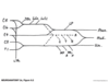
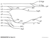
Figure 9.3 Lumbosacral Plexus

Figure 9.4 Lumbosacral Plexus: Simplified Schematic
Most Clinically Relevant:
F = femoral
Obt - obturator
Sc = sciatic
T = tibial
(CP = common peroneal)
SP = superficial peroneal
DP = deep peroneal

Lower Extremity Strength Testing:
https: //drive.google.com/file/d/0B5o1XviBdHwrOC1mNU5ZbEl0cFU/view?usp=sharing
https: //drive.google.com/file/d/0B5o1XviBdHwrc21rcHRBTlFEX00/view?usp=sharing
Important Nerves of the Leg

Important Nerves of Leg

Cervical Plexus
CN XII and C1 - C5
Phrenic nerve;
C3,4,5 keeps the diaphragm alive.
Brachial Plexus
- Axillary Nerve: C5, C6.
- Musculocutaneous Nerve: C5, C6, C7.
- Radial Nerve: C5, C6, C7, C8,T1.
- Median Nerve: C6, C7, C8, T1.
- Ulnar Nerve: C8, T1.
Five Important Nerves in the Arms

Five Important Nerves in the Arm

Upper Extremity Strength Testing:
https://drive.google.com/file/d/0B5o1XviBdHwrbU00cm9IaW9fMkE/view?usp=sharing
Thumb Strength Testing & Nerves:
https://drive.google.com/file/d/0B5o1XviBdHwrTGMxYWZNUlNnVGM/view?usp=sharing
Intrinsic Hand Muscles
innervated by ulnar nerve except LOAF
Lumbricals 1 and 2
Opponens pollicis
Abductor pollicis brevis
Flexor pollicis brevis
- Thunor eminence.
- Opponens pollicis, ABD pollicis brevis, flexor pollicis brevis.
- Hypothenar eminence.
- Opponens digiti minimi, flexor digiti minimi, ABD digit minimi.
- Lumbricals.
- Interossei.
Figure 9.6 Three Nerves Acting on the Thumb

Muscles Contributing to Flexion and Extension at Finger Joints

Upper Extremity Nerve Injuries
- Brachial plexus, upper trunk injury = (Erb-Duchenne palsy).
- Traction of infants shoulder.
- Motorcycle accident.
- Loss of C5C6 = weak biceps, deltoids, infraspinatus & wrist extensors.
Figure 9.7 “Bellman’s,” or “Waiter’s Tip,” Pose Assumed in Upper-Plexus Lesions

Upper Extremity Nerve Injuries
- Brachial plexus, lower trunk injury = Klumpke’s palsy.
- Grabbing a branch during a fall, TOS, Pancoast tumour.
- Weakness C8, T1 = hand & finger weakness, atrophy of hypothenar, sensory loos ulnar side of hand & forearm.
- If T1 is damaged proximal to sympathetic trunk; Horner’s syndrome: triad of miosis (constricted pupil), partial ptosis, and loss of hemifacial sweating (anhidrosis).
Thoracic Outlet Syndrome
Lower brachial plexus compressed between clavicle & 1st rib

*Cervical rib?
*ABD with ext. rot. increases symptoms & maybe decrease arterial pulse.
*EMG & X-Ray
Pancoast Syndrome
- Apical lung tumour (usually small cell carcinoma.)
- Affects lower brachial plexus.
- Sometimes Horner’s syndrome.
- Sometimes hoarseness (recurrent laryngeal nerve.)

Axillary Neuropathy
- Dislocation of proximal humerus compressing axillary nerve.
- Weak deltoid.
- Shoulder numbness.
- Differential dx – C5 radiculopathy.
(biceps).
Brachial Neuritis
- Unknown cause, inflammation?
- Burning shoulder or lateral neck pain.
- Weakness of muscles innervated by brachial plexus.
- Recovery usually 6-12 weeks.
Radial Neuropathy
- Saturday night palsy.
- Crutch palsy.
- Humeral fx at spiral groove.
- Weakness in all extensors of arm, hand, fingers.
- Weakness in supinator.
- Loss of triceps reflex.
- Sensory loss radial nerve distribution.
- Wrist drop.
- Handcuff neuropathy; sensory loss in dorso- lateral hand.
Ulnar Neuropathy
- Compression of ulnar nerve in hand / passing over hamate in Guyon’s canal.
- Prolonged leaning forward, resting on handlebars.
- Weakness in finger ADD or Abduction but no sensory loss.
Figure 9.8 Classic Hand Poses in Lesions of the Radial, Median, and Ulnar Nerves

Five Important Nerves in the Arm

Carpal Tunnel Syndrome
- Differential Dx:
- C6 C7 radiculopathy.
- Compression of median nerve proximal to carpal tunnel.
- Tinnel’s Sign (percuss median nerve.)
- Phalen’s Sign (compress dorsal surfaces of hands together.)
Ulnar Neuropathy
- Entrapment in cubital canal.
- Post traumatic, degenerative or congenital increased carrying angle at the elbow.
- Acute fx of medial epicondyle.
- Habitual resting on hard surface.
- Weakness wrist flexion, adduction, finger add &abduction, flexion of 4th & 5th digits.
- Hypothenar atrophy.
Median Neuropathy
- Humeral fx or distal radial dislocation.
- Pronator teres entrapment.
- Weakness in wrist flexion, ABDuction & opposition, flexion of 2nd & 3rd digits = make a fist/preacher’s hand.
- Median nerve sensory distribution loss.
Ulnar Neuropathy
- Entrapment in cubital canal.
- Post traumatic, degenerative or congenital increased carrying angle at the elbow.
- Acute fx of medial epicondyle.
- Habitual resting on hard surface.
- Weakness wrist flexion, adduction, finger add &abduction, flexion of 4th & 5th digits.
- Hypothenar atrophy.
Simian Hand
- Combination of chronic median and ulnar lesions.
- Thenar & hypothenar atrophy.
- Lack of opposition.
= Simian hand or monkey’s paw.
Simian Hand
- Combination of chronic median and ulnar lesions.
- Thenar & hypothenar atrophy.
- Lack of opposition.
- = Simian hand or monkey’s paw.
Lower Extremity Nerve Injury
- Femoral Neuropathy.
- Pelvic surgery.
- Pelvic Mass.
- Retroperitoneal hematoma.
- Weakness thigh flexion & knee extension.
- Loss of patellar reflex.
- Sensory loss anterior thigh.
Figure 9.8 Classic Hand Poses in Lesions of the Radial, Median, and Ulnar Nerves

Carpal Tunnel Syndrome
- Differential Dx:
- C6 C7 radiculopathy.
- Compression of median nerve proximal to carpal tunnel.
- Tinnel’s Sign (percuss median nerve.)
- Phalen’s Sign (compress dorsal surfaces of hands together.)
Important Nerves in the Leg

Figure 9.8 Classic Hand Poses in Lesions of the Radial, Median, and Ulnar Nerves

Lower Extremity Nerve Injur
- Femoral Neuropathy.
- Differenetial Dx.
- L3 or L4 radiculopathy.
- L3 or L4 may have thigh adduction weakness/not in femoral nerve neuropathy.
Important Nerves in the Leg

Lower Extremity Nerve Injury
- Peroneal palsy.
- Fibular head.
- Laceration.
- Stretch injury.
- Forcible foot inversion.
- Compression (tight stockings).
- Trauma.
- Foot drop.
- Weakness dorsiflexion, eversion & sensory loss dorsolateral foot & shin.
Important Nerves in the Leg

Lower Extremity Nerve Injury
- Sciatica.
- Painful paresthesias in a sciatic distribution.
- Compression of L/S nerve roots.
- Disc.
- Osteophytes.
Lower Extremity Nerve Injury
- Obturator palsy.
- L2-4.
- Compression in complicated deliveries.
- Pelvic trauma or surgery.
- Gait instability .
- Pain & numbness in medial thigh.
Important Nerves in the Leg

Lower Extremity Nerve Injury
- Meralgia Paresthetica.
- Lateral femoral cutaneous nerve (L2L3).
- Entrapment under inguinal ligament or fascia lata.
- Paresthesia & loss of sensation in lateral thigh.
-Pregnancy, obesity, weight loss, heavy equipment belts, worse with prolonged walking, sitting or standing
Lower Extremity Nerve Injury
- Morton’s metatarsalgia.
- Tight fitting shoes compressing digital nerves (esp. 3rd & 4th toes.)
- Patches of numbness & paresthesias.
Lower Extremity Nerve Injury
- Meralgia paresthetica.
- Differential dx.
- L2 or L3 radiculopathy.
- Usually has motor changes or decreased patellar reflex.
Distinguishing PNS from CNS Deficits
- Electrodiagnostic testing can be useful.
- Can help determine nerve & muscle disorders.
- Electromyography (EMG).
- Nerve conduction studies (NCV).
Figure 9.9 Nerve Conduction Study

NCV (nerve conduction velocity)
- CMAP recorded over muscle belly innervated by nerve, get summated electrical activity of muscle cells.
- SNAP if distal sensory nerve branch is stimulated getting summated electrical activity in sensory neuron axons of the nerve.
NCV
- Lesions proximal to DRG leave cell bodies & axons intact so SNAP is normal.
- Proximal lesions of motor nerve roots cause degeneration of distal motor neuron axons & decrease CMAP.
- Standard values for SNAP and CMAP latencies or conduction velocity for each major nerve at certain points.
NCV
- Slow conduction – demyelination.
- Decreased SNAP amplitude – axonal damage.
- CMAP – evaluate function of NMJ with repetitive stimulation.
EMG (electromyography-eval and recording electrical activity by skeletal muscles
- Electrode inserted into a muscle and MUP is recorded and evaluated.
- EMG patterns distinguish weakness of
- Neuropathic disorders (nerve or motor disease).
- Increased spontaneous activity (fibrillations & (+) sharp waves), large MUPs and duration & see fasciculations.
EMG
- Myopathic disease (muscle disease).
- Reduced MUP
- Continuous or increased recruitment patterns.
- Decreased amplitude.