Cerebral Hemispheres & Vascular Supply Flashcards
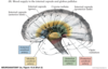
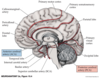
Main Functional Areas of Cerebral Cortex
Circle of Willis
- Arterial supply to the cerebral hemispheres.
- An anastomotic ring giving collateral circulation.
- Anterior circulation.
- From brachial cephalic trunk or aorta > internal carotid aa.
- Posterior circulation.
From subclavian aa. > transverse foramen / vertebral aa. > basilar arter
Anterior & Posterior Circulation

Anterior & Posterior Circulation

Main Arteries of the Circle of Willis
- Anterior cerebral artery (ACA).
- Anterior communicating aa. (AComms) link the ACAs.
- Middle cerebral artery (MCA).
- Posterior cerebral artery (PCA).
- Arise from the top of the basilar a.
Main Arteries of the Circle of Willis
- The anterior & posterior circulations are linked via the posterior communicating aa. (PComms).
- PComms connect internal carotids to PCA.
- Brainstem branches & cerebellar aa. arise from basilar a.
Figure 10.3 Circle of Willis and Its Main Branches

Internal Carotid Artery
- Named segments.
- Cervical segments.
- Petrous segment.
- Cavernous segment (carotid siphon).
- Passes anterior clinoid process to pierce dura & bed posterior to subarachnoid space as intracranial segment.
Circle of Willis and Its Main Branches

Circle of Willis and Its Main Branches
Main Branches of Intracranial Segment of Internal Carotid Artery
- OPAAM
- Ophthalmic a. - enters optic foramen with optic n.
- Posterior communicating a.
- .Anterior choroidal a.
- Anterior cerebral a.
- Middle cerebral a.
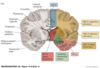
Territories of 3 Main Cerebral Aa.
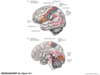
Superficial Cerebral Structures
- ACA (anterior cerebral artery) travels in interhemispheric fissure and back over corpus callosum.
- Supplies most of cortex on anterior medial surface from frontal > parietal lobes & medial sensorimotor cortex.
Anterior Cerebral Artery
Arteries of the Brain

Territories of 3 Main Cerebral Aa.
Superficial Cerebral Structures
•MCA turns laterally & enters Sylvian fissure.

Territories of 3 Main Cerebral Aa.
Superficial Cerebral Structures
- MCA turns laterally & enters Sylvian fissure ad then bifurcates into
- Superior division
- Inferior division
- (varies, sometimes into 3 or 4).
The branches form a loop as they pass over the insula and back out Sylvian fissure.
Middle Cerebral Artery -MCA

Arteries of the Brain

Territories of 3 Main Cerebral Aa.
Superficial Cerebral Structures
- PCA curves back with branches over:
- Inferior & middle temporal lobe
- Occipital cortex.
Arteries of the Brain

Arteries of the Brain

Territories of 3 Main Cerebral Aa.
Superficial Cerebral Structures
- MCA turns laterally & enters Sylvian fissure ad then bifurcates into
- Superior division
- Inferior division
- (varies, sometimes into 3 or 4).
- The branches form a loop as they pass over the insula and back out Sylvian fissure.
Territories of 3 Main Cerebral Aa.
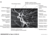
Deep Cerebral Structures
- **MOST IMPORTANT penetrating vessels at the base of brain = lenticulostriate aa.
- Arise from MCA.
- Penetrate anterior perforated substance.
- Supply large part of basal nuclei (ganglia) & internal capsule.
- Prone to narrowing in HTN > infarct > rupture
> hemorrhage.
Figure 10.7 Lenticulostriate Arteries