Muscle Weakness Module Flashcards

1
Q

Intermediary Metabolism

A

All the chemical changes that are involved in the occurance and continuance of life.

Ability to accomplish these changes at constant body temperature requires enzymatic catalysis & thermodynamic coupling of endergonic and exergonic processes.

2
Q

Pathway

A

The series of steps involved in the breakdown or synthesis of major biological constituents.

Cycle = pathway that regenerates the initial substrate

Catabolic and anabolic pathways linked via ATP.

3
Q

Catabolism

A

Degradative (complex to simpler)

Oxidative

Exergonic

ATP generating

Often requires NAD+ or FAD

4
Q

Anabolism

A

Sum of the pathways that are involved in synthesis and growth.

Reductive

Energy consuming

ATP utilization

Often requires NADPH

5
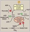
Q

Metabolic rate

A

Expression of enthalpic change.

Gives the normalized total heat production per unit time.

6
Q

Basal metabolic rate

A

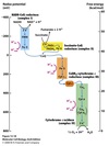
Measured in a resting state (awake laying still)

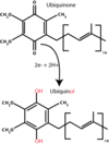
Energy requirements for:

  1. involuntary muscle work
  2. maintenance of osmotic gradients
  3. maintenance of body temperature
  4. turnover and synthesis of cell constiuents
7
Q

Gibb’s free energy

(ΔG)

A

ΔG = ΔH - TΔS

H= enthalpy S= entropy T= temp in kelvins

The energy change occuring under conditions of constant pressure and temperature.

Additive function and independent of pathway.

Total ΔG of a process can be expressed as the sum of ΔG changes of individual steps.

8
Q

Thermodynamic Coupling

A

Coupling of an exergonic and endergonic reaction so that net ΔG is negative and reaction can occur.

A common intermediate must exist.

In an enzyme catalyzed reaction, the common intermediate may not be free and may only exist on the enzyme.

9
Q

Standard reduction potentials

(Eº)

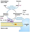
A

The relative affinity of a molecule, atom, or ion for electrons taken under standard conditions where reactants and products are at unit activity (~1 M) then compared to the proton/hydrogen electrode (H+ and ½ H2)

Often expressed as values corrected to pH 7 (Eº’)

Interpreted as the relative affinity of the system for electrons as compared to that of a proton:

  • Negative reduction potential indicates a weaker affinity for electrons than a proton.
  • Positive reduction potential indicates a stronger affinity.
10
Q

Cellular reduction potential

A

Dependent on the ratio of oxidant to reductant agent concentration in a cell.

11
Q

Nernst equation

A

E = Eº’ + 2.3 RT/nF log [oxidant] / [reductant]

R = gas constant

F = Faraday constant

Relationship of reduction potential to ratio of oxidant to reductant agents in a cell.

12
Q

High energy bonds

A

Describes a bond which has a large negative standard Gibb’s free energy (ΔGº’) of hydrolysis

Minimum of -7.0 kcal/mol

Indicated with a ~

13
Q

Adenosine triphosphate

(ATP)

A
  • Contains two phosphoanhydride bonds which have a ΔGº’ of hydrolysis of approximately -7.3 kcal/mol each.
  • Biological utilization of these high energy bonds require the ATP form.
  • One or both bonds may be used in a reaction.
  • Phosphoryl group(s) of ATP can be transferred to acceptor molecules to generate activated intermediates for metabolism.
  • Can function as coenzyme-cosubstrates.
14
Q

Pyrophosphate

(PPi)

A

Use of both phosphoanhydride bonds of ATP is the functional equivalent of using 2 ATP’s.

Reactions coupled to the hydrolysis of the pyrophosphate (PPi) product to 2 orthophosphates (Pi) by pyrophosphatase which drives reactions forward.

15
Q

Adenylate kinase

A

Catalyzes the reversible reaction:

AMP + ATP ⇔ 2 ADP

16
Q

Adenine nucleotide cosubstrate metabolic pool

A

Total concentration of adenine nucleotide pool essentially constant, however, ratio of adenine nucleotides vary with metabolic state of the cell.

Concentrations of ATP, ADP, and AMP in a cell are in rapid equilibrium due to activity of adenylate kinase.

The cosubstrate pool communicates between and significantly influences the different pathways of the cell which utilizes those cosubstrates.

AMP level most sensitive parameter to change in pool and usually initiates the responses to decreased ATP levels.

17
Q

Energy charge

A

Energy charge = ½ • ( [ADP] + 2[ATP] ) / ( [AMP] + [ADP] + [ATP])

A stoichiometric expression of the mold fraction of high energy phosphate bonds present relative to the maximal high energy bonds possible.

0 = nucleotides are totally in the form of AMP

1 = nucleotides are totally in the form of ATP

18
Q

ΔG for ATP hydrolysis

A

ΔGATP→ADP = ΔGº’ATP→ADP + RT ln ( [ADP] x [Pi] / [ATP] )

Actual ΔG for ATP hydrolysis in a cell is dependent upon and may be calculated from the concentrations of ATP, ADP, and inorganic phosphate.

May be very different from the ΔGº’.

In a resting cell may be as high as = 15 kcal/mol which is equivalent to an energy charge of approximately 0.9.

“High energy charge”, “large negative ΔG of ATP hydrolysis”, and “resting cell” all indicate that ATP is plentiful.

19
Q

Pathway regulation

A
  • Pathways for synthesis and breakdown of the same constituent are never the same, although individual steps may be reversible and utilized in both pathways.
  • Opposing pathways typically occur in seperate cellular compartents and/or different tissues or organs.
  • Regulation of pathway enzyme activity by:
    • product inhibition
    • allosteric regulation
    • covalent regulation i.e. phosphorylation
  • Changes in the rate of synthesis/degradation of an enzyme or its mRNA.
20
Q

Equation for glycolysis

A

glucose + 2 NAD+ + 2 ADP + 2 Pi

2 pyruvate + 2 NADH + 2 ATP + 2 H2O + 2 H+

21
Q

General stages of glycosis

A
  1. Priming steps
  • Two ATP-linked phosphorylations
  • Functions to prevent diffusion of the intermediates of the pathway out of the cell.
  1. Oxidation/reduction with the production of NADH
    * Results in the generation of ATP
  2. Re-oxidation of the NADH produced.
22
Q

Substrate-level phosphorylation

A

The synthesis of ATP involving two coupled reactions linked by a common intermediate containing a high-energy bond.

23
Q

Steps of glycolysis

A
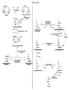
  1. Irreversible phosphorylation of glucose at carbon-6 by hexokinase using ATP•Mg2+ to produce glucose-6-phosphate.
    • Hepatic isozyme is glucokinase.
    • Traps glucose because cell membrane is impermeable to phosphate esters.
    • Commits glucose to intracellular metabolism but NOT glycolysis.
  2. Freely reversible aldose-ketose isomerization of glucose-6-phosphate to fructose-6-phosphate by phosphoglucose isomerase.
  3. Irreversible phosphorylation of fructose-6-phosphate to fructose-1,6-bisphosphate using ATP by phosphofructokinase (PFK1).
    • True commitment step and primary regulatory site for glycolysis.
  4. Fructose-1,6-bisphosphate split into two triose phosphates: dihydroxyacetone phosphate (DHAP) and glyceraldehyde-3-phosphate (GAP) by aldolase.
    • DHAP freely interconverted to GAP by triose phosphate isomerase.
    • Only GAP enters next stage of glycolysis.
  5. Glyceraldehyde-3-phosphate dehydrogenase catalyzes the reversible oxidation of glyceraldehyde-3-phosphate to 1,3-bisphosphoglycerate.
    • NAD+ converted to NADH **
  6. Phosphate from the carboxyl group from 1,3-bisphosphoglycerate is reversibly transferred to ADP to produce ATP and 3-phosphoglycerate by 3-phosphoglycerate kinase.
    • First substrate-level phosphorylation.
    • Additional mutase found in erythrocytes which transfers carbonyl phosphate of 1,3-bisphosphoglycerate to carbon-2 to produce 2,3-bisphosphoglycerate (2,3-BPG).
  7. 3-phosphoglycerate converted to 2-phosphoglycerate by phosphoglycerate mutase.
  8. 2-phosphoglycerate converted to phosphoenolpyruvate (PEP) by enolase with loss of H2O.
  9. Phosphate of PEP is transferred to ADP by pyruvate kinase (PK) to produce pyruvate and ATP.
    • Irreversible
    • Has a ΔGº’ of -14 kcal/mol
  10. NADH produced is recycled back to NAD+
    • In aerobic conditions via malate shuttle.
    • In anerobic conditions pyruvate converted to lactate via lactate dehydrogenase.
24
Q

Malate shuttle

A

Used for the indirect transport of the electrons from glycolysis in the cytoplasm to the respiratory chain in mitochondria.

  1. Cytosolic oxidation of NADH coupled with reduction of oxaloacetate to malate.
  2. Malate transported across the inner mitochondrial membrane.
  3. Malate oxidized back into oxaloacetate as mitochondrial NAD+ is reduced to NADH.
25
Q

Pyruvate kinase (PK) deficiency

A
  • PK-LR gene encodes both RBC and liver isozymes for pyruvate kinase through alternative promoters.
  • PK deficiency is an autosomal recessive disorder that primarily affects the mature RBC isozyme.
    • Mutations in coding, splice site, or promoter regions described resulting in an abnormal protein.
  • Abnormal PK protein has lower affinity for PEP or it’s allosteric effectors.
  • Results in premature distruction (hemolysis) of RBC’s leading to hemolytic anemia due to diminished ATP production.
26
Q

Glycolytic isozymes

A

Transcriptional regulation of glycolytic isozymes used to regulate the flow of glycolysis in order to meet the metabolic needs of rapidly dividing cells (ex. embryonic stem cells or tumor cells).

  • GLUT-3 transporter and hexokinase (HK-2) up-regulated - both have lower Km for glucose.
  • Use of pyruvate kinase (PKM2) and lactate dehydrogenase (LDH-A) increases flow of glucose through glycolysis.
27
Q

Fermentation

A

Glycolysis occurring under anaerobic conditions.

28
Q

Lactate fermentation

A

Occurs in mammalian tissues specifically skeletal muscle.

Pyruvate + NADH → lactate + NAD+

Catalyzed by lactate dehydrogenase.

Production of NAD+ allows glycolysis to continue.

29
Q

Alcoholic fermentation

A

Occurs in microorganisms such as yeast.

1. Pyruvate → CO2 + acetaldehyde

Catalyzed by pyruvate decarboxylase.

2. Acetaldehyde + NADH → ethanol + NAD<strong>+</strong>

Catalyzed by alcohol dehydrogenase.

30
Q

Mitochondrial pyruvate carrier

(MPC)

A

Transports the pyruvate made via cytoplasmic glycosis through the inner mitochondrial membrane.

31
Q

Stages of cellular respiration

A
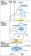
  1. Carbohydrates, proteins, and fatty acids are oxidized to two-carbon fragments in the form of acetyl~CoA.
  2. The acetyl groups enter the tricarboxylic acid (TCA) cycle which produces CO2 and the reduced energy carriers NADH and FADH2.
  3. NADH and FADH2 produced are oxidized in the electron transport chain (ETC) producing H+ and electrons. Electrons are transferred to O2 producing water. Protons used in oxidative phosphorylation to produce ATP.
32
Q

Coenzyme A

(CoA)

A
  • Derived from ATP and pantothenic acid (Vit B5)
  • Contains a reactive thiol group (-SH) that is covalently linked to the acetyl group via a thioester bond
  • Acetyl~CoA readily donates acetyl groups to other acceptors
33
Q

Pyruvate dehydrogenase complex

(PDH)

A
  • Member of the α-ketoacid dehydrogenase family
  • Large multienzyme complex which contains 3 enzymes in multiple copies
  • Very efficient due to close spatial proximity of complexes

Enzyme subunits

  • E1: pyruvate dehydrogenase (aka pyruvate decarboxylase) #30
  • E2: dihydrolipoyl transacetylase #60
    • Acts as a swinging arm between E1 & CoA preventing substrate from diffusing away ⇒ substrate channeling
  • E3: dihydrolipoyl dehydrogenase #12
  • Contains two stoichiometric coenzymes/cosubstrates
    • NAD
    • CoA
  • Contains three catalytic coenzyme prosthetic groups
    • thiamine pyrophosphate (TPP)
    • FAD
    • lipoic acid
  • Contains two regulatory enzymes associated with but not part of the complex
    • PDH kinase
    • PDH phosphatase
34
Q

Steps for oxidative decarboxylation of pyruvate

A
  1. Pyruvate is decarboxylated and the acetyl group is attatched to thiamine pyrophosphate (TPP) coenzyme of pyruvate decarboxylase (E1).
  2. Acetyl group transferred to the lipoic acid covalently bound to dihydrolipoyl transacetylase (E2),
  3. The acetyl group, bound as a thioester to the side chain of lipoic acid, is transferred to free CoA.
  4. The sulfhydryl form of lipoic acid is oxidized by FAD-dependent dihydrolipoyl dehydrogenase (E3) to regenerate the oxidized lipoic acid.
  5. FADH2 on E3 is reoxidized to FAD as NAD+ is reduced to NADH2 + H+.
35
Q

Regulation of PDH complex

A
  • Allosteric regulation
    • Acetyl~CoA and NADH can inhibit complex via allosteric end-product inhibition (feedback)
  • Covalent modification via phosphorylation and dephosphorylation - main mechanism
    • E1 inactivated by phosphorylation by PDH kinase
    • E1 activated by dephosphorylation by PDH phosphatase
  • Both PDH kinase and PDH phoshatase are in turn allosterically activated by a number of molecules that signal the energy state of the cell
  • PDH kinase
    • activated by high-energy signals
      • ATP
      • acetyl~CoA
      • NADH
    • inhibited by pyruvate
  • PDH phosphatase
    • activated by rise in intracellular [Ca++]
      • important in skeletal muscle
36
Q

TCA cycle

aka

Citric Acid Cycle

aka

Krebs cycle

General

A

Acetyl~CoA + 3 NAD+ + FAD → 2 CO2 + 3 NADH + FADH2

  • 8 steps
    • 4 oxidations that produce NADH or FADH2
    • one substrate-level phosphorylation producing GTP
  • Except for succinate dehydrogenase which is embedded in the inner mitochondrial membrane all enzymes are located in the matrix
37
Q

Steps of the TCA cycle

A
  1. Formation of citrate.
    • Condensation of acetyl~CoA and oxaloacetate (OAA) catalyzed by citrate synthase.
    • Reaction made irreversible by the hydrolysis of the thioester bond of acetyl~CoA.
    • Facilitated by an enzyme-bound intermediate, citroyl~CoA.
    • Citrate synthase inhibited by citrate via competitive inhibition.
  2. Isomerization of citrate to isocitrate.
    • Citrate isomerized to isocitrate by aconitase.
    • ΔGº’ = 6.3 kJ/mol but reaction pushed to the right in vivo because product rapidly consumed in next step.
  3. Oxidation of isocitrate to α-ketoglutarate and CO2.
    • Irreversible decarboxylation of isocitrate to α-ketoglutarate by isocitrate dehydrogenase.
    • Loss of CO2 make the reaction irreversible.
    • NADH produced.
    • Isocitrate dehydrogenase inhibited by ATP and NADH. Stimulated by ADP and Ca++.
  4. Oxidation of α-ketoglutarate to succinyl~CoA and CO2.
    • Catalyzed by α-ketoglutarate dehydrogenase complex.
      • Same family as PDH dehydrogenase.
      • Uses same coenzymes/cosubstrates (NAD and CoA) and catalytic coenzymes (TPP, FAD, and lipoic acid)
      • Inhibited by succinyl~CoA and NADH.
      • Activated by Ca2+.
      • Not regulated by phosphorylation/dephosphorylation.
    • NADH produced.
    • Loss of CO2 makes reaction irreversible.
  5. Conversion of succinyl~CoA to succinate.
    • Succinate thiokinase catalyzes the hydrolysis of the thioester bond in succinyl~CoA paired to the conversion of GDP to GTP.
    • GTP interconvertible to ATP by nucleoside diphosphate kinase.
  6. Oxidation of succinate to fumarate.
    • Catalyzed by succinate dehydrogenase with production of FADH2.
    • Enzyme located on inner mitochondrial membrane.
    • ΔGº’ = 0 so reaction can go either way but pushed to the right in vivo because fumarate used in next step.
  7. Regeneration of oxaloacetate.
    1. Fumarate converted to malate by fumarase.
    2. Malate converted to oxaloacetate by malate dehydrogenase with production of NADH.
      • Rxn has a ΔGº’ = 29.7 kJ/mol but pulled to the right because OAA used by citrate synthase in step 1.
38
Q

Regulation of the TCA cycle

A

Regulated almost exclusively at the three irreversible steps.

  1. Citrate synthase competitively inhibited by citrate.
  2. Isocitrate dehydrogenase
    • Inhibited by ATP and NADH
    • Stimuated by ADP and Ca++
  3. α-ketoglutarate dehydrogenase
    1. Inhibited by succinyl~CoA and NADH
    2. Activated by Ca++
39
Q

Additional roles of TCA cycle

A

TCA cycle intermediates can be used for other reactions such as:

  • amino acid synthesis
  • fatty acid synthesis
  • gluconeogenesis
40
Q

TCA cycle intermediate replenishment

A

Intermediates of the TCA cycle can be provided for by other metabolic pathways, specifically amino acid metabolism.

41
Q

Electron transport chain

(ETC)

aka Respiratory chain

Basics

A

Inner mitochondrial membrane bound complex.

Consists of four seperate protein complexes.

Each complex accepts or donates electrons to/from mobile electron carriers (coenzyme Q and cytochrome C).

ETC pumps protons from matrix to intermembrane space to form a proton gradient.

42
Q

ETC complexes

A
  • Complex 1: NADH dehydrogenase
    • Flavin mononucleotide (FMN) coenzyme accepts two electrons from NADH to become FMNH2.
    • FMNH2 transfers electrons to CoQ to form CoQH2.
    • Enzyme contains iron-sulfur (Fe-S) centers which acts as intermediate electron carriers.
  • Complex 2: Succinate dehydrogenase
    • Electrons from succinate of TCA cycle transferred via Fe-S centers to FAD to form FADH2.
  • Complex 3: Cytochrome bc1 reductase
    • Electrons from CoQH2 (from complex 1) are used to reduce cytochrome c which acts as electron carrier.
    • Contains Fe-S centers which act as intermediates.
    • Contains heme group which shifts from Fe3+ to Fe2+ and back as electrons move through.
  • Complex 4: Cytochrome oxidase
    • Contains two different heme groups
      • Heme a
      • Heme a3
    • Contains two Cu ions which act as intermediates
    • Electrons from cytochrome c transferred to heme a via one of the Cu centers, then from heme a to heme a<em>3</em> via other Cu center, and finally to O2 to form H2O.
43
Q

Coenzyme Q

A

Oxidized form ubiquinone reduced to ubiquinol after accepting electron.

Accepts electrons from complex I and II of the ETC.

44
Q

Cytochromes

A

Contain a heme group in which the iron shifts from Fe3+ to Fe2+ oxidations states and back as electrons move to and from the heme group.

All contain the porphyrin ring with differing side chains.

45
Q

Redox potentials

(E’º in volts)

A

Measures the ease with which an electron can be added or removed.

The more positive the value of E’º, the greater the tendancy of the oxidant in the redox pair to accept electrons.

Electrons flow from the redox pair with the more negative E’º to one with a less negative/more positive E’º.

46
Q

Relationship of redox potential and free energy change

A

The redox potential (E’º) is related to the free energy change in the reaction by the Farady constant (F).

ΔG’º = -nFΔEº

n = number of electrons transferred

ΔEº = difference in reduction potentials of the overall reaction

47
Q

Effective reduction potential

(E)

A

In the cell, the effective reduction potential (E) depends on the concentration of reactants.

48
Q

Electron flow in ETC

A

Due to the reduction potentials of each complex the electrons always flow downhill.

49
Q

ETC site-specific inhibitors

A

Used to block electron flow in ETC.

Carriers befor the block become reduced and those after remain oxidized.

50
Q

Oxidative phosphorylation

A

ATP synthase uses the proton gradient set up by ETC to produce ATP.

Dependent on the integrity of the inner mitochondrial membrane.

No high-energy intermediate exists (as with substrate-level phosphorylation).

51
Q

Reactive oxygen species

(ROS)

A

When more electrons enter the ETC than can be immediately passed to O2 a state of oxidative stress exists.

Highly reactive superoxide free radicals such as superoxide (•O2-) are generated.

Mitochondria have systems to eliminate free radicals:

  1. Superoxide (•O2-) is converted to peroxide (H2O2) by superoxide dismutase.
  2. Peroxide converted to water by glutathione peroxidase.

NADPH used by glutathione reductase to regenerate glutathione peroxidase.

52
Q

Respiratory control

A

The coupling of ATP synthesis and electron transport.

Neither process can occur without the other.

53
Q

P:O ratio

A

The stoichiometry of ATP synthesis relative to the substrate that is oxidized.

The amount of ATP formed per ½ O2 consumed (or per pair of electrons.

Approaches 3 for NADH.

Closer to 2 for FADH2 (because succinate oxidation bypasses complex I)

54
Q

Proton pumping by ETC

A

For each pair of electrons that travel through the ETC and transferred to O2:

Complex 1 pumps out 4 protons

Complex 3 pumps out 4 protons

Complex 4 pumps out 2 protons

Total of 10 protons per electron pair.

NADH + 11 H+M + ½ O2 → NAD+ + 10 H+IM + H2O

55
Q

Proton motive force

(PMF)

A

Proton pumping by ETC across the inner mitochondrial membrane sets up a pH gradient (ΔpH) and a transmembrane potential (Δψ) with matrix side negative.

Can be calculated as

ΔG = 2.3 RT ΔpH + F Δψ

In general, transport of a pair of electrons through the ETC generates ~ 53 kcal.

56
Q

Chemiosmotic Hypothesis

A

The proton motive force drives ATP synthesis as the protons flow passively back through the inner mitochondrial matrix down their concentration gradient through a proton pore in the ATP synthase.

The ~ 53 kcal produced per pair of electrons can drive the synthesis of 3 ATPs (using ~ 22 kcal) with the remaining energy used to drive ancillary reactions or dissipated as heat.

ADP + Pi + nH+IM → ATP + H2O + nH+M

Value of n depends on the structure of the ATP synthase and varies between species (~3.3 - 5 protons per ATP)

57
Q

Mitochrondrial ATP synthase

(Complex V)

A

F1Fo ATPase

O stands for oligomycin which blocks the proton pore

F-type ATPase which functions as a reversible ATP-driven proton pump

  • F1 portion contains the ATPase domain.
    • Contains 9 subunits α3β3γδε
    • α and β subunits for the knoblike catalytic section
    • γ subunit forms a shaft that connects with Fo
  • Fo complex consists of a, b, and c subunits
    • 8 c-subunits form the c-ring in vertebrates
      • Rotation of C-ring induces conformational changes in the β subunits of F1 that drive ATP synthesis
      • Requires 8 protons moving across the membrane with each rotation and produces 3 ATP’s per turn
58
Q

Yield of ATP production

A
  • Takes 11 H+ to generate 3 ATP’s
    • 8 H+ to give a complete c-ring rotation
    • Additional 3 H+ to bring 3 phosphates into the matrix via the mitochondrial phosphate transporter
  • From 1 NADH: 10 H+ pumped so (10/11) x 3 ATPs produced = 2.75 ATPs per NADH
  • From 1 FADH2 (Succinate): 6 H+ pumped so (6/11) x 3 ATPs produced = 1.64 ATPs per FADH2
  • Glycolysis generates 2 net ATPs and 2 NADH
  • PDH generates 1 NADH/pyruvate → 2 NADH/glucose
  • TCA generates:
    • 3 NADH/pyruvate → 6 NADH/glucose
    • 1 FADH2/pyruvate → 2 FADH2/glucose
    • 1 ATP/pyruvate → 2 ATP/glucose
  • OxPhos:
    • 10 NADH → 27.5 ATP
    • 2 FADH2 → 3.3 ATP

Going all the way through oxidative phosphorylation generates:

  • ~ 31 ATP by OxPhos
  • 2 ATP for glycolysis
  • 2 ATP for TCA

TOTAL OF ~35 ATP/GLUCOSE

59
Q

Respiratory inhibitors

A

CN- or CO

blocks electron transport and subsequently ATP synthesis

60
Q

Oligomycin

A

Phosphorylation inhibitor

Blocks ATP synthesis and thus electron transport

61
Q

Uncouplers

A

dinitrophenol (DNP)

uncoupling protein 1 (UCP1 aka thermogenin)

  • Causes collapse of the proton motive force releasing energy in the form of heat rather than ATP.
  • Electrons free to move through the ETC.
  • Increases oxygen consumption.
62
Q

Regulation of oxidative phosphorylation

A

ETC and ATP synthesis coupled so increasing or decreasing one does the same to the other.

63
Q

Ancillary reactions

A

All the transport processes associated with oxidative phosphorylation which utilize the proton motive force or transmembrane potential.

Mostly used to transport molecules across the impermeable inner mitochondrial membrane.

  • Agents that disrupt the PMF will reduce transport.
  • Transport of these substances will conversely reduce the PMF available for ATP synthesis.
  • Electroneutral transport systems driven by concentration gradients alone.
  • Electrogenic transport systems utilize concentration gradients as well as the transmembrane potential.
    • positively charged molecules enter matrix more easily
  • Adenine nucleotide translocase
  • Phosphate translocase
  • Transporters for other charged metabolites such as pyruvate, malate, citrate, etc.
  • Ca++ and asparate also transported in a PMF-dependent fashion
64
Q

Adenine nucleotide translocase

A

Antiporter that exchanges ADP3- from the intermembrane space for ATP4- from the matrix.

Net transport of 1 negative charge out of the matrix (electrogenic) stimulated by the matrix-negative transmembrane potential.

Inhibited by atractyloside.

65
Q

Phosphate translocase

A

Symporter that moves one phosphate (H2PO4- ) and one proton (H+) into the matrix.

Driven by the proton gradient established by ETC.

Electronically neutral.

66
Q

ATP synthasome

A

Complex containing ATP synthase, adenine nucleotide translocase, and phosphate translocase can be isolated by gentle disruption of the inner mitochondrial membrane suggesting that these three proteins are spatially integrated.

67
Q

Nicotinamide nucleotide transhydrogenase

A

A transmembrane protein embedded in the inner mitochondrial membrane.

Uses the proton gradient to drive:

NADH + NADP+ + H+IM → NAD+ + NADPH + H+M

NADH is used by ETC so its levels are related to the potential for ROS generation.

Production of NADPH by transhydrogenase will increase as more electrons travel down ETC.

NADPH indirectly used in ROS neutralization.

68
Q

Malate-Aspartate Shuttle

A

Found in most tissues.

  1. Electrons from NADH transferred to cytosolic oxaloacetate using cytosolic malate dehydrogenase to form malate.
  2. Malate enters the matrix via malate-α-ketoglutarate transporter.
  3. Inside the matrix, malate converted back to oxaloacetate by mitochondrial malate dehydrogenase with concomitant reduction of NAD+ to NADH.
  4. Matrix oxaloacetate converted to aspartate and sent back to the cytosol where it is metabolized to oxaloacetate, completing cycle.
69
Q

Glycerol-3-phosphate shuttle

A

Used in skeletal muscle and brain.

  1. NADH generated in glycolysis used by cytosolic glycerol-3-phosphate dehydrogenase to convert dihydroxyacetone phosphate to glycerol-3-phosphate.
  2. Glycerol-3-phosphate enters the matrix where it is converted back to DHAP by mitochondrial glycerol-3-phosphate dehydrogenase producing FADH2 which enters ETC at CoQ.

Since FADH2 produced instead of NADH the yield of ATP is diminished.

70
Q

Mitochondrial genes

A

Mitochondria contain their own genome which contains 37 genes.

13 encode subunits of respiratory chain proteins.

71
Q

Mitochondrial diseases

A

Maternally inherited.

Defects in oxidative phosphorylation most commonly associated with mutations in mitochondrial genes.

Presumably due to generation of reactive oxygen species.

Affects tissues with high requirement for ATP such as brain, liver, and skeletal/cardiac muscle.

72
Q

Leber’s hereditary optic neuropathy

(LHON)

A
  • Caused by a mutation in one of the subunits of Complex I of ETC which renders it unable to transfer electrons from NADH to CoQ.
  • Patients with LHON can only use electron transfer via succinate (complex II) thus produce less ATP.
  • Leads to bilateral loss of vision in early adulthood.
73
Q

Mitochondria and apoptosis

A
  • Triggered by:
    • External signals acting via a receptor
    • Oxidative stress
    • Heat shock
    • Viral infection
  • Exposure to stimulus induces formation of large pores in the outer mitochondrial membrane called permeability transition complex
    • Allows release of cytochrome c into the cytosol
  • Cytochrome c in associated with apoptosis protease activating factor 1 (Apaf-1) activate a family of cytosolic proteases (the caspases) that degrade proteins and lead to cell death.
74
Q

Glycogen

Structure & Function

A
  • Major short-term storage form of carbohydrates in animals.
    • For times of metabolic need.
  • Branched chain homopolysaccharide of α-D-glucose
    • Primary bond is α-1,4-glycosidic linkages.
    • Every 8-10 glucose residues there is a branch attached via a α-1,6-glycosidic linkage
  • Stored in the cytoplasm as large hydrated granules.
    • Each granule contains as many as 55,000 glucose units.
  • Found in the cytoplasm of liver and skeletal muscle cells primarily.
75
Q

Sources of blood glucose

A
  1. Diet
  2. Degradation of glycogen
  3. Gluconeogenesis
76
Q

Liver Glycogen

A
  • Functions to maintain the blood glucose concentration particularly during the early stage of a fast.
  • Glucose rapidly released from liver glycogen.
    • Glucose able to enter systemic circulation due to presence of glucose-6-phosphatase in the liver.
  • Hepatic glycogen stores ~24 hour supply of glucose.
  • Liver also able to synthesize glucose via gluconeogenesis.
77
Q

Skeletal Muscle Glycogen

A
  • Serves as a fuel reserve for synthesis of ATP that will power muscle contraction.
  • Muscle glycogen is not available to other tissues because muscle lacks glucose-6-phosphatase.
78
Q

Methods of Activating Sugars

A
  1. Phosphorylation
    • Glycolysis utilizes glucose-6-phosphate.
  2. Create a nucleotide sugar
    • Glycogenesis utilizes UDP-glucose.

Alternate activation methods allows for both pathways to occur at the same time.

79
Q

Glycogenesis

Step 1: Chain Synthesis

A
  • Occurs in the cytoplasm and can be divided into two stages:
    1. Chain synthesis
    2. Chain branching

I. Chain Synthesis

A. Synthesis of UDP-glucose.

  1. Glucose is phosphorylated to glucose-6-phosphate.
    - By glucokinase in hepatic tissue.
    - By hexokinase in peripheral tissue.
  2. Glucose-6-phosphate converted to glucose-1-phosphate by phosphoglucomutase.
  3. Glucose-1-phosphate reacts with UTP to form UDP-glucose and PPi which is catalyzed by glucose 1-phosphate uridylyltransferase (aka UDP-Glc pyrophosphorylase)
    - Hydrolysis of PPi2 Pi by pyrophosphatase makes the reaction energetically favorable and irreversible.

B. Requirement of primer to initiate glycogen synthesis.

⇒Glycogen synthase cannot initiate glycogen synthesis de novo and can only add glucose to an existing chain, therefore, glycogenesis requires a primer.

  1. Glycogen fragment can serve as a primer for glycogen synthase which attaches glucosyl residues to existing chain using UDP-glucose.
  2. The protein glycogenin (homodimer) can prime glycogen synthesis and attach glucose residues through auto-glucosylation ⇒ serves as both substrate and enzyme in its role as primer.
    a. The glycosyltransferase activity of glycogenin transfers the first molecules of glucose from UDP-glucose to a specific tyrosine side-chain (tyr-194) on itself.
    b. After at least 4 (about 7) glucose residues have been added, glycogen synthase takes over.
    - Glycogenin remains within the glycogen granule.

C. Elongation of glycogen chains.

  1. Glycogen synthase transfers glucose from UDP-glucose to the non-reducing end of the growing chain via α-1,4-linkages between the -OH group on C-1 of the activated sugar and the C-4 of the accepting sugar.

Glycogen synthase is the rate-limited and regulated enzyme of glycogenesis.

⇒ There are liver & muscle isozymes.

UDP released when α-1,4-glycosidic bond is formed can be converted to UTP by nucleoside diphosphate kinase + ATP.

80
Q

Glycogenesis

Step 2: Chain Branching

A
  • Occurs in the cytoplasm and can be divided into two stages:
    1. Chain synthesis
    2. Chain branching

II. Chain branching

A. Catalyzed by “branching enzyme” called glucosyl 4:6 transferase.

  1. Glucosyl 4:6 transferase cleaves an α-1,4-glycosidic bond from the non-reducing end of the glycogen chain producing a 6-8 glucosyl residues fragment.
  2. Enzyme then transfers the fragment to another residue on the linear chain via an α-1,6-glycosidic bond.
  3. Resulting new non-reducing end and old non-reducing ends can be further elongated by glycogen synthase.
  4. After further elongation, new chains of 6-8 residues can be transferred to make additional branches.
81
Q

Functions of branching in Glycogen

A
  1. Increases solubility of glycogen molecule.
    • Stored as hydrated granules.
  2. Increases number of non-reducing ends to which new glucosyl residues can be added to or removed from glycogen.
    • Facilitates fast breakdown of glycogen into glucose when energy needed.
82
Q

Glycogenolysis

Overview

A
  • Occurs primarily in the cytoplasm of liver and skeletal muscle cells.
  • Involves 2 stages:
    1. Shortening of chains
    2. Removal of branches
83
Q

Glycogenolysis

Step 1: Shortening of Chains

A
  1. Glycogen phosphorylase uses Pi to cleave the α-1,4-glycosidic bonds between glucose residues at the non-reducing ends of the glycogen chains releasing glucose-1-phosphate.
    ⇒Enzyme is a homodimeric exoglucosidase
    ⇒Requires pyridoxal phosphate (PLP) coenzyme (derivative of Vit B6)
    ⇒Liver and muscle isozymes
    • Glycogen phosphorylase stops attacking α-1,4-glycosidic bonds four glucosyl residues from an α-1,6-branch point.
    • Resulting structure is called a limit dextrin.
  2. Glucose-1-phosphate is converted to glucose-6-phosphate by phosphoglucomutase.
  3. Next step in glycogenolysis depends on the tissue:
    • In liver: glucose-6-phosphate is hydrolyzed to free glucose and Pi by glucose-6-phosphatase.
      ⇒Free glucose able to leave the liver and enter the blood stream.
    • In peripheral tissues: glucose-6-phosphate will be oxidized in the glycolytic pathway to produce energy.
84
Q

Glycogenolysis

Step 2: Removal of branches

A

Catalyzed by a debranching enzyme which is a bifunctional protein with two catalytic activities.

  1. 4-α-D-glucantransferase activity transfers the outer three glucosyl residues of the limit dextrin to a non-reducing end by breaking and formation of α-1,4 bonds leaving one glucosyl residue in α-1,6 linkage.
    ⇒ 4,4 transferase activity
  2. α-1,6 linkage is then cleaved hydroytically by the amylo-α-1,6 glucosidase activity of debranching enzyme releasing free glucose (non-phosphorylated).
  3. Co-operative and repetitive action of phosphorylase and debranching enzymes results in complete hydrolysis of glycogen to yield glucose-1-phosphate and free glucose in a 12:1 ratio.

⇒Free glucose is quickly phosphorylated in muscle for intracellular use.
⇒Phosphorylated glucose is already trapped inside muscle cells.

85
Q

Regulation of Glycogen Metabolism

A

Key regulatory enzymes of glycogen metabolism:

Glycogen synthase (synthesis)

&

Glycogen phosphorylase (degradation)

Each is controlled by:

  1. Hormone-induced covalent modification through phosphorylation or dephosphorylation of ser residues.
    * Way of responding to the needs of the body as a whole.
  2. Allosteric effectors
    * Way of responding to the need of a particular tissue at a particular time.
86
Q

Glycogen Phosphorylase

Covalent Regulation

(Control of glycogen breakdown)

A

I. Regulation by covalent modification:

Glycogen phosphorylase exists in two forms:

A or active form: phosphorylated

B or inactive form: dephosphorylated

A and B forms are interconverted by:

Phosphorylase kinase
Produces active phosphorylated A-form.

&

Phosphoprotein phosphatase-1

Produces inactive dephosphorylated B-form.

*The A form of glycogen phosphorylase is more active because phosphorylation causes a conformational change in the enzyme which shifts the equilibrium of the enzyme between its T-state (taut and inactive) and R-state (relaxed and active) towards the active R-state. The B-form is inactive because the taut state is favored.

Phosphorylase kinase (regulatory enzyme) is itself regulated by phosphorylation/dephosphorylation.

A and B forms are interconverted by:

Protein Kinase A (PKA)
Produces active phosphorylated A-form.

&

Phosphoprotein phosphatase-1

Produces inactive dephosphorylated B-form.

87
Q

Hormonal Control

of

Glycogen Breakdown

A

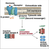
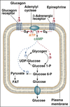
Hormonal signals that activate PKA include Glucagon and Epinephrine.

Phosphorylase kinase (regulatory enzyme)

&

Glycogen phosphorylase (regulated enzyme of glycogenolysis)

are phosphorylated in response to hormonal signals that are transduced via cAMP which activates protein kinase A.

PKA also results in the inhibition of phosphoprotein phosphatase-1 by phosphorylating and activating protein phosphatase inhibitor (A-form).

This enzyme is used to maintain the level of phosphorylation until hormone signal changes.

Active protein kinase A has a short half-life because seperation of regulatory and catalytic subunits revealed PEST sequences which marks protein for degradation.

88
Q

Glucagon

A

Peptide hormone released from the alpha-cells of the pancreas when blood glucose is low.

Glucagon binds to plasma membrane receptors on liver cells but not muscle.

Stimulates glycogen degradation via PKA-mediated activation of phosphorylase kinase.

Activated during periods of fasting thus making glucose available to tissues.

89
Q

Epinephrine

A

Hormoned released by the adrenal medulla in response to physiological stress.

Epinephrine binds to β-adrenergic receptors on the plasma membrane of both liver and muscle cells.

Stimulates glycogen degradation via cAMP and PKA mediated phosphorylase kinase activation.

90
Q

Glycogen Synthase

Covalent Regulation

(Control of glycogen synthesis)

A

Glycogen synthase exists in two forms:

A or active form: dephosphorylated

B or inactive form: phosphorylated

Several kinases phosphorylate glycogen synthase A to the inactive B form including:

  • cAMP dependent PKA*
  • Phosphorylase kinase*
  • Glycogen synthase kinase-3*
  • AMP-dependent kinase*

B-form converted back into the active A-form by

Phosphoprotein phosphatase-1.

PKA also results in the inhibition of phosphoprotein phosphatase-1 by phosphorylating and activating protein phosphatase inhibitor (A-form).

91
Q

Reciprocal Regulation of Glycogen

Synthesis & Degradation

A

cAMP regulates glycogen metabolism through the simultaneous activation of glycogenolysis and inhibition of glycogenesis.

Also displays amplification: 1 hormone activates many adenyl cyclase, makes many cAMP, each activates many PKA, each phosphorylates many phosphorylase kinase, each phosphorylates many phosphorylase ect.

92
Q

Insulin

A

Released by pancreas in response to high blood glucose levels.

Has the opposite affect of glucagon/epinephrine.

Activates glycogen synthesis and inhibits degradation in liver and muscle.

  1. Promotes inhibition of several protein kinases and activation of phosphoprotein phosphatase.
    • Causes subsequent dephosphorylation of glycogen synthase (activating) and dephosphorylation of phosphorylase kinase and phosphorylase (inactivating).
  2. Promotes conersion of cAMP to 5’ AMP by activating a phosphodiesterase.
    • Causes subsequent decrease in active PKA.
93
Q

Allosteric Regulation of Glycogen Synthesis

A
  • Allosteric effectors are superimposed onto covalent regulation in order to meet the needs of the tissue.
  • Enzymes of glycogen metabolism including glycogen phosphorylase kinase, glycogen phosphorylase, and glycogen synthase are regulated in an allosteric effectors acting in a non-covalent manner.
    • ​Postive allosteric effectors bind to a regulatory site on the R (active) form of the enzyme stabilizing it and pulling the equilibrium towards the R-form.
    • Negative allosteric effectors bind to the T (inactive) form and stabilizes that pulling equilibrium towards T-form.
  • Effectors include:
    • Ca2+ and AMP which are signs of low energy
    • Glucose and glucose-6-phosphate which are signs of high energy
94
Q

Calcium Mediated

Regulation of Glycogen Metabolism

A

Released in times of energy need.​

Ca2+ binds to the calmodulin-like δ-subunits of dephosphorylated (B-form) phosphorylase kinase causing conformational change which activates the catalytic γ-subunits in the absence of phosphorylation ⇒ stabilizes the R-state

Ca2+ also required for maximal activation of phosphorylase kinase a.

Phosphorylase kinase subsequently phosphorylates and inhibits glycogen synthase.

End result of a rise in [Ca2+]in​ is increased degradation and decreased synthesis of glycogen.

  • Contracting muscle
    • Ca2+ released in in response to nerve impulses.
    • [Ca2+]in​ activates sarcoplasmic glycogenolysis by activating phosphorylase kinase.
  • Liver cells
    • Epinephrine binds to α-adrenergic receptors, activating phospholipase-C, and generating IP3 and DAG from PIP2.
      • IP3 causes release of Ca2+​ from the SER.
        • [Ca2+]in​ activates glycogenolysis by activating phosphorylase kinase⇒ activates glycogen degradation.
      • DAG activates PKC which phosphorylates and inactivates glycogen synthase ⇒ inhibites glycogen synthesis.

Side note: Ca2+​ also activates mitochondrial events:

  • Activates PDH phosphatase thereby activating PDH and pyruvate degradation.
  • Allosteric effector of isocitrate dehydrogenase and α-ketoglutarate dehydrogenase thereby activating TCA cycle.
95
Q

AMP Mediated

Regulation of Glycogen Metabolism

A
  • AMP to ATP ratio reflects the energy state in muscle cells.
    • Increase in AMP signals low energy and need for glycogen degradation.
  • AMP functions as an allosteric activator of muscle phosphorylase b.
    • Directly activates the dephosphorylated form of myophosphorylase b.
96
Q

Glucose Mediated

Regulation of Glycogen Metabolism

A
  • When glucose is plentiful, heptatic glycogenolysis is decreased by glucose itself acting as an allosteric inhibitor of hepatic phosphorylase a.
  • The glucose-6-phosphate fromed from glucose is an allosteric activator of glycogen synthase b in both liver and muscle, thus increasing glycogenesis.
    • For this reason, glycogen synthase b is sometimes designated “D” because it is dependent on glucose-6-phosphate for activity while synthase a is designed “I” for independent.
97
Q

Hypoxic Response

A
  • Under normal oxygen conditions:
    • Prolyl hydroxylase domain (PHD) enzyme hydroxylates two prolines in the oxygen-dependent degradation domain (ODDD) of HIF-1α
      • Rxn requires Fe2+ and Vit C
      • Hydroxylated HIF-1α is a candidate for von-Hippel-Lindau (VDL) protein which ubiquitonates it
      • Ubiquinated HIF-1α degrated by proteasomes
    • Factor Inhibiting HIF-1 (FIH) hydroxylates C-domain of HIF-1α
      • Hydroxylation prevents binding of HIF-1α transcription coactivator p300
  • Under hypoxic conditions:
    • PHD and FIH enzymes inactive
    • HIF-1α has NSL which allows entry into nucleus
    • Dimerizes with HIF-1β
    • HIF-1 response elements (HREs)
    • Complex binds to HIF-1 response elements (HREs)
      • Upregulates vascular endothelial growth factor (VEGF) and other factors to stimulate angiogenesis
      • Upregulates erythropoietin (EPO) to stimulate erythropoiesis
      • Transcriptionally re-programs glycolysis to increase glucose uptake and increase the output of ATP.
      • Up-regulation of lactate dehydrogenase to allow for the regeneration of NAD+
98
Q

GSD Type Ia

A

Von Gierke Disease

  • Glucose-6-phosphatase deficiency
    • Liver and kidney
  • Severe fasting hypoglycemia hallmark
  • Major Findings
    • Hepato/renomegaly
    • Fasting hypoglycemia
    • Lactic acidemia
    • Hyperuricemia
    • Hyperlipidemia
99
Q

GSD Type Ib

A

Von Gierke Disease

  • ER glucose-6-phosphate transporter deficiency
    • Liver and kidney
  • Recurrent infections as a result of neutropenia
  • Major Findings
    • Hepato/renomegaly
    • Fasting hypoglycemia
    • Lactic acidemia
    • Hyperuricemia
    • Hyperlipidemia
100
Q

GSD Type II

A

Pompe Disease

aka

Generalized Glycogenosis

  • Lysosomal acid α-glucosidase deficiency
  • Infantile, juvenile, and adult-onset forms
  • Affects all organs but skeletal/cardiac most
  • Cardiomegaly/myopathy in infantile forms
  • Muscle weakness in later forms
  • Enzyme replacement therapy has reduced mortality
101
Q

GSD Type III

A

Cori Disease

aka

Limit Dextrinosis

  • Debranching enzyme deficiency (both actions)
  • IIIa : affects liver and muscle
  • IIIb: affects liver only
  • Milder hepatomegaly
  • Muscle weakness
  • Accumulated glycogen has abnormal structure with shorter chains
  • May cause liver fibrosis or cirrhosis
102
Q

GSD Type IV

A

Andersen Disease

aka

Amylopectinosis

  • Branching enzyme deficiency in liver
  • Progressive hepatomegaly
  • Accumulated glycogen has abnormal structure with longer chains and no branches
  • Progressive liver cirrhosis in infantile form can be lethal
103
Q

GSD Type V

A

McArdle Disease

  • Muscle glycogen phosphorylase deficiency
  • Infantile and Adult forms
  • Exercise intolerance and muscle cramps
  • Symptoms usually first appear in adolescence
  • Failure of blood lactate to rise after anaerobic exercise
104
Q

GSD Type VI

A

Hers disease

  • Liver glycogen phosphorylase deficiency
  • Hepatomegaly
  • Benign in general
  • Mild fasting hypoglycemia
105
Q

GSD Type VII

A

Tarui Disease

  • Muscle PFK-1 deficiency
  • Affects muscle and RBC’s
  • More severe exercise intolerance and muscle cramps
  • RBC’s show some percentage of normal activity